The Cardiovascular System Flashcards

1
Q

WHEN DOES A MYOCARDIAL INFARCTION OCCUR?

A

When blood circulation to the muscle of the heart itself is blocked

2
Q

WHAT IS DIFFUSION?

A

The net movement of solute down a concentration gradient, brought about by the innate, randomly-directed jumping around of the solute due to its thermal energy

3
Q

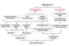
FUNCTION OF THE CARDIOVASCULAR SYSTEM

  • Delivery of O2 and nutrients to each cell
  • Removal of CO2 and waste products from each cell
  • Communication between organs through transport of hormones and other extracellular mediator
  • Temperature regulation
  • Crucial hydrodynamic device in sexual reproduction!
A
4
Q

WHAT THREE FACTORS DETERMINE BLOOD PRESSURE?

A

Blood pressure is determined by three main factors – cardiac output (pumping of the heart), the blood vessels or vasculature which not only carry the blood but are responsible for the resistance that creates the blood pressure and the various fluid compartments.

5
Q

Cardiovascular circulation can be divided into the systemic and pulmonary (lung) circuits with the heart as the central pump

A
6
Q

WHAT TWO CIRCUITS IS CARDIOVASCULAR CIRCULATION DIVIDED INTO?

A

Systemic and Pulmonary (lung)

7
Q

WHAT IS THE COMPOSITION OF BLOOD?

A
8
Q

BLOOD DISTRIBUTION AT REST

At rest….most blood is flowing through abdominal organs and _.

Majority of pumping is in parallel which means that all flow through organs is not linked. Some exceptions including intensines which run into liver….carry food. However if blood flow is occulled then reduction in blood to liver

Heart flow is quite small and any small change in flow and therefore oxygen can lead to reduce O2 and pain (called angina).

A

Kidneys

9
Q

AT REST, WHERE DOES MOST BLOOD FLOW THROUGH?

A

Abdominal organs

Kidneys

10
Q

BLOOD VESSELS CONTAIN MULTIPLE CELL LAYERS THAT REGULATE THEIR FUNCTION

Lumen

Endothelial cells (Tunica Intima)

Smooth muscle cells (Tunica Media)

Connective Tissue (Tunica Adventita)

All blood vessels contain endothelial cells but vary in the _ of the smooth muscle and connective tissue.

A

Thickness

11
Q

BLOOD VESSELS INVOLVED IN THE CIRCULATION

A
12
Q

THICH LAYER OF ELASTIC SMOOTH MUSCLE ACTS AS A PRESSURE RESERVOIR

A
13
Q

ARTERIOLES CONTAIN A THIN MUSCULAR WALL AND SMALL LUMEN

Contraction of the smooth muscle regulates the diameter of the lumen:

  • Determine blood flow to organs
  • Major determinant of mean arterial pressure
A
14
Q

CAPILLARIES HAVE A SINGLE LAYER OF ENDOTHELIAL CELLS

Exchange of nutrients, oxygen and _ across the capillary wall but NOT proteins.

Intercellular clefts and fused vesicles channels assist the exchange.

A

Waste

15
Q

WHAT SUBSTANCES ARE EXCHANGED ACROSS CAPILLARY WALLS?

A

Nutrients

Oxygen

Waste

16
Q

THE SKELETAL MUSCLE PUMP

Return of venous blood to the heart is facilitated by valves and the skeletal muscle pump.

Veins run close to the _ muscle .

A

Skeletal

17
Q

SUMMARY OF STRUCTURE AND FUNCTION OF BLOOD VESSELS

A
18
Q

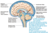
OVERALL STRUCTURE OF THE HEART

A
19
Q

MAJOR BLOOD VESSELS TO THE HEART

A
20
Q

INTERNAL STRUCTURE OF THE HEART

A
21
Q

WHAT DO VALVES DO?

A

Prevent the backflow of blood (ensures that blood flows in one direction).
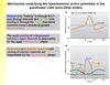
22
Q

BLOOD FLOW

A
23
Q

BLOOD SUPPLY TO THE HEART

  • Heart is has an extensive network of blood vessels supplied with oxygenated blood via the coronary arteries.
  • Coronary arteries branch off the _ (coming from left ventricle)
  • Most deoxygenated blood drains back into the right atrium via a single vein (coronary sinus)
A

Aorta

24
Q

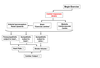
CORONARY ARTERIAL DISEASE CAN LEAD TO ISCHEMIA AND HEART ATTACKS

Coronary Arterial Disease

  • insufficient blood flow (ischemia) is associated with chest pains (angina) often radiating down left arm
  • severe blockage leads to damage (death) of the heart region and myocardial infarction or heart attack
  • ventricular fibrillation and death (heart attack)

Causes of Coronary Arterial Disease

  • Atherosclerosis (thickening of the coronary arteries)
  • Blood clot (coronary thrombosis)
  • Drugs
  • Surgery
A
25
Q

WHAT DO YOU CALL A BACTERIAL INFECTION IN THE CEREBRAL SPINAL FLUID?

A

Meningitis

26
Q

CEREBRAL SPINAL FLUID (CSF)

  • CSF cushions the brain against damage (brain is floating in CSF).
  • CSF produced in specialised epithelial cell called choroid _ (500ml/day)
  • Circulation around brain and spinal cord driven by changes in circulation, respiratory and posture.
  • Passes into vein via valves at the top of the skull (arachnoid villus)
  • Bacterial infection in CSF is called meningitis (increased pressure in brain leading to seizures and loss of consciousness)
A

Plexus

27
Q

WHERE IS CSF PRODUCED?

A

In a specialised epithelial cell called the choroid plexus

28
Q

BLOOD VESSELS AND BLOOD-BRAIN BARRIER

  • Blood vessels ‘dive’ down into the brain.
  • Capillaries contain tight junction and are less permeable to many substances (blood brain barrier)
  • Difficult to get drugs and proteins into brain.
  • Exception is lipophilic molecules such as anaesthetics, alcohol etc .
  • Brain has no stored glycogen and requires constant supply of glucose and oxygen (damage within minutes) – 15% of resting blood flow
  • Loss of blood supply and death of neurons - stroke
A
29
Q

WHAT CAUSES A STROKE?

A

Loss of blood supply and death of neurons in the brain

30
Q

TWO MAJOR FLUID COMPARTMENTS: EXTRACELLULAR AND INTRACELLULAR

THIS OCCURS IN THE CAPILLARIES

A
31
Q

HOW ARE THE VOLUMES IN THESE COMPARTMENTS MAINTAINED?

A
32
Q

WHAT IS OSMOSIS?

A

Net diffusion of water across a selectively permeable membrane from a region of high water concentration to one that has a lower water concentration (low particle concentration to high particle concentration)

33
Q

OSMOLES

One osmole (osm) = 1 mole (6.02 x 10²³ ) of solute particles in 1L.

FOR EXAMPLE:

1 M glucose = 1 Osm

1 M NaCl = 2 Osm
1 M MgCl2 = 3 Osm

Osmolarity is INDEPENDENT of molecular weight

A
34
Q

CELLULAR MEMBRANES

  • Permeable to water
  • Impermeable to solutes (ions) such as Na+, Cl-, K+ etc
  • Osmosis determines distribution of water (i.e. size of intracellular and extracellular compartments)
A
35
Q

WHAT IS OSMOTIC PRESSURE?

A

The pressure required to prevent osmosis

36
Q

WHAT WOULD HAPPEN IF YOU PUT A CELL INTO A HYPOTONIC SOLUTION?

A

The cell would swell (due to water moving into the cell).

37
Q

WHAT WOULD HAPPEN IF YOU PUT A CELL INTO A HYPERTONIC SOLUTION?

A

The cell would shrink (due to water moving out of the cell)

38
Q

WHAT IS THE COLLOID OSMOTIC PRESSURE?

Capillary membrane is semi-permeable.

  • Permits diffusion of ions, water, oxygen, nutrients and waste
  • NOT PROTEINS.
  • Pressure exerted by the higher levels of protein in the plasma compared with the interstitial fluid= draws water back into plasma by osmosis (absorption)
  • 28mmHg (plasma) – 3mmHg (interstitium)

= 25mmHg

A
39
Q

WHAT IS HYDROSTATIC PRESSURE?

A

The force exerted by the blood on the capillary walls.

Hydrostatic pressure drives blood from plasma into interstitial space

40
Q

CAPILLARY NET FILTRATION PRESSURE VARIES BETWEEN THE ARTERIAL AND VENOUS END OF THE CAPILLARIES

A
41
Q

BULK FLOW OF FLUID FROM PLASMA

A
42
Q

LYMPHATIC SYSTEM

  • Lymph system is parallel vascular system with two major functions:
  • Draining fluid from the tissues and returning to the cardiovascular system
  • Maintenance of the immune response
A
43
Q

WHAT ARE THE TWO MAJOR FUNCTIONS OF THE LYMPH SYSTEM?

A
  • Draining fluid from the tissues and returning to the cardiovascular system
  • Maintenance of the immune response
44
Q

LYMPHATIC SYSTEM: DRAINAGE

  • Fluid (plasma) passes (8L/day) from blood into the interstitial area (surrounds cells in the tissues)
  • Collects fats from the intestines/liver and deposits into veins
  • Excess fluid passes into lymph capillaries, through lymph nodes (detection of infection) before passing back to blood stream at the neck (largest is thoracic duct that drains into subclavian vein)
  • Lymph vessels contain valves and fluid is forced along by action of muscles and breathing (respiration). Larger lymph vessels are surrounded by smooth muscle that contract spontaneously and driven by pacemaker cells (~ heart)
A
45
Q

LYMPHATIC SYSTEM: IMMUNITY

  • Lymph fluid contains white immune blood cells (lymphocytes, macrophages, dendritic cells)
  • Collects antigens (proteins produced by pathogens)
  • Antigens recognised by B-lymphocytes in lymph nodes leading to activation of immunity.
  • B-cell proliferate to produce antibodies. Lymph nodes also contain multiple other immune cells (swelling can occur)
A
46
Q

HEART FAILURE

Excessive kidney retention of water
Increased arteriolar resistance

High venous pressure

A
47
Q

DECREASE IN COLLOID OSMOTIC PRESSURE

Reduction in plasma proteins

Loss of proteins in urine (kidney failure)

Loss of protein in denuded skin areas (burns)

Malnutrition

A
48
Q

WHAT IS INTRACELLULAR OEDEMA?

A

Depression of metabolic systems of the tissues and lack of adequate nutrition to the cells e.g. ischaemia: reduced activity of Na+ pumps leads to accumulation of Na+ in cells, causing osmosis of water into cells

49
Q

WHAT IS IT THAT DRIVES HEART RATE?

A

Heart rate is driven by waves of electrical activity that induce the cardiac muscles to contract

50
Q

HEART- BLOOD FLOW

A
51
Q

COMPONENTS OF THE CONDUCTION SYSTEM OF THE HEART

If you cut the nerves the heart continues to beat …..Shows that heart has its own intrinsic beat.

Heartbeat is driven by specialised myocytes composed of the sino-arterial node (at the top of the right atrium) and the artrioventricular node.

AV node connects atria and ventricles and delays the signal for 0.1ms….allow atrial to contract before ventricle

Bundles of his are fast conducting myocytes that connect to the Purkinje fibers. Purkinje fibers are very wide allowing rapid conduction throughout the ventricle and simultaneous contraction.

A
52
Q
A
53
Q

WHAT ARE THE BUNDLE OF HIS?

A

Fast conducting myocytes that connect to the Purkinje fibers. Purkinje fibers are very wide allowing rapid conduction throughout the ventricle and simultaneous contraction.

54
Q

SEQUENCE OF CARDIAC EXCITATION

Delay at AV node allows atria to contract before ventricles

A
55
Q

WHAT CAUSES THE PACEMAKER CELLS OF THE S-A NODE TO TRIGGER AN ACTION POTENTIAL?

Low resting membrane potential (-60 to -70 mV)

Na+ leakage that leads to depolarization

Graph for the SA node cell

A
56
Q

MECHANISM UNDERLYING THE ‘SPONTANEOUS’ ACTION POTENTIALS IN THE PACEMAKER CELLS (SINO-ATRIAL NODES)

A
57
Q

MECHANISM OF CONTRACTION OF THE VENTRICULAR CARDIOCYTES

A
58
Q

WHAT IS IT THAT REGULATES THE CONTRACTION OF CARDIAC MUSCLES?

A

Calcium ions

59
Q

HOW DOES CALCIUM PRODUCE CONTRACTION OF THE CARDIAC MUSCLES?

The entry of extracellular calcium ions causes the release of calcium from the sarcoplasmic reticulum, the source of about 95% of the calcium in the cytosol.

A
60
Q

WHAT IS THE REFRACTORY PERIOD?

A

Time required before it is possible to re-stimulate muscle contraction

61
Q

Cardiac muscle has a prolonged refractory period which allows for ventricles to fill with blood prior to pumping.

A
62
Q

WHAT SYSTEM REGULATES THE SINO-ATRIAL?

A

The autonomic nervous system

63
Q

WHAT HAPPENS WHEN YOU STIMULATE THE VAGUS NERVE?

A

Heart rate decreases

64
Q

HEART RATE REGULATION

Sympathetic system innervates the WHOLE heart

Parasympathetic innervates just the SA and AV nodes

Both systems are tonically active but parasympathetic dominants at rest

A
65
Q

HEART RATE REGULATION: DECREASING HEART RATE

Parasympathetic and sympathetic nervous system regulate the rate of depolarisation in the sino-atrial node

A
66
Q

HEART RATE REGULATION: INCREASING HEART RATE

Parasympathetic and sympathetic nervous system regulate the rate of depolarisation in the sino-atrial node.

Both the parasympathetic and sympathetic nerves are tonically active.

At rest – parasympathetic dominates and heart rate reduced from 100 to 70

A
67
Q

WHAT DO YOU CALL A LOW HEART RATE?

A

Bradycardia

68
Q

WHAT DO YOU CALL A FAST HEART RATE?

A

Tachycardia

69
Q

WHAT ARE THE EVENTS THAT OCCUR TO DECREASE HEART RATE?

A
70
Q

WHAT ARE THE EVENTS THAT OCCUR TO INCREASE HEART RATE?

A
71
Q

WHAT DOES AN ELECTROCARDIOGRAM DO?

A

Measures the electrical activity of the heart.

72
Q

EINTHOVEN’S TRIANGLE

A
73
Q

AN ECG IS A SUMMATION OF THE SPREAD OF ACTION POTENTIALS THROUGH THE VARIOUS SECTIONS OF THE HEART.

TRUE OR FALSE?

A

TRUE

74
Q

ELECTRICAL EVENTS OF THE CARDIAC CYCLE

A
75
Q

SUMMARY OF THE MEANING OF THE DEFLECTIONS OF THE ELECTROCARDIOGRAM

P - Atrial depolarization

QRS - Ventricular depolarization

T - Ventricular repolarization

PQ segment – Atrial contraction

QT segment – Ventricular contraction

A
76
Q

ECG IS A POWERFUL TOOL FOR DIAGNOSING HEART DISEASE

Provides valuable information concerning the electrical but NOT _ activity of the heart

Heart rate

Heart rhythm

Disturbances of rhythm and conduction

(arrhythmia, pacemaker)

Conduction velocity

Anatomical orientation of the heart

Relative size of chambers

Condition of tissue within the heart

Damage to the myocardium

Influence of certain drugs

A

Mechanical

77
Q

AN ECG PROVIDES VALUABLE INFORMATION CONCERNING THE MECHANICAL ACTIVITY OF THE HEART BUT NOT ELECTRICAL.

TRUE OR FALSE?

A

FALSE

Provides electrical but not mechanical

78
Q

WHAT IS THE RESULT OF VENTRICULAR FIBRILLATION, AND WHAT IS IT CAUSED BY?

A

RESULT:

  • Random Firing of hearts
  • Fibrillating ventricles cannot pump blood
  • Fatal after a few minutes

CAUSED BY:

  • Myocardial Infarction (heart attack)
  • Electrical shock
  • Drug intoxication
  • Impaired cardiac metabolism
79
Q

WHAT IS THE TREATMENT FOR VENTRICULAR FIBRILLATION?

A

CPR followed by electronic defibrillator

80
Q

HOW DO YOU CALCULATE CARDIAC OUTPUT?

A

Heart rate x Stroke volume

81
Q

HEART STRUCTURE

A
82
Q

WHAT TWO PHASES CAN THE CARDIAC CYCLE BE DIVIDED INTO, AND WHAT HAPPENS IN EACH OF THESE PHASES?

A

Systole – ventricular contraction and blood ejection

Diastole – ventricular relaxation and blood filling

.

83
Q

BLOOD PRESSURE IS INCREASED AS IT PASSES FROM ARTERIES TO VEINS.

TRUE OR FALSE?

A

FALSE

It decreases

84
Q

STAGE 1: SYSTOLE- VENTRICULAR CONTRACTION AND BLOOD EJECTION

A
85
Q

STAGE 2: DIASTOLE- VENTRICULAR RELAXATION AND BLOOD FILLING

A
86
Q

SEARCH UP HOW THE HEART WORKS (THE SERIES OF CONTRACTIONS AND RELAXATION SEQUENCE)

A
87
Q

WHAT CAUSES THE HEART SOUNDS THAT YOU HEAR THROUGH A STETHOSCOPE?

A

The opening and closing of valves

88
Q

HEART SOUNDS THROUGH A STETHOSCOPE

1st sound – closure of the atrioventricular (AV) valves ‘lub’- onset of systole (contraction)

2nd sound – closure of pulmonary and aortic valves ‘dub’- onset of diastole (relaxation)

A
89
Q

WHAT IS A SEPTAL DEFECT?

A

A hole in the heart

90
Q

WHAT IS STENOSIS?

A

Narrowed vessels

91
Q

REGULATION OF STROKE VOLUME

A
92
Q

FACTORS THAT INFLUENCE THE END DIASTOLIC VENTRICULAR VOLUME (PRELOAD)

A
93
Q

VENTRICLES CONTRACT WITH MORE FORCE WHEN THEY CONTAIN MORE BLOOD.

TRUE OR FALSE?

A

TRUE

94
Q

FRANK-STARLING MECHANISM MAINTAINS BALANCE BETWEEN RIGHT AND LEFT SIDES OF THE HEART- STOP ACCUMULATION OF BLOOD IN THE LUNGS

  • If there is an _ in venous return to right ventricle then there will also be an _ in contraction= more blood to lungs
  • If there is an increase in venous return to left ventricle then there will also be an increase in contraction= higher stroke volume= blood doesnt accumulate in lungs
A
95
Q

OVERALL REGULATION OF CARDIAC OUTPUT

A
96
Q

WHAT IS CONGESTIVE HEART FAILURE (CHF) CHARACTERISED BY?

A

Reduced cardiac output

97
Q

WHAT ARE SOME SYMPTOMS OF CONGESTIVE HEART FAILURE?

A

Tiredness

Shortness of breath

Coughing

Swelling in ankles and legs

Excess of fluid in lungs (Pulmonary edema)

Swollen abdomen

Excess of fluid around the lungs (Pleural effusion)

98
Q

CHRONIC LEFT VENTRICULAR FAILURE (MOST COMMON)

RISK FACTORS

  • Coronary artery disease leading to ischaemic heart disease and myocardial infarction (~ 65%) – systolic dysfunction
  • Hypertension (high blood pressure) (~ 10%) – diastolic dysfunction
  • Cardiomyopathy (viral infection, heavy drinking)
  • Getting old!!!
A
99
Q

HEART FAILURE: SYSTOLIC DYSFUNCTION (VENTRICULAR EJECTION)

Myocardial infarction –> Damage to heart muscle –> _ in ventricular contractility –> Decrease in stroke volume

A

Decrease

100
Q

HEART FAILURE: DIASTOLIC DYSFUNCTION (VENTRICULAR FILLING)

High Blood Pressure (Hypertension) –> Increase in arterial _ –> Increase in cardiac resistance –> _ in ventricular muscle (hypertrophy) –> stiffening of ventricular wall –> End-diastolic ventricular volume –> _ in stroke volume.

A

Pressure

Increase

Decrease

101
Q

HOW DO YOU CALCULATE MEAN ARTERIAL PRESSURE (BLOOD PRESSURE)?

A

Cardiac output x Total Peripheral Resistance

102
Q

WHAT DOES POISEUILLE’S LAW DESCRIBE?

A

Describes the factors that determine the flow through a tube in terms of pressure, flow and resistance

103
Q

POISEUILLE’S LAW

(THIS IS ABOUT TUBES, NOT SPECIFICALLY BLOOD VESSELS)

A
104
Q

POISEUILLE’S LAW: BLOOD VESSELS

Flow (Q) is directly proportional to the pressure gradient (P) vessel and the radius4 (r4) of the tube and inversely proportional to the length (L) of the vessel and to the viscosity (η) of the fluid.

A
105
Q

THE CARDIOVASCULAR SYSTEM CAN BE EQUATED TO POISEUILLE’S LAW

Flow (Q) is proportional to the pressure gradient (ΔP ) and inversely proportional to the resistance:

Q = ΔP / R

Flow (Q) = cardiac output (L/min)

Pressure gradient (ΔP ) = Blood pressure (mmHg)

Resistance (R) = Peripheral resistance

Cardiac output = Blood pressure/Peripheral resistance

Blood pressure = Cardiac Output x Peripheral resistance

A
106
Q

HOW CAN YOU CALCULATE CARDIAC OUTPUT NOT USING HEART RATE AND STROKE VOLUME?

A

Cardiac output = Blood pressure/Peripheral resistance

107
Q

BLOOD IS PROPORTIONAL TO PRESSURE DIFFERENCE, BUT INDEPENDENT OF THE ABSOLUTE PRESSURE.

TRUE OR FALSE?

A

TRUE

108
Q

FLOW IS INVERSELY PROPORTIONAL TO THE LENGTH AND VISCOSITY

Length (L)

  • Longer the tube = more resistance = less flow
  • remains constant and therefore does NOT control blood flow

Viscosity (η)

Higher the viscosity (i.e. treacle) = more resistance = less flow

Viscosity is related to the hematocrit (red blood cells/erythrocytes)

  • Dehydration
  • High altitude
  • Erythropoietin (EPO) – stimulate production RBC

Remains constant under physiological conditions and therefore does NOT control blood flow

A
109
Q

WHAT IS THE MOST IMPORTANT FACTOR/CHARACTERISTIC OF A VESSEL IN DETERMINING (FLOW) RESISTANCE?

A

The radius of the vessel

110
Q

RADIUS OF THE VESSEL (THE MOST IMPORTANT FACTOR WHEN DETERMINING (FLOW) RESISTANCE)

Radius (r)

  • Caused by the _ of the fluid against the vessel wall
  • The effect of changes in the radius is raised to the power of 4
  • Reducing radius by two fold from 2 to 1 reduces flow by sixteen fold (i.e. resistance increased sixteen fold)
  • Possible to regulate the radius of blood vessels using smooth muscle.
A

Friction

111
Q

WHAT IS TOTAL PERIPHERAL RESISTANCE?

A

The sum of the resistance of all the blood vessels and determines blood pressure

112
Q

VASCULAR SMOOTH MUSCLE

A
113
Q

WHAT IS TOTAL PERIPHERAL RESISTANCE PRIMARILY REGULATED BY?

A

Arterioles

114
Q

WHERE DOES BLOOD MOSTLY FLOW THROUGH WHEN AT REST?

A

Abdominal organs

Kidneys

115
Q

ARTERIOLES DETERMINE BLOOD FLOW TO DIFFERENT ORGANS.

TRUE OR FALSE?

A

TRUE

116
Q

DETERMINING BLOOD PRESSURE

A
117
Q

MEASURING ARTERIAL BLOOD PRESSURE

A
118
Q

WHAT CAUSES THE PULSE?

A

Vibration of the arteries caused by the ejection of the blood from the heart (left ventricle) into the systemic circulation.

119
Q

WHAT IS THE TYPICAL RESTING PULSE RATE?

A

70/min

120
Q

WHAT IS THE TYPICAL PULSE RATE AFTER EXERCISE?

A

220/min

121
Q

WHAT ARTERY IN THE WRIST CAN YOU MEASURE YOUR PULSE FROM? WHAT ABOUT IN YOUR NECK?

A

Wrist= radial artery

Neck= corotid artery

122
Q

HOW DO YOU CALCULATE PULSE PRESSURE?

A

It’s the difference between your systolic and diastolic pressure (e.g. 120-80=40mmHg)

123
Q

MEAN ARTERIAL PRESSURE (MAP)

Mean arterial pressure (MAP) is the average pressure over the cycle

  • since diastole is twice as long as the systole

= diastolic pressure (DP) + 1/3 pulse pressure (PP)

= 80 + 1/3 (40) = 93mmHg

A
124
Q

BLOOD PRESSURE IS INFLUENCED BY HEIGHT

A
125
Q

BLOOD PRESSURE AND POSTURE

Heart = ‘zero pressure point’

Standing

Increased pressure of mean arterial blood pressure in legs (from 90 to 195 mmHg) leads to pooling of blood in the veins

  • Feet swelling on flights
  • Fainting in stationary soldiers

However, since arterial and venous blood are at the same height.

Blood gradient Delta-P = constant = blood flow continues

A
126
Q

HYPERTENSION IS ASSOCIATED WITH INCREASED MORTALITY

A
127
Q

WHICH RECEPTORS REGULATE MEAN ARTERIAL PRESSURE (MAP)?

A

Baroreceptors

128
Q

WHERE ARE ARTERIAL BARORECEPTORS LOCATED?

A

In the aortic arch and carotid sinuses

129
Q

ARTERIAL BARORECEPTORS

Baroreceptor neurons function as sensors in the homeostatic maintenance of mean arterial pressure (MAP).

Located in the aortic arch and _ sinuses where they monitor pressure..

Located in thinner walls that can be stretched (by pressure)

Baroreceptor nerve endings are sensitive to stretch and _ in artery

A

Carotid

Pressure

130
Q

ARTERIAL BARORECEPTORS ACT AS PART OF A NEGATIVE FEEDBACK PATHWAY THAT REGULATES MEAN ARTERIAL PRESSURE VIA THE BRAIN (CENTRAL CONTROL)

A
131
Q

AUTONOMIC NERVOUS SYSTEM

Sympathetic nerves

  • mediated by noradrenaline via beta-adrenergic receptors
  • alpha-1-adrenergic receptors in arterioles (except skeletal muscle which is a2-adrenergic )
  • increase cardiac output and peripheral resistance

Parasympathetic nerves

  • mediated by acetycholine via muscarinic receptors
  • inhibit heart rate
A
132
Q

STANDING UP (ORTHOSTASIS)

  • Standing and effect of gravity leads to blood pooling in legs (up to 500ml)
  • Reduced blood volume and lowering of central venous pressure
  • Reduced venous return, reduced end-diastolic pressure, reduced stroke volume and reduced blood pressure
  • Baroreceptor reflex will compensate
  • Standing and effect of gravity leads to blood pooling in legs (up to 500ml)
  • Reduced blood volume and lowering of central venous pressure
  • Reduced venous return, reduced end-diastolic pressure, reduced stroke volume and reduced blood pressure
  • Baroreceptor reflex will compensate
A
133
Q

AUTONOMIC NERVOUS SYSTEM

A
134
Q

WHAT CAN CAUSE HYPOTENSION?

A

Blood loss

Loss of salts

Stress or emotions

135
Q

WHERE IS MOST BLOOD DISTRIBUTED AT REST?

A

In the veins

136
Q

BLOOD VOLUME IS AN IMPORTANT LONG-TERM REGULATOR OF MEAN ARTERIAL PRESSURE

  • Baroreceptors can regulate short term (seconds-minute) changes in blood pressure
  • Changes in blood volume regulated by the kidneys are responsible for long-term regulation of blood pressure (renin-angiotensin system)
A
137
Q
A
138
Q

WHAT THREE FACTORS REGULATE BLOOD FLOW? (ARTERIOLE RADIUS)

A

Local

Neuronal

Hormonal

139
Q

INTRINSIC (LOCAL) CONTROL OF ARTERIOLE VASODILATION AND VASOCONSTRICTION

Local factors

Myogenic Response

  • Vasoconstriction of arterioles caused by stretch of smooth muscle
  • Brain, kidney and heart – not skin

Vasodilation induced by metabolites:

  • O2 ↓, CO2 ↑,
  • H+ ↑, adenosine ↑, K+ ↑, osmolarity↑

Autocoids

  • Mainly release as a result of inflammation and bleeding
  • Histamine, bradykinin, prostaglandins etc

Endothelial cells (sheer stress)

  • Release local factors (paracrine)
  • Nitric oxide and prostacyclin (vasodilation)
  • Endothelin-1 (vasoconstriction)
A
140
Q

WHEN WOULD AUTOCOIDS MAINLY BE RELEASED?

A

As result of inflammation or bleeding

141
Q

INTRINSIC (LOCAL) REGULATION OF BLOOD FLOW

  • Increase metabolic activity leading to ACTIVE HYPEREMIA
  • Reduce blood flow in organ leading to FLOW AUTOREGULATION
A
142
Q

NEURAL CONTROL OF ARTERIOLES VASODILATION AND VASOCONSTRICTION (FROM BRAIN)

Skin

Room Temperature

•Arterioles constricted by moderate rate of sympathetic discharge

Cold, Fear or Loss of blood

  • Sympathetic discharge increased and arterioles vasocontriction
  • Divert blood to essential organs

Increased Body Temperature

  • Sympathetic discharge reduced and arterioles vasodilation
  • Blood flow to skin to facilitate body cooling
A
143
Q

HORMONAL CONTROL OF ARTERIOLE VASODILATION AND VASOCONSTRICTION

A
144
Q

WHAT HAPPENS WHEN YOUR BODY TEMPERATURE INCREASES?

A
  • Sympathetic discharge reduced and arterioles vasodilation
  • Blood flow to skin to facilitate body cooling
145
Q

WHAT HAPPENS WHEN YOU’RE COLD, IN FEAR OR HAVE LOST A LOT OF BLOOD?

A
  • Sympathetic discharge increased and arterioles vasocontriction
  • Divert blood to essential organs
146
Q

WHAT HAPPENS WHEN YOUR BODY IS AT ROOM TEMPERATURE?

A

Arterioles constricted by moderate rate of sympathetic discharge

147
Q

EXERCISE AND THE RESPONSE OF THE CARDIOVASCULAR SYSTEM

Major problems to address:

  1. Boost O2 uptake and CO2 removal (increased cardiac output)
  2. Increase blood flow to muscles, heart and skin (changes peripheral resistance)
  3. Stabilise arterial blood pressure (despite changes in cardiac output and peripheral resistance)
A
148
Q

DURING EXERCISE, WHICH PARTS OF THE BODY RECEIVE AN INCREASED BLOOD FLOW, AND VICE VERSA?

A

Increased blood flow= Skeletal muscle, skin, heart

Decreased blood flow= Kidneys, abdominal organs

149
Q

EXERCISE: DIVERT FLOW TO THE SKIN, MUSCLES AND HEART

A
150
Q

VALSALVA MANOEUVRE

TURN OVER TOO

A
151
Q

SUMMARY

A