The Acute Red Eye Flashcards

1
Q

how do contact lenses affect infectious presentation

A

delay it - act as a bandage to the eye
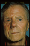
2
Q

blepharitis

A
  • lid inflammation
  • the eyes have burning, itchy red margins with scales on the lashes
  • gritty eyes
  • FB sensation
  • mild discharge
  • dry eyes
3
Q

which part of the lid is redder in anterior blepharitis

A

lid margin redder than deeper part of lid

4
Q

anterior blepharitis: seborrheic dermatitis

A

squamous

there are red lid margins, greasy sclaes on lashes, which are stuck together

dandruff

there is no ulceration

5
Q

anterior blepharitis: staphylococcal

A

ulcerative - infection involving lash follicle

there is a red lid margin

eyelashes are distorted, some trichiasis

there is matted hard crusts around lashes, with scarring and hypertrophy if long standing. removal of crusts leaves small ulcers which bleed/ooze

styes are seen

6
Q

trichiasis

A

in growing of eye lashes

7
Q

stye

A

red/tender lump caused by an infection of an oil gland of the eyelid - hordeolum

8
Q

which type of stye is seen in staphylococcal anterior blepharitis

A

hordeolum externum - abscess/infection in the glands of Moll in the lash follicle

points outwards and may cause much inflammation

9
Q

what effect does trichiasis have on the cornea

A

irritates it - corneal staining and marginal ulcers

10
Q

posterior blepharitis

A

due to meibomian gland dysfunction

secretions of the gland thicken and block the gland, there is scarring and the glands fill up with secretion

there is insipissated (dried) secretion at the gland openings

11
Q

describe the appearance of posterior blepharitis

A

redness in the deeper part of the lid, margins appear quite normal

12
Q

what type of stye is seen in posterior blepharitis

A

hordoleum internum - abscess of tarsal glands, point inwards opening onto conjunctiva and cause less of a local reaction but leave residual swelling called a chalazion/Meibomian cyst when they subside

pic: chalazion

13
Q

hordeolum internum and externum

A
14
Q

what is blepharitis a common cause of

A

secondary infections eg conjunctivitis, keratitis, episcleritis

15
Q

what is the most common manifestation of ocular rosacea (acne rosacea)

A

anterior and posterior blepharitis

16
Q

treatment of blepharitis

A

lid hygiene is essential, crusts must be cleaned off lashes

supplementary tear drops

oral doxycycline for 2-3 months

difficult to eradicate

17
Q

general symptoms of conjunctivitis

A

red and inflamed conjunctiva

eyes itch burn and lacrimate

pain on eye movement

FB sensation - gritty eye

may be photophobia

often bilateral with eyelids sticking together

18
Q

what is unaffected in conjunctivitis

A

vision (acuity), pupillary responses and corneal lustre

19
Q

chemosis

A

odema of conjunctiva

non specific sign of eye irritation

20
Q

allergic conjunctivitis

A

stringy discharge

itch

papillae present

21
Q

what is allergic conjunctivitis treated with

A

anti-histamine drops

22
Q

3 most common causes of bacterial conjunctivits in neonates

A
  • Staph. aureus
  • Neisseria gonorrhoea
  • Chlamydia trachomatis
23
Q

3 most common causes of bacterial conjunctivitis

A
  • Staph. aureus
  • Strep, pneumoniae
  • H. influenzae (especially in children)
24
Q

bacterial conjunctivitis

A

red sticky eye

purulent discharge

papillae - red, small and circular

25
treatment of bacterial conjunctivitis
often self limiting, swab eye topical ABx used, 1. **chloramphenicol** * 2. Fusidic acid* * 3. Gentamicin*
26
treatment of bacterial conjunctivitis in pregnant women
fusidic acid - chlormaphenicol can cause grey baby syndrome
27
when is chloramphenicol avoided
* history of anaplastic anaemia (can cause bone marrow aplasia - type B reaction!) * allergy
28
what is used to treat *Pseudomonas aeruginosa* bacterial conjunctivitis
not covered by chloramphenicol covered by Gentamicin
29
viral conjunctivitis
more pink than bacteiral no pus, more of a thick watery secretion often have URTI, and enlarged pre-auricular and submandibular lymph nodes not serious but v contagious red velvety apperance of conjunctiva and raised follicle (rice grains)
30
adenovirus causing viral conjunctivitis
often follows URTI
31
HSV causing viral conjunctivitis - appearance - enquiries - treatment
ask about ulcers treat with **acyclovir**
32
HZ opthalmicus - epidemiology - signs
Pain and neuralgia in the distribution of CNV1 dermatome precedes a blistering, inflamed rash often so significant it can track over to the other eye typically occurs in older adults Hutchison's sign
33
how can HZ opthalmicus present
34
Chlamydial Conjunctivitis
suspected in unresponsive, chronic conjunctivitis (may last for months) most common in newborns and young adults
35
classical Chlamydial Conjunctivitis presentation
unresponsive, chronic unilateral conjunctivitis in young adults symptoms of urethritis and vaginitis follicular conjunctivitis
36
chronic follicular Conjunctivitis
subtarsal scarring, the scarring abrades the cornea on blinking. the cornea eventually becomes opaque commonly cause by Chlamydial Conjunctivitis and trachoma
37
treatment of Chlamydial Conjunctivitis
topical oxytetracycline may need oral azithromycin for a genital infection
38
trachoma
* caused by repetitive infection with *Chlamydia Trachomatis* and most commonly seen in hot climates, spread by flies * there is lacrimation, redness and rupturing of conjunctival follicles and replacement with scar tissue * scar tissue distorts the lids and may lead to entropion * eye lashes scratch cornea - may ulcerate * can lead to blindness
39
treatment of trachoma
Treatment is with Tetracycline and oral Azithromycin. Lid surgery may be required.
40
what medication can patients typically become sensitised to
preservatives eg Benzalkonium
41
drug sensitivity
typically causes peri-orbital oedema once drug is stopped skin will subside in 2-3 weeks
42
keratitis
corneal inflammation, indicated by a hypopyon moderate to intense pain, impaired eyesight, photophobia, red eye and a gritty sensation
43
examination of keratitis
* Use of anaesthetics (eg 1% tetracaine) if photophobic * Corneal reflex * Use of fluorescein and bright blue light to examine the cornea and identify area of epithelial loss * Vascularisation * Opacity * Oedema
44
layers of the cornea
45
central vs peripheral ulcers
central tend to be infective (eg bacterial, viral, fungal) peripheral tend to be autoimmune (eg RA, hypersensitivity)
46
name 4 autoimmune causes of a corneal ulcer
**rheumatoid arthritis - 30%** SLE IBD ANCA vasculitis
47
peripheral corneal ulcer
48
symptoms of corneal ulcer
**pain - needle like and severe, due to nerve exposure due to epithelium disruption - NOTE** photophobia profuse lacrimation reduced vision red eye - circumcorneal
49
signs of a corneal ulcer
circumcorneal redness corneal reflex (reflection abnormal) corneal opacity hypopyon
50
hypopyon
collection of inflammatory cells seen in the anterior chamber - also called sterile pus
51
bacterial keratitis
round hypopyon usually only found with other corneal pathology (eg corneal ulcer) or contact lense wearer
52
which pathogen may progress rapidly
Pseudomonas
53
treatment of bacterial keratitis
hourly drops **4-quinolone (Ofloxacin)** (doesn't treat *Strep. Pneumoniae*) or Gentamicin or Cefuroxime
54
HSV keratitis
* classic **dendritic ulcer** * very painful and may relapse and recur, eventually resulting in reduced corneal sensation (as infection is in the nerves) * gritty red eyes * slight photophobia and reduced vision
55
classical history of HSV keratitis
very fatigued patient, recent cold sore
56
treatment of HSV keratitis - investigation - what to avoid
can be viewed with fluroscein and bright blue light avoid steroids!! can cause corneal melt and perforation acyclovir - topical if on the epithelium, otherwise need oral
57
adenoviral keratitis
bilateral and usually follows UTI contagious small white flecks (subepithelial infiltrates) seen may affect vision
58
treatment of adenoviral keratitis
topical ABx to prevent 2y infection steroids can speed recovery in chronic cases
59
fungal keratitis
* usually seen in contact lense wearers * Acanthamoeba or Pseudomonas aeruginosa usually * chronic, slow progessing history where the signs are more significant than the symptoms * deep severe ulcer * often history from trauma/vegetation
60
acanthamoeba
* causes a fungal keratitis almost exclusive to contact lense wearers (often from pools) * very difficult to eradicate * prevention: dont over wear/sleep/swim in contacts
61
diagnosis of keratitis
corneal scrapes for Gram stain and culture
62
typical causes of anterior uveitis
63
clinical features of anterior uveitis
pain reduced acuity significant photophobia lacrimation circumcorneal redness
64
describe the pain in anterior uveitis
dull ache, boring pain may refer to eyebrow
65
signs of anterior uveitis
ciliary (circumcorneal) injection synechiae - smal/irregular pupil (iris inflammation) hypopyon cells/flare in anterior chamber
66
treatment of anterior uveitis
*the aim of treatment is to prevent damage from prolonged inflammation leading to disruption of aqueous flow, and glaucoma and iris and lens adhesions* topical steroids eg Prednisolone forte 1% mydriatrics eg tropicamide, cyclopentolate 1%
67
episclera
layer between sclera and conjunctiva
68
episcleritis
*inflammation of episclera* relatively common and has no serious associations, often recurrent and self-limiting. bilateral in 30% often seen with inflammatory nodule Sclera may look **blue** below a focal, cone shaped wedge (thin and towards pupil) of engorged vessels that can be moved around, unlike in scleritis where they are deeper. Eye aches dully and is tender (especially over inflamed area).
69
what is associated with gout
episcleritis
70
treatment of episcleritis
self-limiting lubricants/topical NSAIDs/sometimes steroids
71
scleritis
significant, deep boring pain. has been known to wake patients up from sleep erythema - can be limited to area of eye photophobia generalized inflammation and chemosis, scleral thinning injection of deep vascular plexus causes **violaceous hue**
72
what is often assoicated with scleritis
anterior uveitis
73
how can one distinguish between episcleritis and scleritis
**phenyephrine test** (vasoconstrictor) - the episcleral vessels will blanch with it, scleritis will not
74
treatment of scleritis
oral NSAID oral steroids steroid sparing agents
75
orbital cellulitis
Divided into pre and post septal. Pre-septal cellulitis is not that severe. Post-septal cellulitis is **inflammation of eye tissues behind orbital septum,** it is sight-threatening
76
where does infection typically spread from to cause orbital cellulitis
adjacent sinuses (paranasal) or through the blood can also be an extension of a focal orbital infection or post-operative
77
if left untreated, what can orbital cellulitis cause
it can lead to the development of orbital abscesses or can spread posteriorly to cavernous sinus
78
classical presentation of orbital cellulitis
child with lid swelling, fever, orbital inflammation, decreased eye mobility (pain on eye movement) as it is often associated with paranasal sinusitis there is often mucous coming out of the nose - check the ears and pupils
79
orbital cellulitis - uni or bi lateral
inflammatory causes tend to be unilateral, whereas allergic causes tend to be bilateral
80
investigation and treatment of orbital cellulitis
CT scan to identify and drain orbital abscesses broad spectrum ABx and close monitoring abscess may require drainage
81
ABx treatment of orbital cellulitis
* ceftriaxone * flucloxacillin * metronidazole
82
Endophthalmitis
devastating infection of the inside of the eye very painful and associated with decreased vision - can be sight threatening very red eye
83
what is the most common causative organism of Endophthalmitis
Staph. Epidermidis
84
treatment of Endophthalmitis
Intravitreal amikacin/ceftazidime/vancomycin and topical antibiotics
85
what classically causes chorioretinitis in AIDS
CMV
86
toxoplasma gondii causing chorioretinitis
it is a protozoan infection, typically from cats and raw meat creates a mild flu like illness that rarely causes any further problems and is fairly common however, it can be sight threatening if it affects the macula can reactivate
87
toxocara canis casuing chorioretinitis
parasitic nematode (round worm) affects cats and dogs. is unable to replicate in humans so remains in an immature form of worm, this means it is often self limiting can form **granulomas** which can lead to irreversible vision loss
88
what is a common contaminant of contaminated bottles
Psuedomonas