Anatomy: Eye Autonomics Flashcards
describe the sensory innervation of CNV1
upper eyelid
cornea and conjunctiva
skin of root/bridge/tip of nose

name 2 branches of the CNV1 and their areas of innervation

what notch does the supraorbital nerve (CNV1) pass through
supraorbital foramen

describe the sensory innervation of CNV2 (maxillary nerve)
skin of lower eyelid
skin over maxilla
skin over ala of nose
skin/mucosa of upper lip

infraorbital nerve
what CNV2 is called after it enters the infraorbital foramen
describe the sensory innervation of CNV3 (mandibular nerve)
skin over mandible and TMJ
(apart from angle of mandible - C2, 3 spinal nerves)

what is the only branch of CNV that has motor innervation
mandibular branch - muscles of mastication
describe the corneal reflex
- involuntary blinking of eyelids, stimulated by tactile, thermal or painful stimulation of cornea
- CNV1 is the afferent branch - conducts APs from the cornea to the trigeminal ganglion, it then goes along CNV to pons
- there are central CNS connections between CNV and CNVII, which is the motor efferent branch
- causes contraction of orbicularis oculi

describe the route of sympathetic axons
originate from autonomic centres in the brain
pass down spinal cord and exit with T1-L2 nerves (thoracolumbar outflow)
travel so sympathetic chains and into all spinal nerves, and then splanchnic nerves to eventually supply organs

describe the presynaptic sympathetic axons from CNS
exit spinal cord in T1 spinal nerve and ascend within sympathetic trunk
synapse in superior cervical sympathetic ganglion

describe the postsynaptic sympathetic axons
enter internal and external carotid nerves and pass onto the surface of internal and external carotid arteries
they are carried to the organs of the head on the surface of the branches of these arteries
ophthalmic artery carries the sympathetic axons into the orbit

describe the parasympathetic outflow
all parasympathetic axons leave the CNS via cranial nerves III, VII, IX and X and via sacral spinal nerves (craniosacral outflow)

describe the course of CNIII in the cranial cavity
originates from the anterior aspect of the midbrain
passes through cavernous sinus
exits cranial cavity via superior orbital fissure and divides into superior and inferior branches

what does the superior branch of CNIII do

motor innervation to superior rectus and levator palpebrae superioris
what does the inferior branch of CNIII do
motor innervation to inferior rectus, medial rectus and inferior oblique
parasympathetic fibres to ciliary ganglion, which ultimately innervates sphincter papillae and ciliary muscles

where is the ciliary ganglion located
within the bony orbit
anteriorly to the superior orbital fissure, between the lateral rectus muscle and the optic nerve
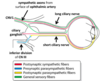
describe the sympathetic innervation with ciliary ganglion
preganglionic sympathetic fibres relay in superior cervical ganglion
postganglionic fibres are carried by long ciliary nerves (and some short) from sympathetic plexus around ICA to supply dilator pupillae muscle

describe the parasympathetic innervation with ciliary ganglion
preganglionic fibres originate from Edinger-Westphal nucleus in midbrain and run with CNIII (inferior division) to relay in ciliary ganglion
postganglionic fibres are carried by short ciliary nerves to innervate ciliary muscles and sphincter pupillae muscles

what are pre-ganglionic fibres of ciliary ganglion associated with
inferior division of CNIII

which ciliary nerves form the first part of the corneal reflex
long ciliary nerves - sensory fibres from the surface of CNV1
what are some autonomic reflexes of the eye
maximal eyelid elevation/wide eye opening
pupillary dilation/constriction to adjust light entry
focussing lens far and near vision
lacrimation reflex tear production
vestibulo-ocular reflex
stabilizes gaze on an object during head movement by turning the eyes in the opposite direction of the head
involves connections between CN VIII and CNIII, IV and VI
oculocardiac reflex
reflex bradycardia in response to tension on extraocular muscles/pressure on eye
CNS connections betwen CNV1 and CNX
does sympathetic innervation cause one to focus on far or near objects
far
parasympathetic - near


