Anatomy: The Eye and Raised ICP Flashcards
what is the normal intraocular pressure
12-22mmHg
what can cause raised ICP
brain tumour
head injury
hydrocephalus
meningitis
stroke
Monro-Kellie hypothesis
the cranial compartment is incompressible and the volume inside it is fixed - made up of brain tissue, blood and CSF
the cranium and its constituents create a state of volume equilibrium, such that an increase in the volume of one constituent must be compensated for by a decrease in the volume of another

what are the principle buffers in the IC cavity to increased volume
CSF and, to a lesser extent, blood volume
what damage can raised ICP cause in the cranial cavity
damage to tissues, shifts in tissues, herniation and constrictionof blood vessels
what are the signs and symptoms of raised ICP
- Headache – worse on awakening and may wake them from sleep, exacerbated by cough, bending, sneezing etc.
- Vomiting without nausea
- Ocular palsy
- Back pain
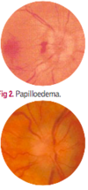
- Papilloedema
- Altered level of consciousness
what effect can raised ICP have on the eye
65-75% patients with ICP report visual problems
transient blurred vision
double vision
loss of vision
papilloedema
pupillary changes
is the effect of raised ICP on the eye uni or bilateral?
either
how is raised ICP transmitted to the eye
via the optic nerves - these are surrounded by cranial meninges, and found in the subarachnoid space
this becomes abnormally filled with CSF
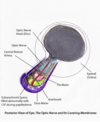
what is seen on fundoscopy of papilloedema
bulging optic disc
disc margins are blurred and in places retinal vessels are concealed, because oedema has impaired the translucency of disc tissues.
optic cup is lost
describe the 3 layers of the meninges

which nerve innervates the dura mater
sensory supply from CNV
what is enclosed between the 2 layers of the dura mater
dural venous sinuses

what do the dural venous sinuses drain to
IJV at jugular foramen
where is the jugular foramen


describe the vascualture and innervation of arachnoid mater
avascular and no innervation
arachnoid granulations
small projections of arachnoid mater into the superior sagittal sinus in dura that allow CSF to re-enter the venous circulation via the dural venous sinuses

subarachnoid space
contains circulating CSF that acts to cushion the brain
also contains blood vessels

pia mater
‘faithful mother’
thin and tightly adhered to the surface of the brain and spinal cord - follows the contours of the brain (gyri and sulci)
highly vascularised


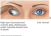
which other nerve is susceptible to damage
CNIII
what happens if CNIII is damaged
paralysis of motor innervation - LPS, SR and IR, MR, IO
paralysis of parasympathetic innervation of sphincter of pupil (ciliary ganglion and short ciliary nerve)
results in lose/slowness of pupillary light reflex, dilated pupil, ptosis and eye turned inferolaterally (down and out)
think, this is because LR and SO are not innervated by CNIII, LR pulls laterally, SO pulls down and laterally
name the septas that dura mater forms
also, diaphragma sellae over pituitary fossa
the brain can herniate through these

what is the purpose of the tentorial notch
to allow passage of brain stem
