Anatomy: The Orbit and the Eye Flashcards


where is the optic canal


how many bones make up the orbit
6
what is the thinnest bone of the optic canal
ethmoid - medial wall
where is the apex of the bony orbit located
nasal side
what bones form the orbital margins

what forms the orbital rim
combiend orbital margins
what function does the orbital rim have in respect to orbital trauma
nothing wider than diameter of orbital rim can hit the eye
describe the angle of the orbital rim

orbital blowout fracture
force on orbital rim damages medial wall (ethmoid) and orbital floor (maxilla) which are extremely thin
can damage the infraorbital NVB resulting in general sensory deficit of facial skin
typically entraps the inferior rectus - patients will complain of pain on upward gaze
which direction does a fractured zygoma tend to rotate, and what can this result in clinically
medially towards orbit floor
as the suspensory ligament of the eye attaches to the zygoma laterally, the eye may be lowered towards the orbital floor
results in diploplia (double vision)

what is the difference between notch and foramen
notch is an incomplete circle
outer eyelid:
name the 2 parts of the orbicularis oculi


describe the functions of the orbital and paplebral parts of the orbicularis oculi
orbital part closes the eye tightly
palpebral part is involved in normal blinking

what is the orbicularis oculi innervated by
CNVII
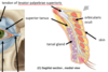
what is another name for the superior tarsal muscle (connective tissue?)
Muller’s muscle
medial eyelid layer:


what is the orbital septum made of and what is its function
sheet of fascia
helps to prevent spread of infection from superficial to deep
inner eyelid layer:
tarsal glands
also called Meibomian glands
glands embedded in the tarsal muscles, responsible for lipid secretion
they secrete a layer of oil on top of tears to stop them from evaporating


tears:
- antibacterial properties
- pH
antibacterial properties due to action of lysozyme
pH of around 7.6
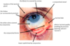
describe the conjunctiva - 2 types and where they meet
the inner surface of the eyelid is lined by palpebral conjunctiva
the eyeball is lined by bulbar conjunctiva
meet at the fornix

conjunctival fornix
connects the conjunctiva lining the inside of the eyelid with that covering the eyeball

describe the features of the palpebral conjunctiva
has follicles and papillae
contains goblet cells which secrete part of the tear film

which conjunctiva is more vascular
palpebral
describe the movement the eyelid makes when shutting
sweeping action - lateral to medial
this helps to wash debris towards the lacrimal sac
what is the iris covered by
cornea
what is the sclera covered by and what is it’s function
conjunctiva
- forms a defensive barrier to foreign bodies penetrating deep to the orbit


describe the lacrimal apparatus
lacrimal gland produces lacrimal fluid (parasympathetic CNVII)
- this washes medially over eye toward medial angle
- drains through lacimral puncta (openings in lacrimal papillae)
- goes through canaliculi to lacrimal sac
- and then nasolacrimal duct to reach inferior meatus in nose

what is the parasympathetic innervation of the lacrimal gland
CNVII


what can corneal light reflections be used for clinically
clinically to track the symmetry of bilateral eye positions/movements
what forms the outer layer of the eye
sclera and cornea

what holds 2/3 of the eye’s refractive (focusing) power
the cornea
describe the layers of the cornea

how is the transparency of the cornea maintained
the endothelium maintains transparency by pumping fluid out of the stroma

where is the cornea thickest
peripherally
what is the stroma of the cornea formed by
reguarly spaced collagen fibres
what are the outer and inner parts of the lens known as
cortex and nucleus

what forms the middle (uvea - vascular layer) of the eye
the uveal tract structures are continuous with eachother and pigmented

function of iris
controls pupil diameter
function of ciliary body
controls iris, shape of lens (ciliary muscle) and secretion of aqueous humour (ciliary epithelium)

function of choroid
- nutrition and gas exchange
- supplies the outer retina

what forms the inner layer of the eye
retina - photosensitive layer

describe the chambers of the anterior segment of the eye
anterior chamber between cornea and iris contains aqueous humour
posterior chamber between iris and suspensory ligaments contains aqueous humour

how much of the eye does the posterior segment occupy
2/3rds
what does the posterior segment of the eye contain
vitreous body/humour
- clear gel

what is a common location for floaters in the eye
vitreous body in posterior segment

cataract
clouding of the lens


define limbus
border of cornea and sclera
describe the circulation of aqueous

how does the composition of aqueous fluid allow for optical clarity
low protein content
what angle is being referred to in ‘open’ and ‘closed’ glaucoma
the iridocorneal angle
what does raised intraocular pressure cause
ischaemia of the retina and glaucoma
what is in the cavernous system
cavernous sinus
internal carotid artery
CNIII, IV, V1, V2, VI


Kiesselbach’s area

what type of artery is the central artery of the retina
end artery - insufficient anastomoses to maintain viability of the tissue supplied if arterial occlusion occurs
what are the central artery and vein of the retina contained in
optic nerve (CNII)
what is the ophthalmic artery a branch of
internal carotid artery

what supplies the outer layers of the retina
the choroid


where does the inferior ophthalmic vein drain into
superior ophthalmic vein
- to cavernous sinus
where does the superior ophthalmic vein leave the eye
via the superior orbital fissure

which cranial nerves exit the cranial cavity via the superior orbital fissure
CNIII, IV, V1 and VI

danger triangle
infection from here can spread to brain via cavernous sinus
eg from popping spot
(unlikely though)

fundus
interior surface of the eye
includes retina, fovea, macula and optic disc
can be visualised with fundoscopy

optic disc
point of CNII formation - the only point of entry and exit for blood vessels and CNII axons

where is one’s blind spot
optic disc - there are no photoreceptors
15 degrees temporally in the visual field

where is the greatest density of cones
macula
where is the fovea
centre of macula
it is a small depression in the macula which provides the most acute vision

describe the layers of the retina


what is the optic nerve formed from
the axons of the ganglion cells

how does monocular blindness occur
complete interruption of flow of central artery (end artery) or vein
how does loss of an area of visual field occur
complete interruption of flow in a retinal artery branch/retinal vein

Right eye:

which optic nerves cross at the optic chiasma
nasal retina nerves

where is light from objects in the right visual field processed by
left primary visual cortex

where do the rectus muscles originate from and insert onto
originate from common tendinous ring
insert onto sclera

name the extraocular muscles


where do the oblique muscles insert
onto sclera
what does the levator palpebrae superioris do
lift the upper eye lid
describe the somatic innervation of the EO muscles

which EO muscles dont have secondary movements
medial and lateral rectus
what is the primary position of the eye
gaze directed forward
which EO muscle passes through the trochlea
superior oblique (supplied by CNIV)

lateral rectus movement and clinical testing
can only abduct (CNVI)
- bring line of gaze into that of sup and inf rectus

superior rectus movement and clinical testing
when in abduction, can only elevate
(CNIII)

inferior rectus movement and clinical testing
when in abduction, can only depress
(CNIII)

medial rectus movement and clinical testing
can only adduct the eyeball
brings line of gaze into that of sup and inf oblique

inferior oblique movement and clinical testing
when in adduction, can only elevate
(CNIII)

superior oblique movement and clinical testing
when in adduction, can only depress
(CNIV)

which muscles produce the movement of pure elevation
superior rectus and inferior oblique
- SR adduction and IO abduction cancel each other out

which muscles produce the movement of pure depression
superior oblique (abduction) and inferior rectus (adduction)

what is the strongest eye muscle
medial rectus - maybe due to the ammount of accomodation performed