Surgical Paediatrics Flashcards
What is this lesion?

Things to DDx:
- congenital - feels part of scalp because its so hard and looks like a whole on XR. Periosteum holds it tight.
- inflammatory
- cancer
External angular dermoid cyst
- Abnormality of embryogenesis
- Abnormality of the fusion of the frontonasal + maxillary processes
- During the fusion of these processes an epidermal cell get trapped in the fusion line
- skin divides + peels in this cyst (cyst of peeling skin)
- Often beneath the periosteum
- takes 3-6 months to notice it.
- treatment via simple excision, cut in the eyebrow.
Spot the Diagnosis?

Infantile capillary haemangioma (strawberry naevus)
- regresses in 2nd year of life.
- 1-2% of population
- Common tumour of childhood (vascular malformation)
- Only rx if:
- in the visual plane (i.e. eyebrow), obstructs vision, 2o blindness
- Other critical area (e.g. face)Lesions vulnerable to skin breakage → troublesome bleeding
- Between toes
- Around anus
- Should be Ix to ensure Ø involving any deeper structures
What is this disease? Talk through examination findings?

Hydrocephalus
- Large head
- Setting sun eyes = rare pathognomonic sign of enlarged 4th ventricle
- This child has a medulloblastoma
- Obstructs outlet of 4th ventricle
- Compresses oculomotor nucleus (controls eyelid) → retraction of upper lid
What is the diagnosis? talk through the origin?

Sacrococcygeal Teratoma
- Teratoma arising from + attached to sacral tip / front of coccyx
- Mixed solid + cystic areas arising from all embryological layers
- Obvious at birth – can sometimes be larger than the baby
- Excised within first week of life
- Usually benign
- Will quickly become malignant (5-15%)
- IMMEDIATE referral
What is this condition? Talk through the classification

Cleft Palate - Pierre Robin Syndrome (mandible too small)
- rare variant of cleft palate. not failure to join they can’t even get close because the tongue is in the way.
- small lower jaw, receeding chin.
Description:
- no hard palate
- nasal septum visible (sphenoid)
Presentation:
- nasal voice
- normal swallowing just abnormal sucking so can still feed
Classification:
- Primary cleft = lip + anterior hard palate
- Failure of mesoderm to merge between frontonasal process + maxillary process of first brachial arch
- 4-7/52 gestation
- Secondary cleft = hard + soft palate behind incisive foramen
- Failure of fusion of 2 hemispheric shelves
- 7-10/52 gestation
Classification
- Clefts of lip
- Complete
- Incomplete
- Uni/bilateral
- 2/3 will also have secondary palate
Spot the Diagnosis

Ambiguous genitalia
- Phallus too large for clitoris, too small for penis
- Proximal urethral opening, near labioscrotal (genital) folds
- Genital folds remain unfused - gives appearance of labia / cleft scrotum
- Testes are undescended / impalpable
May be an outward sign of life threatening internal disorder
Spot the Diagnosis?

Neck Lump - yet to be diagnoses.
- inflammatory or hot its and infection. If not inflammed its cancer.
- sentinel lymph nodes on the junction of the thoracic duct and the jugular system. Pus up thoracic duct is dead people.
- Hodgkins and neuroblastoma in children.
- in adults - sympathetic chain or lymphoma.
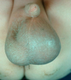
What is the problem shown in this picture?

Testicular Torsion
- Unilateral pain in abdomen or testicle
- Referred pain from scrotum.
- get torsion of the:
- epididymis
- testis
- hydatis of morgani
- high riding testicle
- +/- bellclapper deformity (congenital but happens after)
- scrotal reddness = stops at the edge of the hemiscrotum
- limited by the tunica vaginalis
- if redness extends consider something else
- blue appearance is because its ischaemic (haemorrhagic infarct)
- PHx of resolving episodes of pain
- two peaks in babies and >13. <11 in most common.
- treat immediately - <6 hours to save it. and fix other testis so it cannot twist
- torsion of the hydatid of morgnagni - can’t tell due to red skin and sore - common in onset of puberty as testerone at 10-11, armomatase as estrogen stimulates it.
DDx:
- epididymitis - don’t do ultrasound, this is secondary to the ischaemic.
- blue dot sign seen below there will be immediate system exploration of the appendix. Haemorrhage.
- glands penis is generally the same size of the testis of a young boy.
- torsion commonest at 13-15 - enlarged due to androgens in puberty.
*

What is the problem shown in this picture?

Perinatal torsion - different from torsion in adolescent boy where testis is on longer mesentery
- occurs in utero as testis are descending in tunica vaginalis
- will be painless and hard at birth
- baby is usually restless in womb that day - 35-36weeks. Gubernaculum eats its way through and starts making fibrous tissue and collagen to attach.
- will involute over time
DDx:
- teratoma can be form in the uterus.
- US to make sure its not a tunour.
- speramtic cord all twisted up.
‘vanishing testis’ - 6 months later the dead testis disapears. No one realises until afterwards.
Spot the Diagnosis

Idiopathic Scrotal Oedema
- Cellulitis from perianal infection and sores on the bum.
- Early childhood from a flea bite causing an allergic reaction
- extends past the hemiscrotum
WHat is the diagnosis?

Varicocele
- abnormal enlargement of the pampiniform venous plexus in the scrotum. Plexus of veins drains the testis.
- common abnormality in adolescent boys (1 in 10). Anatomical variant.
- asymmetric scrotum
- ‘bag of worms’ will only be seen when vertical - will feel non-descript and squishy when horizontal
- transillumination - 3rd world XR - easy way to see through the tissue.
- sometimes painful due to too much blood, dragging feeling.
- treated by tying of the veins and affects fertility by cooking the testis so sperm counts goes down.
- indication for surgery - smaller testis will interfere with fertility, need an operation.
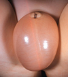
What is the diagnosis from this picture?

Nephrotic Syndrome
- outside of the processus vaginalis (outside tunica vaginalis)
- presents at 3am in the morning with:
- generalised oedema - ‘can the child open their eyes?’
- HTN
- spherical scrotum (hydrocele with dumbbell shaped scrotum which is surgical)
- may obliterate penis
- looks transparent
What is the diagnosis from this picture?

Undescended Testis
- 1 is normal sized (based on glans) and 1 is absent
- the other will have compensatory hypertrophy
- other testis is not palpable but left in the inguinal canal - under the fat, search for a moving target (in mesentery).
- treatment is orchipexy at 6-12 months
- can happen in cerebral palsy due to increased tone of cremaster muscle.
What is the diagnosis based on this history?

- left hemiscrotum too big = hydrocele
- on the other side:
Associated strangulated inguinal hernia:
- undescended testis but encarcerated hernia
- give pain relief and reduce it
- fix undescended testis at the same time.
What is the Diagnosis? Explain the condition?

Branchial cleft defect - sinus or fistula
- commonly 2nd cleft (connecting to tonsillar fossa) but sometimes 3rd (posterior o piriform fossa)
- elective surgical excision to prevent recurrent salivary discharge.
What is the diagnosis?

Tongue Tied - ankylosglossia
- abnormally short and thick frenulum
- Complications:
- cosmetic
- function (speech and dental hygeine
- interfere with breast feeding (controversy)
What is the Diagnosis?

Meconium Ileus
- sticky mucus blocks the ileum
- occurs in Cystic Fibrosis
- if distension from birth must have blocked above the ileocecal valve because otherwise the colon would resorb fluid in utero.
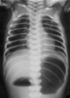
What is the diagnosis?

Duodenal Atresia
- wrinkly skin often on outside
- congenital absence of complete closure of a portion of duodenum
- see duoble bubble on USand AXR
- 30% risk of Down’s syndrome
What is the Diagnosis?

Malrotation Volvulus
- commonly a few days after birth
- S shape on contrast study (barium swallow)
- offered LADDs procedure where untwisted and mesentery base is extended.
What is the Diagnosis?

Gastroschisis
- failure of full retraction of the umbilical hernia during development.
- Genetically normal foetus
- no membrane around the bowel like in omphalocele
- inflammed appearance of the bowel
- sterile urinary + fecal peritonitis from bowel + mesentery bathing in urine + meconium
- wrap in glad wrap with thermal insulation
- don’t twist bowel
- dry gauze to stop it twisting
Diagnosis?

Omphalocele (aka exomphalos)
- failure of embryogenesis
- failure of three layers to fully unite around the umbilicus during migration
- very likely genetic abnormality
- 50% other anomalities - often cardiac.
- severe heat and water loss
- wrap in glad wrap with insulation.
What is the diagnosis based on the enema?

Hirschsprung’s disease
- progressive distansion after birth
- aganglionosis of the rectum which is in permanent spasm without neural control.
- no submucosal plexus causing overgrowth of extrinsic nerves
- treated with excision of the aganglionic distal gut
What is the Diagnosis based on this picture?

Malrotation Volvulus
- first few months of life.
- S sign or corkscrew.
- mesentery doesn’t appropriately migrate (two bases of attachment) are too close together.
- Degree of twist is important
- 720 causes arterial ischaemia
- Requires surgery, Ladd’s procedure


