SM 110a - Cardiovascular Embryology Flashcards
Describe the development of the interventricular septum
Where does it grow from?
What does it fuse with?
The interventricular septum grows to separate the left and right ventricles of the developing heart
Grows from the floor of the ventricles toward the fused endocardial cushions
Eventually fuses with the endocardial cushions with the help of the spiral septum
Which fetal structure gives rise to the ascending aorta and the pulmonary trunk?
Truncus arteriosus
Which fetal structures give rise to the smooth back of the right and left atria?
- Smooth back of right atrium is derived from the right horn of the sinus venosus (Left horn -> Coronary sinus)
- Smooth back of left atrium is derived from the fetal pulmonary veins
Which structure partitions the atrioventricular canal into the left and right sides?
Dorsal and ventral endocardial cushions
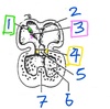
At this stage of the embryonic heart, which structure is labeled by #2?

Septum primum

What is the ductus arteriosus?
Where is it located
One of the two shunts between fetal pulmonay and systemic circulations (other = foramen ovale)
Connects the pulmonary trunk to the arch of the aorta
Does the umbilical vein carry oxygenated or deoxygenated blood?
What about the umbilical arteries?
Umbilical vein: Oxygenated
Umbilical artery: Deoxygenated
(from fetal heart -> fetal circ -> placenta)
(from placenta to fetal heart)
List the sequence of cardinal vein development
- Anterior and posterior cardinal veins (Common cardinal veins)
- Subcardinal system (kidney level)
- Supracardinal system (thoracic wall)
What does the subcardinal system of veins give rise to?
Middle inferior vena cava
What is the fate of the fetal bulbus cordis?
The bulbis cordis becomes the smooth outflow part of the right and left ventricles
- RV: Conus arteriosus
- LV: Aortic vestibule
What is Tetralogy of Fallot?
A combination of four congenital abnormalities caused by a faulty spiral septum
- Ventricular septal defect (VSD)
- Pulmonary valve stenosis
- Misplaced (overriding) aorta
- Thickened right ventricular wall (right ventricular hypertrophy)
Which component of the fetal circulation has the highest oxygen content?
Umbilical vein
List the 3 shunts in fetal circulation and their locations
Lungs
- Foramen ovale
- Shunts blood from the right atrium -> Left atrium (bypass pulmonary circulation
- Ductus arteriosus
- Shunts blood from pulmonary trunk -> Aortic arch (bypass pulmonary circulation)
Liver
- Ductus venosus
- Shunts blood from the umbilical vein -> inferior vena cava (bypass liver)
List the two shunts between the fetal pulmonary and systemic circulatiosn
Foramen ovale
Ductus arteriosus
Why do congenital heart defects become more problematic as a child ages?
After birth, systemic pressure increases as body size increases, and pulmonary pressure decreases as airways mature.
Heart defects become more problematic as the pressure differential between pulmonary and systemic circulation increasees
Which structure(s) contribute to division of the atrium?
Describe their growth and important features
Septum primum
- First structure to begin dividing the atrium
- Grows down toward the endocardial cushions
- Contains…
- Transient foramen primum
- Foramen secundum (high in the septum primum)
Septum secundum
- Appears later
- Not as tall as the septum primum
- Contains the foramen ovale (lower down in the septum secundum)
What do the anterior and posterior cardinal veins give rise to?
Anterior (portions above the heart persist into adult hood)
- Brachiocephalic vein
- Superior vena cava (with common cardinal vein)
Posterior
- Pelvic and leg veins
Which fetal structure gives rise to the coronary sinus?
Left horn of the sinus venosus
(Right -> smooth back of the right atrium)
At this stage of the embryonic heart, which structure is labeled by #3?

SA Valves

What are the 6 changes to fetal circulation that are triggered by the baby’s first breath?
- Blood begins to flow to the lungs and return to the left atrium
- Increase in left atrial pressure due to return from pulmonary veins - this pushes the septum primum against the septum secundum to close the foramen ovale
- Oxygenated blood flows to the umbilical arteries causes smooth muscle contraction
- Umbilical vein collapses due to lack of blood
- The ductus venosus begins becoming a fibrous ligamentum venosum
- Increased blood flow to the left ventricle increases pressure in the aortic arch. This decreases blood flow through the ductus arteriosus, which begins to become the fibrous ligamentum arteriosum
Describe the shunt between the right and left atrium that is present in embryonic developpment
What is its purpose?
The shunt consists of the foramen ovale (in the seputm secundum) and the foramen secundum (in the septum primum)
The foramen ovale is located inferior to the foramen secundum; this allows for…
- Fluid flow from the right atrium to the left atrium in embryonic development
- Rapid closure of the foramen when left atrial pressure increases upon first breath; there is no overlap in the holes when the two septums are pressed together

Describe the steps in ventricular division
- Interventricular septum
- Grows from apex of the heart toward the endocardial cushions
- Spiral septum (aka aortico-pumlmonary septum)
- Grows from endocardial cushions to fuse with the interventricular septum
- Allows for independent exit of blood from RV and LV
- When this is finished, ventricular division is complete
What does the primitive interventricular septum give rise to?
The thick, muscular interventricular seputm
Why do babies born with transposition of the great arteries typically survive with surgery?
Transposition of the great arteries is usually accompanied by an IV septal defect that allows for oxygenated blood in the left ventricle to mix with deoxygenated blood in the right ventricle before it is pumped through the aorta
This amount of oxygen is enough to keep the baby alive if surgery is performed immediately after birth
List the layers of the embryonic heart tube and the structures they give rise to
- Endocardial tube (innermost)
- -> Stratified squamous epithelium that lines the heart chambers
- Cardiac jelly
- Disappears
- Myocardial mantel (outermost)
- Will form cardiac muscle
At this stage of the embryonic heart, which structure is labeled by #5?

Dorsal endocardial cushion

At this stage of the embryonic heart, which structure is labeled by #4?

Atrioventricular canals

What is the fate of the fetal pulmonary veins?
Smooth back wall of the left atrium
What is the fate of the primitive atrium and ventricle?
Rigid muscle
- Pectinate muscle in the atria (auricles)
- Trabecular muscle walls (ventricle)
At this stage of the embryonic heart, which structure is labeled by #1?

Sinoatrial orifice

What does the supacardinal system of veins give rise to?
Azygous system
Lower inferior vena cava
(Supplies the thoracic wall)
What is the clinical manifestation of persistent truncus arteriosus?
Persistent truncus arteriosus is an interventricular septal defect that results in incomplete separation of the pulmonary trunk and the aorta
Mixed blood is sent to the pulmonary, systemic, and coronary circulations
There is increased volume of blood to the lungs that bakcs up, resulting in fluid in the lungs and heart failure
Describe the heart defect: transposition of the great arteries
The pulmonary trunk and aorta are switched
The right ventricle pumps deoxygenated blood into the aorta -> systemic circulation
The left ventricle pumps oxygenated blood into the pulmonary trunk -> pulmonary circulation
Usually accompanied by an IV septal defect that allows mixing of blood; this allows the baby to survive if it recieves immediate surgery after birth
What do the vitelline veins gie rise to?
Supply the transverse abdominal wall
- Interahepatic inferior vena cava
- Hepatic veins
- Hepatic portal system
What is the fate of the fetal truncus arteriosus?
The fetal truncus arteriosus becomes the ascending aorta and the pulmonary trunk
Describe the pathway of fetal blood flow starting from…
The placenta
The fetal brain
From Placenta - High O2 blood
- Umbilical vein
- Inferior vena cava
- Right atrium
- Through foramen ovale
- Left atrium
- Left ventricle
- Aortic arch
- 3 great arteries
- Brain (highest O2 content; branches before mixing from ductus arteriosus)
- Interior iliac arteries
- Left and right umbilical arteries
- Placenta
- 3 great arteries
From brain - Low O2 blood
- Superior vena cava
- Right atrium
- Right ventricle (due to angle, mostly goes here instead of through foramen ovale)
- Pulmonary trunk
- Though ductus arteriosus
- Aortic arch (after 3 great vessels have branched off)
- From here, see above for fate after aortic arch
What is the function of the bulbus cordis in the early embryonic heart?
The bulbus cordis is the only outflow of blood from the embryonic heart
It is located in the right ventricle
At this stage of the embryonic heart, which structure is labeled by #6?

Left ventricle

What is the fate of the fetal sinus venosus?
- Right horn -> Smooth back (inflow) of the right atrium
- Left horn -> Coronary sinus
Which fetal structure gives rise to the pectinate muscles (atria) and the trabecular muscles (ventricles)?
Primitive atrium and ventricle
Why might prolonged crying spells in newborns be dangerous?
Crying increases intrathoracic pressure, which can divert unoxygenated blood from the pulmonary trunk through the ductus arteriosus (not fully closed yet) instead of to the lungs
This is dangerous because blood does not have the opportunity to get oxygenated, and therefore the systemic circulation does not recieve oxygenated blood
Which fetal structures gives rise to the adult interventricular septum?
The muscular IV septum is derived from the primitive interventricular septum
The (upper) membranous IV septum is derived from the fusion of the spiral septum with the endocardial cushions and the muscular IV septum
At this stage of the embryonic heart, which structure is labeled by #7?

Interventricular septum
What happens to the ductus arteriosus after birth?
At birth:
- Vascular beds of the lung open
- Pulmonary BP decreases, and aortic BP increases
- The path of least resistance for blood leaving the right atrium is now through the pulmonary trunk, instead of through the ductus arteriosus
- The ductus arteriosus becomes the ligamentum arteriosum a few weeks after birth
