104- Cardiac Muscle Contraction Flashcards
The intracellular concentration of which ion is most important in determining contractility?
Ca2+
What is lusitropy?
Myocardial relaxation
Which structure is labeled by #8?

M Line

Which section is labeled by #2?

A band

Which structure is labeled by #9?
Which molecules make up this structure?

Thick filament
Made from myosin light and heavy chains

What is the effect of afterload on the strength of cardiac contraction?
Why?
Afterload decreases the velocity of contraction, which decreases the strength of contraction

What is digoxin?
How does it work?
Digoxin is a drug prescribed to patients with heart failure to increase cardiac output
- It blocks the membrane Na+/K+ ATPase
- Per ATP: 3 Na+ out, 2 K+ in
- If the ATPase cannot pump Na+ out of the cell, intracellular Na+ increases
- Increased Na+ decreases the activity the Na+/Ca2+ exchanger
- Normallly pumps Na+ in and Ca2+ out
- Less Ca2+ out = increased intracellular Ca2+ concentration
- Increased inotropy -> Stronger heart contractions
Note: Digoxin can also slow the HR
Does systolic blood pressure approximate preload or afterload?
Afterload
We are measuring how hard the heart must work to overcome the pressure in the aorta
Describe the troponin complex
The troponin complex is attached to tropomyosin
It is made up of…
- Troponin T (TnT), which binds to tropomyosin
- Troponin C (TnC), which binds to Ca2+
- Troponin I (TnI), which inhibits contraction when it is bound to actin
- It covers up the myosin binding site on actin
If systolic blood pressure increases, what happens to the volume of blood ejected from the heart?
The volume decreases
Increased systolic blood pressure -> slower velocity of contraction -> decreased strength of contraction -> less blood ejected
How would inhibiting cardiac K+ channels affect the cardiac action potential?
Inhibiting cardiac K+ channels would lengthen the cardiac action potential, because it woud take longer for K+ to leave the cell and repolarize the membrane
Why is an increase in intracellular calcium necessary for cardiomyocyte contraction?
Ca2+ must bind to Troponin C in the Troponin complex in order for the tropomyosin + troponin complex to swing out of the way and expose the myosin binding site
The myosin binding site must interact with actin for cross bridge cycling and contraction to occur
Which structure is labeled by #5?

T Tubule

What is the sarcolemma?
The tubular sheath which envelops skeletal and cardiac muscle fibers
It basically acts as the extracellular membrane

Which ion is most responsible for phase 3?

K+
K+ rushes out of the cell (IK1), taking the positive charge with it
This allows the membrane to repolarize

List 3 things that regulate contractility in cardiomyocytes
(All have an effect on Ca2+ concentration in the cytosol)
- Andrenergic signaling acts on B1 receptors in the heart. B1 activation = increased contractility
- Digoxin = increased contractility
-
Heart rate
- Increased HR = increased contractility (Ascending staircase effect aka Bowditch effect aka Treppe)
- Postextrasystolic potentiation: increased contractility of the beat after the extra heart beat
A patient recieves a blood transfusion.
Would you expect the strength of their cardiac contractions to increase or decrease?
Why?
Increase
More blood will increase preload, becaues it will cause the ventricles to fill more during diastole
Increasing preload increases the strength of cardiac contraction
Describe the configuration of actin and myosin in the attached state of cross-bridge cycling
The myosin head is attached to the thin filament after the previous power stroke completion

What is the main mediator of cardiomyocyte relaxation?
The SERCA Pump
(SERCA = Sarcoendoplasmic reticulum Ca2+ ATPase)
How would inhibiting cardiac Ca+ channels affect the cardiac action potential?
Inhibiting cardiac Ca+ channels would prevent Ca2+ from entering the cell. There would be less Ca2+ to oppose the K+ currents, and repolarization would occur more quickly.
Shorter action potentials
What is Ca2+ induced Ca2+ release?
How does it work? Describe the steps
The mechanism for excitation-contraction coupling; this is responsible for muscle contraction in response to recieving an action potential
- When the myocyte recieves an action potential, Ca2+ rushes into the myocyte though L-type Ca2+ channels
- Ryanodine receptors (RyR) in the terminal cisternae of the sarcoplasmic reticulum (SR) sense the small increase in cytoplasmic [Ca2+] (trigger Ca2+) and stimulate Ca2+ release from the SR through the RyR
- Ca2+ from the SR causes a x10 to x100 increase in intracellular calcium concentration
- Ca2+ binds to Troponin-C on the thin filament of the sarcomere
- Cross-bridge cycling ensues
What is the ascending staircase effect?
How does it impact cardiac contractility?
Ascending staircase effect = Bowditch effect = Treppe
- Increasing heart rate ->
- Increase in intracellular Ca2+ in SR ->
- Increase in contractility
- More is released when RyRs are activated

Without sufficient ATP, in which step of cross-bridge cycling is the sarcomere arrested?
The attached state
If ATP cannot bind to the actin/myosin complex, the myosin head cannot be released from actin, and it cannot get to the released state.
(It will not be able to release and move to the next actin monomer, 2 positions over)
What is inotropy?
Contractility
Describe the interaction of actin and myosin in the power stroke state of cross-bridge cycling
Power stroke state - Pi dissociates from myosin, leaving the muscle fiber in a rigid state

Describe the interaction of actin and myosin in the cross-bridge state of cross-bridge cycling
Cross-bridge state - The cocked myosin head binds actin
Which structure is labeled by #7?

Intercalated disk
Intercalated discs separate myocytes

What causes preload?
How do we measure it?
Increased ventricular filling or transfusion can increase preload
Stretch (preload) is measured by LV end diastolic pressure
Which mechanism increases cardiac contractility without altering intracellular Ca2+ levels?
Andrenergic signaling via B1 receptors
Via the Gs-alpha cascade, PKA phosphorylates Troponin-I, which directly increases the amount of force generated at the myofilament level
Which structure is labeled by #7?
Which molecules make up this structure?

Thin filament
Actin + Tropomyosin + Troponin T, C, and I

Which ion is most responsible for phase 2?

Ca2+

Why is it important for the cardiomyocyte to lower cytoplasmic Ca2+ concentration after contraction?
If Ca2+ concentration is to high, the myocyte cannot relax
Which molecule is labeled by #6?
What is its function?

Titin
A length-sensing giant molecule that acts as a stiff spring
It prevents over-stretching of the sarcomere, and recoils after stretch to pull the sarcomere back to its normal length

What are the 3 mechanisms for lowering cytoplasmic Ca2+ after cardomyocyte contraction?
- SERCA pump sequesters Ca2+ into the SR
- Ca2+ pump expells Ca2+ through the sarcolemma
- Ca2+/Na+ exchanger moves Na+ into the cytoplasm and expells Ca2+ through the sarcolemma
If a sarcomere is relaxing, would you expect cytoplasmic Ca2+ concentration to be low or high?
Low
Low Ca2+ prevents contraction of the sarcomere
What is SERCA?
Why is it important?
SERCA = Sarcoendoplasmic reticulum Ca2+ ATPase
SERCA is an ATP-dependent pump that sequesteres cytoplasmic Ca2+ into the sarcoplasmic reticulum.
It lowers cytoplasmic Ca2+ concentration, allowing for relaxation of the sarcomere, and storage for the next contraction
(Ca2+ pump, Ca2+/Na+ exchanger also work to lower cytoplasmic Ca2+)
Where are Ryanodine Receptors (RyRs) found?
What do they do?
RyRs are found in the terminal cisternae fo the SR
They sense the “trigger Ca2+” that enters the cardiomyocyte through L-type Ca2+ channels during the caridac action potential
In response, they facilitate the release of lots of Ca2+ from the SR to increase intracellylar calcium x10 to x100. This allows Ca2+ to bind to troponin C, which allows cross-bridge cycling to commence
What is postextrasystolic potentiation?
How does it impact cardiac contractility?
Postextrasystolic potentiation is the increase in cardiac contractility following an extrasystole (extra heart beat, aka premature ventricular contraction)
If the heart contracts early, it is in the relative refractory period; the contraction will not be very strong
The next contraction will be very strong due to the extra Ca2+ that was not used in the previous contraction

What is a T tubule?
What does it do?
A T tubule is an invagination of the sarcolemma
It invests the myofibrils to effectively bring all of the sarcomeres and mitochondria in close contact with the interstitial fluid; this helps facilitate coordinated contraction
The sarcoplasmic reticulum has terminal cisternae attached to the T tubules
They depolarize when the myocardium depolarizes
Which structure is labeled by #1?

Z disk

Describe the interaction of actin and myosin in the cocked state of cross-bridge cycling
ATP is hydrolyzed to ADP + Pi. The products remain, and the myosin head unhinges 11 nm. It lines up with the acin monomer 2 positions over from the previous binding site

Does LV end diastolic pressure approximate preload or afterload?
Preload - we are measuring stretch, which is greatest at the end of diastole (the ventricle is full)
What is the relationship between intracellular Ca2+ concentration and contractility?
Increased Ca2+ = increased contractility
What is the effect of preload on the strength of cardiac contraction?
Why?
Preload increases the strength of cardiac contraction
Because cardiac muscle has titin, stretching the sarcomere will increase tension. Titin will want to “pull” the sarcomere back to its normal length, which will help toincrease the force of contraction
Also, the sarcomere may have increased Ca2+ sensitivity at greater lengths
(up to a point)

If cyotoplamsic Ca2+ concentration is higher than baseline in a cardiomyocyte, would you expect it to be contracting or relaxing?
Contracting
What causes afterload?
How do we measure it?
Increased systolic blood pressure can increase afterload (must reach a high pressure to overcome pressure in the aorta)
LV afterload is approximated by systolic blood pressure
Which step of excitation-contraction coupling is responsible fora marked increase in intracellular calcium concentration?
Release of large amounts Ca2+ from the Sarcoplasmic reticulum
This is triggered when Ryanodine Receptors in the terminal cisternae of the SR sense a small increase in intracellular calcium due to action potential firing
How does andrenergic signaling affect contractility of the heart?
Describe the process
The heart contains B1 receptors. When activated by andrenergic stimulation…
- Alpha subunit (stimulatory) of Gs binds Adenylyl cyclase
- Increased production of cAMP
- cAMP phosphorylates PKA (protein kinase A)
- PKA phosphorylates many things
- Phosphorylates phospholamban -> increased lusitropy
- Dissociation of phospholamban from SERCA
- Without phospholamban attached, SERCA is faster. Faster lusitropy
- This results in increased myocardial filling and increased contractility
- Phosphorylates Troponin-I -> increased inotropy
- Increases the amount of force generated at the myofilament level
- This is how andrenergic signaling directly increases contractility without the Ca2+ middleman
- Phosphorylates phospholamban -> increased lusitropy
Which section is labeled by #3?

H band

Describe the configuration of actin and myosin in the released state of cross-bridge cycling
ATP binding causes the myosin head to be released from actin

List and describe the steps of cross-bridge cycling
- Attached state - The myosin head is attached to the thin filament after power stroke completion
- Released state - ATP binding causes the myosin head to be released from actin
- Cocked state - ATP is hydrolyzed to ADP + Pi. The products remain, and the myosin head unhinges 11 nm. It lines up with the acin monomer 2 positions over from the previous binding site
- Cross-bridge state - The cocked myosin head binds actin
- Power stroke state - Pi dissociates from myosin, leaving the muscle fiber in a rigid state

Which structure is labeled by #6?
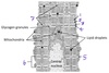
Sarcomere

What is contractility, as it refers to cardiac muscle?
Contractility is the intrinsic strength of contraction, independent of loading conditions
(force of contraction regardless of preload or afterload)
What would high troponin T in the bloodstream indicate?
Heart damage
When damaged, cardiac muscle releases Troponin T into the blood stream
What molecule makes up #5?
(The pink cord encircling the green)
What is its function?

Nebulin
Regulates thin filament length

Which ion is most responsible for phase 0?

Na+

Which segment is labeled by #4?

M band

