Skeletal Muscle Flashcards
White Muscle Disease is caused by
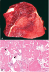
Vitamine E / Se deficiency
Pathogenesis of Malignant Hyperthermia
- Inherited defect in skeletal muscle ryanodine receptor
- Excessive Ca release and contraction when stimulated
- Heat production and myocyte necrosis
Disease
Exertional Rhabdomyolysis
Disease

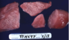
White Muscle Disease
Causes of monophasic lesions of skeletal muscle
Trauma - will be focal
Exertion - Capture myopathy
Toxin - ionophores, plants
MDx
Rhabdomyosarcoma
Myocytes increase in size by
Addition of Myofilaments
MDx

Diffuse eosinophilic myositis
Disease
Splay Leg
Swimmer Syndrome
T/F: Atrophy can be the result of another pathological process affecting the muscle
True
MDx
Polyphasic myocyte degeneration and necrosis with dystrophic calcification
What types of causes incite this pattern of degeneration and necrosis?
Polyphasic lesion causes:
Nutritional deficiency - Vitamin E/Se
Ongoing toxicities
Genetic defects in myocyte structure/metabolic elements
Pathogenesis of Botulism
- Decaying organic matter
- Clostridium botulinum thrives and elaborates into environment
- Ingested
- Toxin inhibits Ach release from nerve terminals at neuromuscular junction
- Progressive generalized paralysis with death by cardiorespiratory failure
Disease

Compartment Syndrome
Muscular Dystrophy
X linked inhereted myopathy reported in dogs and cats (especially goldens)
Defects in dystrophin gene - cytoskeleton protein
No treatment
Pathological process that accounts for this appearance?
Degeneration / Necrosis
Lesions caused by White Muscle Disease are seen in (Active/Inactive) Muscle
Active
MDx

Focal Muscle Degeneration and Necrosis
Disease

Black Leg
Clostridium chauvoie infection
Why is it important to use special stains when diagnosising sarcomas?
To differentiate between sarcomas - each type behaves differently
MDx

Atrophy
Describe morphological problem
Muscle is pale, swollen and dry to the touch
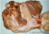
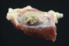
MDx
(Rear leg of dog)

Hemangiosarcoma
MDx

Myocyte Hypoplasia
Pathogenesis of Infarction
- Recumbancy
- Increased intramuscular pressure
- Poor perfusion
- Ischemia
- Infarction
Possible cause of eosinophilic myostis in a dog

Masticatory Myositis
Pathogenesis of Black Leg (Clostridium chauvoei infection)
- Ingestion of spores
- Dissemination to muscle via blood
- Latency
- Tissue hypoxia/acidosis
- Bacterial proliferation
- Production of exotoxins
- Myonecrosis and systemic endothelial damage
- Death from septicemic shock
Exertional Rhabdomyolysis
Necrosis/lysis of skeletal muscle
Ionic events of contraction can produce adverse environment
May have underlying metabolic condition which predispose - polysaccharide storage disease
MDx

Intramuscular hemorrhage
What typically causes this pattern of necrosis?
Acute Toxicity
Exertion
Cause

Damage to the Left Recurrent Laryngeal Nerve
T/F: Cuase of muscle injury can be determined by histopathology alone
False; supplemental tests and clinical history are usually required
Cause

Trauma - penetrating wounds, fractures
What do myofibers do when they are hurt?
Die
Regenerate
MDx

Rhabdomyosarcoma
Morphological Diagnosis?
Muscle Infarct
Describe the lesion

Fibrosis and fiatty infiltration (steatosis) of skeletal muscle
Causes of myocyte hyperplasia
Double Muscling
Inactivation of the regulatory gene myostatin - involved in myoblast cell progression to muscle fibers
MDx

Acute necrotic and hemorrhagic myositis
Disease

White Muscle Disease
Azoturia
Excess nitrogen in urine
MDx
(Rear leg of dog)
Rhabdomyosarcoma
Causes of myositis
Necrotic / hemorhhagic = Clostridium chauvoei, Clostridium septicum
Suppurative - Pyogenic bacteria
Lymphocytic - Immune- Mediated
Eosinophilic - Active protozoal / parasitic infections, immune mediated
Granulomatous
“Biochemical” pathological processes resulting in severe muscular clinical signs
Neuromuscular Junction Disorders
Electrolyte Disorders
Inherited disorders of Muscle Metabolism - Myotonias
What cell type do neoplasms with striaged muscle differentiation (rhabdomyoma/sarcoma) derive from?
Gross morphological diagnosis?

Skeletal muscle degeneration and necrosis
Rhabdomyolysis
Fibrosis and fatty infiltration (steatosis) often develops in what chronic skeletal muscle condition
Atrophy
Causes of myocyte hypertrophy
Exercise conditioning
Compensatory - decreased number/size of functional myocytes, increased load on remaining
Describe the lesion

Muscles are dark red, swollen, soft/friable/ palpable crepitus
MDx
Focal Suppurative myositis
Pathogenesis of Compartment Syndrome
- Rapid muscle growth
- Period of increased circulation (exertional)
- Muscle swelling confined by facia
- Impedes blood supply
- Ischemia
- Infarction
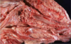
What pattern of necrosis is this?

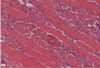
Monophasic
MDx
Chronic eosinophilic myositis and muscle atrophy
What pathological process could account for this appearance?

Degeneration and Necrosis
Inflammation and Repair
Circulatory Disorder
Disease

Splay Leg
Swimmer Syndrome
Causes of skeletal muscle atrophy
Physiologic - disuse/aging
Cachexia/ Malnutrition
Endocrine disease - myocytes have surface receptors for hormones
Denervation - myocyte maintencance requires trophic factors generated at neuromuscular junction - occurs quickly
Histological characteristics of myofiber regeneration
Internalization of nuclei
Macrophage infiltrate
Histologic characteristics of myofibers that die
Vaculation of sarcoplasm
Condensation of sarcoplasm
Nuclear pyknosis
Calcification
MDx

Focal monophasic myonecrosis
Disease

Laryngeal Paralysis
Pathogenesis of Polyphasic Skeletal Muscle Degneration caused by nutritional deficiency
- Vitamine E/Se Deficiency
- Needed for enzymes like glutathione peroxidase / reductase
- Lack of ability to scavange free radicals
- Oxidative damage - lipoperoxidation of cell membranes
- Myocyte injury
What is morphologically abnormal?

Decreased striations
Necrosis
Hypnotic nuclei
Neutrophil infiltration
What 3 Pigments and tissue deposits are observed in skeletal muscle?
Lipofuscin
Exogenous Pigments
Dystrophic Calcification
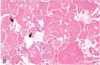
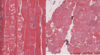
MDx

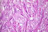
Polyphasic monocyte degeneration and necrosis, chronic, with hypertrophy, atrophy and fibrosis
Capture Myopathy
Zoo and Wild Birds
Due to exertion, stress during capture/handling/transport
Anaerobic glycolysis leads to hyperthermia and metabolic acidosis
Endogenous Pigments seen in skeletal muscle
Hematogenous Pigments (Hemoglobin, Hemosiderin, Bilirubing, Porphyria)
Melanin
Lipofuscin
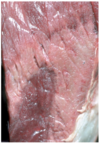
Type of skeletal muscle lesion
Monophasic Lesion
Pathological process to account for this appearance?

Disorders of Growth
T/F: Malignant hyperthermia is an example of a metabolic condition predisposing to necrosis
True
