Inflammation Flashcards
Sources of the multinucleated giant cells
Macrophages
Epithelioid Cell
Large, pale staining macrophages that have an ovoid nucleus and shape resembling epithelial cells
Condition

Johne’s Disease
Focal Inflammation
Single abnormality or inflamed are within a tissue
Morphology of eosinophils
Larger than neutrophils
Affinity of cytoplasmic granules to eosin (acid)
Lysosomal granules contain wide variety of catalytic enzymes similar to neutrophils
T/F: Fibrosis and neovascularization are features of subacute infection
False
Leukocytes
Normal inhabitants of the circulating blood
Total count of leukocytes in circulating blood modified by systemic response to inflammation
Each cell type has distinctive role
Each cell type enters into the inflammatory response in a definite sequence
Classification of inflammation by duration

Subacute Inflammation
Classification of inflammation based on exudate

Mucopurulent - Catarrhal
3 interconnected processes of phagocytosis
Recognition and attachment of the particle to be ingested
Engulfment with subsequent formation of phagocytic vacuole
Killing or degradation of the ingested material
Neutrophils
Crucial to inflammatory process
Constitute the first line of cellular defense
Develop in the bone marrow and the maturation process takes about two weeks
Functions of eosinophils
Modulate hypersensitivity reactions
Defend against helminthic infections
Phagocytic but less active phagocytes than neutrophils
Resolution of inflammation involves
Neutralization of chemical mediators
Return of normal vascular permeability
Cessation of leukocyte infiltration
Removal of edema fluid, leukocytes, foreign agents and necrotic debris
Hemorrhagic Inflammation
Hemorrhage is the main feature of this type of inflammation. Presence of an etiologic agent will indicate that the process is inflammatory rather than a primary circulatory disturbance
Sluggish motile but are responsive to chemotactic influences, they have a long life span (30-60days) and may proliferate at sites of inflammation
Macrophages
Epithelioid cells are specialized for
Extracellular secretion
Multinucleated Giant Cells
Formed by the coalescence of single macrophages
Lesion

Pleural Adhesions
Type of WBC

Lymphocytes and Plasma Cells
Time of onset of subacute inflammation
Depends on the nature of the inciting stimulus, may cover a considerable time span which can vary from a few days to a few weeks
Morphology of lymphocytes
Heterogeneous in size and morphology - smaller than neutrophils
Densely staining nucleus and scant amount of cytoplasm
Traditional division (T and B cells)
Functional division (Helper T, Cytotoxic T Cells)
Specific granules
Seconday granules - small, less dense and more numerous neutrophil granules
Inflammatory cells of peracute inflammation
Not usually numerous
Few leukocytes
Type of WBC

Neutrophil
Roles of inflammation
Dilute, contain and isolate injury
Destroy invading microoranisms and/or inactivate toxins
Achieve healing and repair
Classification of inflammation based on exudate
Fibrinous Exudate
Classification of inflammation based on duration

Chronic inflammation
Two classes of neutrophil cytoplasmic granules
Azurophil granules
Specific granules
Type of inflammatory cell

Multinucleated Giant Cell
Inflammatory cells of subacute infection
Mixed or pleocellular inflammatory infiltrate
Primarily neutrophilic but also has infiltration by lymphocytes, macrophages and plasma cells.
Halmark of chronic inflammation
Fibrosis
Sequelae of chronic inflammation
Destruction of stimuli → resolution of inflammation → repair of tissue
Persitence of stimuli → progression of inflammatory reaction → continuation of disease
Time of onset of fibrinous inflammation
Acute process, can form in seconds
Abscess
Circumscribed collection of pus - localized form of suppurative inflammation
Nuclei of multinucleated giant cells are sometime arranged in what type of pattern
Horseshoe Pattern - Langhan’s Giant Cells
Eosinophilic Cationic Protein
Contributes to parasite killing and also shortens coagulation time and alters fibrinolysis
Four outcomes of acute inflammation
Complete Resolution
Healing by scarring
Abscess formation
Progression to chronic inflammation
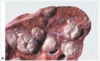
“Cerebroid” appearance of affected intestine is characteristic of what type of inflammation
Granulomatous inflammation
Transudate or Exudate

Transudate
Describe the gross appearance of an abscess
yellow-white to gray white and varies from watery to viscous depending on fluid content
Cells that respond to chemotactic stimuli
Granulocytes
Monocytes
Lymphocytes -lesser extent
Exudation
Escape of fluid, proteins, and blood cells from the vascular system into the interstitium or body cavities. - Alteration of the normal permeability of local blood vessels
Pyogranulomatous Inflammation
Significant number of neutrophils present in the center of a granulomatous reaction
Example of what outcome of acute inflammation?

Abscess formation
Eosinophilic granules
Small granules
Primary granules
Large specific granules
Lymphocytes and Plasma Cells
Involved in immune reactions
Key cellular mediators are immediate antibody response and the delayed cellular hypersensitivity response
Classification of inflammation based on exudate

Suppurative Exudation
Classification of inflammation based on distribution

Multifocal inflammation
Possible stimuli for acute inflammation
Infectious agents
Traugma
Necrotic tissue/cells
Immune reactions
Signs of inflammation
Redness
Heat
Swelling
Pain
Loss of function
Mononuclear phagocyte system (MPS)
Consists of closely related cells of bone marrow origin, including blood monocytes and tissue macrophages
Vascular changes during acute inflammation play a major role in
Maximizing movement of cells and plasma proteins from within circulation to site of injury
What is the purpose of an intense inflammatory response?
Attempt to isolate inflammatory process, formation of a wall
Classification of inflammation based on distribution

Focal inflammation
Type of inflammation

Necrotizing Inflammation
Suppurative Exudation
Consisting of or containing pus, associated with the formation of pus
Histologic hallmarks of chornic inflammation
Infiltration of mononuclear cells (macrophages, lymphoctyes, plasma cells)
Proliferation of fibroblasts and small blood vessels
Increased connective tissue
Tissue destruction
Possible origins of locally extensive inflammation
Local reactions that spread to adjacent normal tissue
Coalescence of foci in a multifocal reaction
Locally extensive inflammation
Involves a considerable zone of tissue within an inflamed organ
Infectious canine hepatitis is an example of what type of inflammation (classified by duration)
Peracute Inflammation
Repair begins during inflammation and it is completed when
Injurious stimuli have been neutralized
Effects of inflammatory stimuli are manifested through
Chemical mediators
Etiology

Mycobacterium avium sp
Epithelioid cells are commonly found where in the cell
Endoplasmic reticulum
Golgi apparatus
Vesicles
Vacuoles
Diffuse Inflammation
Variations in severity may occur, but the eniter tissue is involved
Purpose of neutrophils
Eliminate microorganisms, tumor cells and foreign material
Contents of serous exudation
Outpouring of fluid relatively rick in protein, and derived from blood and locally injured cells
Clinical signs of chronic inflammation
Chronicity is primarily a clinical concept pertaining to prolonged duration of an inflammatory lesion
Major basic protein
Strongly toxic to parasites as well as other kinds of cells found in the granules of eosinophils
Granulomatous Infection
Inflammatory response characterized by the presence of lymphocytes, macrophages and plasma cells with the predominant cell being the macrophage
Clinical signs of peracute inflammation
Shock, sudden death
What happens to neutrophils after phagocytosis
Undergo apoptotic cell death and are ingested by macrophages
Inflammatory cells in acute inflammation
Leukocyte infiltration is variable
Neutrophils usually predominate
Mononuclear cells can also be present
Morphology of Macrophages
15-20um - larger than neutrophils
Prominent central nuclei - folded or bean shaped
In tissues are larger and have variable number of azurophilic granules and remnants of ingested material
Necrotizing Inflammation
Necrosis is the main feature and exudation is minimal. The process is interpreted as inflammatory if infectious etiology is suspected
Clinical signs of acute inflammation
Signs associated with vascular changes
Warm
Red
Swollen
Pain
Loss of function
Gross patterns of chronic inflammation
Diffuse thickening of affected area
Solid, firm, nodular lesions that compress adjacent tissue
Signals for macrophage activation
Lymphokines
Bacterial endotoxins
Contact with fibronectin-coated surfaces
Variety of chemicals
Antibody Dependent Cell Mediated Cytotoxicity
Eosinophils are attracted to sites of helminths invasion in sensitized hosts by chemotactic factors elaborated predominantly as a result of the immune response to products of the parasite
Mild Inflammation
Absent to minimal tissue damage
Few inflammatory cells
Slight vascular involvement
Condition

Lymphadenitis
Inflammatory cells of chronic inflammation
Primarily mononuclear inflammatory cells
Lymphocytes
Macrophages
Plasma cells
Fibroblasts
Tissues damaged by inflammation are replaced with
Regenerated native parenchymal cells
Fibrous tissue
Effect of Inflammation

Edema
Procces and classification of inflammation based on duration

Neutrophil “Paving”
Acute Inflammation
Mediators of rolling event of extravasation
Selectins
Integrins
Origin of chronic inflammation
Follow an acute inflammatory phase
May develop as insidious, low-grade, subclinical process without history of a prior acute episode
Classification of inflammation based on distribution

Locally Extensive Inflammation
Suppurative lesions are often of what origin
Bacterial
Lymphatic involvement in chronic inflammation
Variable
Granuloma
Small, 0.5-2mm, organized collections of modified macrophages (epithelioid macrophages), usually surrounded by a rim of lymphocytes. Another feature is the presence of Langhans giant or foreign body-type cells and presence of fibrous connective tissue
Phagocytosis
Involves the accumulation of white blood cells at the site of injury followed by the release of enzymes by neutrophils and macrophages to eliminate injurous agents
Heterophils
Eosinophilc granules of rabbit, guinea pig, rat, reptile, fish and bird neutrophils
Leukocyte Adhesion Deficiency (LAD)
Disease due to leukocyte adhesion failure, due to type I mutation in Beta-1 integrins CD18
Severe Inflammation
Substantial tissiue damage
Inflammatory cells abundant
Massive edema and hemorrhage seen
Inflammation is initiated by
Exogenous and endogenous stimuli
Azurophil Granules
Primary granules - large, oval and electron dense found within the neutrophil cytoplasm
Neutrophils are capable of killing microorganisms by
Producing oxygen free radicals
Hydrogen peroxide
Lysosomal Enzymes
Lesion

Hepatic granulomatous
Inflammation
Reaction of vascularized living tissues to injury
Eosinophils
Abundant at sites of inflammation in diseases of immunologic, parasitic, or allergic origin
Unique functions as effector cells for killing helminths and thier propensity for both causing and assisting in the regulation of tissue damage in hypersensitivity
Etiologic Diagnosis

Mycotic Airsacculitis
Mucopurulent - Catarrhal
Inflammatory exudate is composed of mucus and pus
Vascular involvement of Acute inflammation
Active hyperemia
Edema
Occassional fibrin thrombi within vessels
Chronic Inflammation
Result of a persistent inflammatory stimulus in which the host has failed to completely eliminate the causative agent
Inflammatory response accompanied by immune response
Evidence of host tissue response - repair
Mediators of margination event of extravasation
Selectins
Cytokines/chemokines
Chronic Inflammation
Type of inflammation resulting from injurious persistant stimuli that leads to a predominantly proliferative, rather than exudative, reaction
What type of organisms are stained pink?
What type of stain is used?

Fungal Organisms
PAS Stain
Inflammation leads to accumulation of fluid (plasma proteins) and WBCs in
Extravascular tissues
Neutrophils regulate inflammatory response by
Releasing chemical mediators such as leukotrienes and platelet activating factor
Inflammation ends when what happens?
The stimulus is eliminated
Transudate or Exudate

Exudate
Serous Exudation
Inflammatory process in which the exudate occurs in tissues in the absence of a prominent cellular response. May be a dominant pattern of exudation for a wide variety of mild injuries
Time of onset of chronic inflammation
variable
Eosinophils are effective in killing helminth parasites by
Antibody-Dependent Cell-Medated Cytotoxicity
Complex Granuloma
Granuloma with a central area of necrosis
Chronic inflamation in various organs arise in what three ways
Following acute inflammation
Repeated bouts of acute inflammation
Insidiously as a low grade smouldering response
Events of acute inflammation
Stimuli for onset of acute inflammation
Vascular changes
Cellular events
Termination of acute inflammatory response
Mediators of Activation and Adhesion event of extravasation
Integrins
Chemokines
Monocytes become macrophages after what occurs
Monocyte migration into tissues
Neutrophils are characterized by
High motility due to rapid amoeboid movement
Response to a wide variety of chemotaxtic compounds
Phagocytic and bactericidal activities - major cellular defense against bacteria
Pus
Inflammatory exudate rich in leukocytes and parenchymal cell debris
Lesion

Multifocal granulomas
Mononuclear cells
Lymphocytes and Plasma Cells
Monocytes and macrophages
Platelets
Gross appearance of fibrinous inflammation
Yellow-white or pale tan, stringy, shaggy meshwork which gives a rough irregular appearance to the tissue surfaces. Casts of material may form in lumen of tubular organ
Functions of macrophages
Phagocytosis
Modulation of inflammatory and repair processes
Regulation of immune response
Production of Interleukin 1
Lymphatic involvement in acute inflammation
Role in moving away exudate.
Transportation of the exudate can lead to acute regional lymphadenitis
Vascular changes that occur during acute inflammation
Increased vascular flow and caliber of blood vessels
Increased vascular permeability
Mechanism of extravasation during acute inflammation
Margination - tethering
Rolling
Activation and Adhesion
Transmigration
Peracute Inflammation
Caused by potent stimulus
Animal has no time to respond
Less common than acute disease
Classify extent of inflammation

Severe
What is the difference between the presence of a fibrinous exudate and fibrosis
Presence of a fibrinous exudate involves an acute process, fibrosis is a chronic process
Acute Inflammation begins in what time
4-6 hours
Polymorphonuclear leukocytes
Neutrophils
Eosinophils
Basophls and Mast Cells
Classification of inflammation based on exudate

Granulomatous Inflammation
Chemical mediators of acute inflammation
Vasoactive amines
Plasma proteases
Lipid mediators
Platelet activating factor
Cytokines
Chemokines
Nitric oxide
Extravasation
Delivery of white blood cells to the site of injury
Classification of inflammation based on exudate

Suppurative Inflammation
Characteristics of inflammation
Involves changes in vascular bed, blood and connective tissue
Intended to eliminate irritant and repair damaged tissue
Outcomes of inflammation
Ideally - Return to normal
Intense inflammatory response - attempt to separate injured tissue
Faiure to eliminate insult - sequel
Mechanism of Leukocyte Adhesion Deficiency (LAD)
Neutrophilia with impaired transmigration because neutrophils are unable to adhere
Describe the morphology of neutrophils
10-12um
Multilobed nucleus
Cytoplasmic granules
Host involvement of chronic inflammation
Parenchymal regeneration or repair by fibrosis
Clinical signs of Leukocyte Adhesion Deficiency (LAD)
Gingivitis
Tooth Loss
Ulcers in oral and enteric mucosa
Cutaneous ulcers
Pneumonia
Classification of inflammation based on exudate

Fibrinous Exudation
Pathogenesis of fibrinous exudation
Severe injury to endothelium and basement membrane results in leakage of plasma proteins which polymerize perivascularly as fibrin
Mediators of transmigration event of extravasation
P-CAM (CD31)
Time of peracute inflammation
0-4 hours
Lesion

Fibrinous peritonitis
Simple classification of inflammation includes
Exudate
Duration
Histologic appearance of what

Fibrinous pneumonia
Macrophages/Monocytes
Derived from circulating blood monocyte of bone marrow origin
May originate from immature resident mononuclear phagocytes in the tissue
Do not have reserve pool in bone marrow
Remain in circulation up to 72 hours
Require activation to become competent macrophages
Cells involved in granulomatous inflammation
Epitheloid cells
Multinucleated giant cells
Lymphocytes
Stop signals for acute inflammatory response include
Switch from pro-inflammatory leukotrienes to anti-inflammatory lipoxins from arachidonic acid
Liberation of anti-inflammatory cytokines such as TGF=beta from macrophages and other cells
Neural impulses resluting in inhibition of TNF production in macrophages
Suppuration
Process by which puss is formed. Use of the term implies that neutrophils and proteolytic enzymes are present, and that necrosis of host tissue cells has occured.
Moderate Inflammation
Some tissue damage
Inflammatory cells evident
Moderate edema and evidence of hemorrhage
Fibrinous Exudation contains
Fibrin
Example of what outcome of acute inflammation
Abscess formation
Inflammation is closely associated with the process of
Repair
Classification of inflammation based on exudate

Granulomatous Inflammation
Diffuse inflammation is often related to what etiology
Viral or toxic
Classification of inflammation based on distribution

Diffuse Inflammation
Describe the gross appearance of serous exudate
Yellow, straw-like color, fluid commonly see in very early stages of many kinds of inflammatory responses
Fusion of epithelioid cells to form multinucleated giant cells is induced by
Cytokines
Describe the histiologic appearance of granulomas
Macrophages clustered in a characterisitc ellipitcal formation around the causative etiologic agent or around a central necrotic area, or simply organized nodules
Large cells with abundant cytoplasm - “Epithelioid cells” “Multinucleated Giant Cells”
Vascular involvment of subacute infection
Decline in the magnitude of vascular changes, compared to acute inflammation
Role of T lymphocytes in granulomatous inflammatory reactions
Produce lymphokines and interferon
Attract and activate macrophages
Induce formation of multinucleated giant cells
Classification of inflammation based on exudate

Fibrinous exudation
Two subdivisions of inflammatory cells
Polymorphonuclear Leukocytes
Mononuclear cells
Classification of inflammation based on duration

Chronic Inflammation
Etiology of granulomatous inflammation
Non-digestible organism or particle which serves as a chronic inflammatory stimulus, delayed type hypersensitivity is often required
Common types of exudate
Suppurative
Fibrinous
Serous
Chemotaxis occurs right after
Extravasation
Classification of inflammation by distribution
Focal
Multifocal
Locally extensive
Diffuse
Chemotaxis
Process where white blood cells emigrate in tissues towards the site of injury
Vascular involvement of peracute inflammation
Hyperemia
Slight edema
Hemorrhage
Neutrophils mediate tissue injury by
Release of oxygen free radicals and lysosomal enzymes
Type of WBC

Eosinophil
Epithelioid cells have (more/less) phagocytic activity than non-specialized macrophages
less
Multifocal Inflammation
Arising from or pertaining to many foci, each focus of inflammation is separated from other by an intervening zone of relatively normal tissue
Subacute inflammation
Gradual change between acute and chronic - used when the inflammatory response does not include reparative responses
Fibrin is composed of
Thread-like eosinophilic meshwork that sometimes forms masses of solid amorphous material
Example of what outcome of acute inflammation

Healing by the formation of scar tissue
Type of inflammation

Hemorrhagic Inflammation
Vascular involvement of chronic inflammation
Proliferation of capillaries and small blood vessels resulting in hemorrhage and congestion
Termination of acute inflammatory response occurs when
Degradation of mediators of inflammation
Stop signals are produced when stimulus is gone
Lymphangitis
Inflammation of lymphatic vessels
Functions of Neutrophils
Phagocytosis
Secretion of proinflammatory substances
Simple Granuloma
Organized accumulation of macrophages and epitheliod cells, often rimmed by lymphocytes
Transudate
Essentially an ultrafiltrate of blood plasma and results from hydrostatic imbalances across the vascular endothelium, has a low protein content and low specific gravity (less than 1.020)
Lymphatic involvement in subacute inflammation
Increased lymphatic drainage
Repaire of endothelial cells
Onset of action of granulomatous inflammation
Always chronic
Fibrinopurulent Exudate
Term used to classify an inflammatory process in which neutrophils and fibrin are abundant
Exudate from an absess consits of what type of inflammatory cells

Neutrophils
Macrophages
Lymphocytes
Exudate
Inflammatory extravascular fluid that has a high protein concentration, cellular debris and high specific gravity (above 1.020)
Macrophage activation occurs in response to
External stimuli that must be presented in an orderly sequence
Major scavengers in the inflammatory response
Macrophages
Edema
Denote an excess of fluid in the interstitial tissue or serous cavities, it can be an exudate or transudate
Effect of inflammation

Pus
Granulomatous Inflammation
Specific type of chronic inflammation characterized by accumulation of modified macrophages (epithelioid cells) and initiated by a variety of infectious and noninfectious agents
Condition

Subacute stomatitis
Condition

Lymphangitis
Example of:

Fribropruluent exudate
Condition

Greasy Pig Diease
