Cell Injury Flashcards
Describe the gross appearance of acute cell swelling
Swollen organ with rounded edges
Pallor when compared to normal
When cut surface - tissue bulges
Heavy “wet” organ
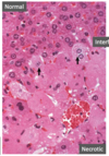
Histologic effects of Liquefactive Necrosis
Loss of cellular detail
Granular cells
Eosinophillic and basophillic debris
Neutrophil nuclei may dominate
No tissue architecture preserved
Niemann Pick Disease
Lysosomal Storage disease
Caseous necrosis is typically related to (acute/chronic) disease.
Caseous necrosis is typically related to (acute/chronic) disease.
Cellular contents in Necrosis vs Apoptosis
Necrosis: Enzymatic digestion; may leak
Apoptosis: Intact; released in apoptotic bodies
T/F: Apoptosis induces inflammation
False
Which (Necrosis or Apoptosis) causes inflammation
Necrosis
Liquefactive Necrosis occurs in
Tissues with high neutrophil recruitment and enzymatic release with digestion of tissue
Tissues with high lipid content
Focal bacterial /fungal infections
MDx

Bilateral, symmetrical encephalomalacia
MDx

Multifocal caseous pneumonia
Sterile Abscess
Process caused by non living irritants such as drugs, likely to turn into firm, solid lumps as they scar
Cell size in Necrosis vs Apoptosis
Necrosis - Enlarged
Apoptosis - Reduced
Example of cell injury
Acute Cell Swelling
Executioner caspases
3 and 6
Abscess
Localized collection of pus in a cavity formed by disintegration of tissues surrounded by fibrous connective tissue
Cell death can occur by what two processes
Necrosis
Apoptosis
Cytochrome C
Essential for life; released into cytoplasm to initiate suicide program of apoptosis
Pyknosis
Nuclear shrinkage - DNA condenses into shrunken basophilic mass
MDx

Hepatitis, multifocal to coalescing, subacute, severe, necrotizing
Cellular changes due to acute cell swelling
Dilution of cytoplasm
Cells enlarged
Increased cytoplasmic eosinophilia
Example of cell injury
Acute Cell Swelling
_________________________
Ballooning degeneration resuling in formation of a vesicle
Apoptosis has a physiologic or pathologic role?
Often physiologic - may be pathologic after some forms of cell injury
Example of cell injury

Liquefactive Necrosis
Changes of necrotic cells in cytoplasm
Increased binding of eosin
Loosing basophillia
Glassy homogeneous
Vacuolation and moth eaten appearance
+/- Calcification
Possible mechanisms resulting in lipid accumulation
Excessive delivery of FFA from fat stores or diet
Decreased oxidation or use of FFAs
Impaired synthesis of apoprotein
Impaired combination of protein and triglycerides to form lipoproteins
Impaired release of lipoproteins from hepatocytes
Apoptotic Bodies
Fragments of apoptotic cells that contain portions of the cytoplasm and nucleus
Intrinisic apoptotic pathway initiated by
withdrawal of growth factors or hormones
Cause

Vitamin E/ Selenium Deficiency
Etiology

Vesicular exanthema of swine virus - Calicivirus
Infarct
localized area of coagulative necrosis
Example of cell injury

Acute Cell Swelling
Name of Disease

Blackhead
Cells that are highly vulnerable to hypoxia and cell swelling
Cardiomyocytes
Proximal Renal Tubule Epithelium
Hepatocytes
Endothelium
CNS Neurons, Oligodendrocytes, Astrocytes
Gangrenous Necrosis
Not a specific pattern of cell death but begins mostly as coagulative necrosis, usually applied to distal extemities and involves multiple planes of tissue
Dry Gangrene
No bacterial superinfection; tissue appears dry
Saponification
Free Fatty Acids + Ca → Ca Soaps
Septic Abscess
Infection, release of enzymes from WBC and infectious agent
Example of cell injury

Fatty Change
Example of cell injury

Necrosis
Acute cellular swelling and fatty chage are considered to be (reversible/irreversible) cell injuries.
Acute cellular swelling and fatty chage are considered to be (reversible/irreversible) cell injuries.
Disease

Caseous lymphadenitis
Dystocia and Recumbent Cattle can cause what type of necrosis
Fat necrosis
_____________________
Traumatic Fat Necrosis
Liquefactive necrosis typically occurs in the ____________ system.
Liquefactive necrosis typically occurs in the central nervous system.
_________ occurs when there is an abnormality of synthesis, utilization and/or mobilization of fat.
Fatty Change occurs when there is an abnormality of synthesis, utilization and/or mobilization of fat.
Etiology

Histomonas melegridis
Only form in which triglycerides can be transported out of hepatocytes
Lipoproteins
Extrinsic Apoptotic Pathway
Death-Receptor Initiated Pathway
Example of cell injury

Liquefactive Necrosis
Necrosis of Abdominal fat in cattle is of what cause
Unknown cause
Acute Cell Swelling
Early, sub-lethal manifestation of cell damage, characterized by increased cell size and volume due to H2O overload
Etiology of Fatty Change
Hypoxia
Toxicity
Metabolic Disorder
Acute cell swelling is typically due to
Acute cell swelling is typically due to loss of ionic and fluid homeostasis
_____________________
Failure of cell energy production
Cell membrane damage
Injury to enzymes regulating ion channels of membranes
MDx

Multifocal hemorrhagic polyomyelitis
Prognosis of acute cellular swelling depends on
Prognosis of acute cellular swelling depends on the number of cells affected and importance of cells
Physiologic causes of hepatic lipidosis
Pregnancy toxemia
Ketosis
Etiology

Sarcocystis neurona
Three types of fat necrosis
Enzymatic Necrosis
Traumatic Necrosis
Necrosis of Abdominal Fat
Describe the morphology of apoptosis
Cell shrinkage, Increased cytoplasmic density
Chromatin condensation
Formation of cytoplasmic blebs and apoptotic bodies
Phagocytosis
Fatty Change
Sub-lethal cell damage characterized by intracytoplasmic fatty vacuolation
__________________
May be preceded or accompanied by cell swelling
Example of cell injury

Wet Gangrene
Lipofuscin in a cell is evidence of
Lipofuscin in a cell is evidence of previous injury
Enzymatic Necrosis
Action of activated pancreatic lipases in escaped pancreatic fluid
Histologic appearance of fatty change
Delineated, lipid filled vacuoles in the cytoplasm
May displace nucleus to the periphery
Karyolysis
Nuclear fading - chromatin dissolution due to action of DNAases and RNAases
Plasma membrane in Necrosis vs Apoptosis
Necrosis: Disrupted
Apoptosis: Intacted, altered structure
Morphologic necrosis due to 2 concurrent processes
Denaturation of proteins
Enzymatic digestion of the cell
Pathogenesis of fatty change
Impaired metabolism of fatty acids → Accumulation of triglycerides → Formation of intracytoplasmic fat vacuoles
DDx
Equine Herpes Virus 1
Rabies
West Nile Virus
Prognosis of fatty change
Initially reversible but can lead to irreversible hepatocyte death
Liquefactive Necrosis
Dead cells are “digested” - transformed from tissue to liquid viscous mass
Ketone Bodies
Alternative fuel for cells
Produced in liver by mitochondria
Example of cell injury

Fatty Change
Possible causes of caseous necrosis
Mycobacterium
Corynebacterium
Fusobacterium
Fungal infections
Example of cell injury

Fatty change
Etiology

Corynebacterium pseudotuberculosis
Bcl Family Proteins
Pro and Anti- Apoptotic
Control release of cytochrome C
Clinical manifestations of lipidosis are most commonly detected as alterations in ________ function because it is most central to lipid metabolism.
Clinical manifestations of lipidosis are most commonly detected as alterations in liver function because it is most central to lipid metabolism.
Increase in cell size can be due to
Cell Swelling
Cell Enlargement
Necrosis
Cell death after irreversible cell injury by hypoxia, ischemia, and direct cell membrane injury
Anti-Apoptotic Proteins
Bcl-2 Bcl-X, Mcl -1
Example of cell injury

Fibrinoid Necrosis
Disorders associated with too little apoptosis
Cells with p53 mutation
Lymphocytes that react against self-Ag
Failure to eliminate dead cells
Fibrinoid necrosis occurs when
Fibrinoid necrosis occurs when Ag-Ab complexes are deposited in walls of arteries
Disease

Polioencephalomalacia
Caseous necrosis is associated with poorly degradable _________ of bacterial origin.
Caseous necrosis is associated with poorly degradable lipid of bacterial origin.
Pro-Apoptotic Proteins
Bim, Bid, Bad
Bak, Bax
Initiator caspases
8 and 9
Example of cell injury

Dry gangrene
A good prognosis is possible with acute cell swelling if
A good prognosis is possible with acute cell swelling if oxygen is restored before the “point of no return”
Common cause of coagulative necrosis
Ischemia in all solid organs except brain
Fibrinoid Necrosis
Special form of necrosis usually seen in immune reactions involving blood vessels
Intrinisic Apoptotic Pathway
Mitochondrial Pathway
Leukoencephalomalacia causes
Necrosis of white matter of cerebral hemispheres, brain stem and cerebellum
Endocrine causes of hepatic lipidosis
Diabetes mellitus
Feline Fatty Liver Syndrome
Fat Cow Syndrome
Caseous Necrosis
Friable white area of necrosis, necrotic debris represents dead WBCs
Coagulative Necrosis
Architecture of dead tissues is preserved, ultimately the necrotic tissue is removed by phagocytosis or digestion by action of lysosomal enzymes of WBC
Apoptosis
Pathway of cell death
Irreversible cell injury is associated morphologically with
Severe swelling of mitochondria
Extensive damage to plasma membrane
Swelling of lysosomes
Hisologic effects of caseous necrosis
Eosinophilic granular cell debris
Rim of inflammatory cells
Obliterated tissue architecture
Dystrophic calcification in center of lesion
Leukoencephalomalacia Pathogenesis
Ingestion of Fusarium moniliforme containing Fumonisin B1 Toxin Producing Moldy Corn → Sphingolipid Synthesis Inhibition → Direct Cellular Toxicity → Leukoencephalomalacia
Disorders associated with too much apoptosis
Neurodegenertive disease
Ischemic injury
Death of virus infected cells
Example of cell injury

Fat necrosis
Example of cell injury

Caseous Necrosis
Wet Gangrene
Bacterial superinfection occured, tissue looks wet and liquefactive by actions of degradative enzymes in the bacteria and the attracted WBC
Example of cell injury

Coagulative Necrosis
The most common and fundamental expression of cell injury is ___________.
The most common and fundamental expression of cell injury is acute cell swelling.
A frequent outcome of necrosis is ___________.
A frequent outcome of necrosis is inflammation.
Describe the gross appearance of fatty change
Diffuse yellow
Enhanced reticular pattern if specific zones affected
Edges rounded and bulge on section
Tissue soft, friable and cuts easily with greasy texture
May float in fix/water
MDx

Multifocal necrohemorrhagic myelitis
Nutritional causes of hepatic lipidosis
Obesity
Protein - Calorie Malnutrition
Starvation
2 types of abscesses
Septic
Sterile
Karyorrhexis
Nuclear fragmentation - pyknoic nucli membrane ruptures and nucleus undergoes fragmentation
Disease

Equine Leukomyelitis
Disease

Leukoencephalomalacia
Disease

Tuberculosis
Most frequent causes of acute cell swelling
Hypoxia
Toxic agents
Patterns of tissue necrosis
Coagulative necrosis
Liquefactive necrosis
Gangrenous necrosis
Caseous necrosis
Fat necrosis
Fribrinoid necrosis
Example of cell injury

Coagulative Necrosis
Example of cell injury

Liquefactive necrosis
Polioencephalomalacia can be caused by
Thiamine deficiency diet
Increased ruminal thiaminase activity
Administration of thiamine analogs
High levels of sulfur in diet
Lead toxicity
Thiaminase containing plants
Lipidosis
Accumulation of triglycerides and other lipid metabolites (neutral fats and cholesterol) within parenchymal cells
Example of cell injury

Enzymatic Fat Necrosis
Apoptosis can occur during what two types of processes
Physiologic
Pathologic
Pus
Necrotic material, frequently creamy yellow because of the presence of dead WBCs
Necrosis has a physiologic or pathologic role?
Invariably pathologic - culmination of irreversible cell injury
Abscesses result due to
Abscesses result due to the body’s defensive reaction to foreign material
Nucleus in Necrosis vs Apoptosis
Necrosis: Pyknosis → Karyorrhexis → Karyolysis
Apoptosis: Fragmented
Describe the gross appearance of necrosis
Pale, soft, friable
Sharply demarcated by zone of inflammation
Example of cell injury

Necrosis
Conversion of acetyl coA from fatty acid oxidation causes
Lipolysis
Extrinisic apoptotic pathway initiated by
Receptor- ligand interactions
____________________
Fas
TNF Receptor
Major classes of lipids that can accumulate in cells
Triglycerides
Cholesterol/ Cholesterol Esters
Phospholipids
Complexes of Lipids and Carbohydrates
Species affected by Leukoencephalomalcia
Horse
Chicken
Pig
Ultrastructural changes of acute cell swelling
Plasma membrane alterations
Mitochondrial changes
Dilation of the ER
Nuclear alterations
Disease

Nutritional Myopathy
White Muscle Disease
