S4) Cellular & Molecular Events in the CVS Flashcards
(26 cards)
In four steps, describe how the resting membrane potential of cardiac cells is generated
⇒ Cardiac myocytes are permeable to K+ at rest
⇒ K+ move out of the cell (down concentration gradient)
⇒ Inside becomes more negative relative to the outside
⇒ As charge builds up an electrical gradient is established
In three steps, briefly explain how excitation leads to action
⇒ Cardiac myocytes are electrically active & fire action potentials
⇒ Action potential triggers increase in [Ca2+]i
⇒ Actin and myosin interact, triggering the contraction mechanism
State the RMP for the following:
- Axon
- Skeletal muscle
- SAN
- Cardiac ventricle

Describe the 4 different stages of the ventricular (cardiac) action potential
- Depolarisation – Na+ influx
- Initial repolarisation – K+ efflux
- Plateau – Ca2+ influx
- Proper repolarisation – K+ efflux

Describe the 3 different stages in the SAN action potential

Describe the mechanisms behind the slow depolarising pacemaker potential
- Turning on of slow Na+ conductance (If – funny current)
- Activated at membrane potentials more negative than - 50mV
- HCN (Hyperpolarisation-activated Cyclic Nucleotide-gated) channels are activated which allow influx of Na+ for depolarisation

Describe how the action potential waveform varies throughout the heart
- SAN is fastest to depolarise, it is the pacemaker and sets rhythm
- Other parts of the conducting system also have automaticity, but it’s slower

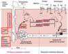
Describe the action potential diagrams for different parts of the heart:
- SAN
- Purkinje fibres
- Atrial muscle
- Ventricular muscle
- AVN

Explain four problems that could occur during the process of excitation leading to contraction
- Action potentials fire too slowly → bradycardia
- Action potentials fail → asystole
- Action potentials fire too quickly → tachycardia
- Electrical activity becomes random → fibrillation
What is the normal range of plasma [K+]?
3.5 – 5.5 mmol L-1
If [K+] is too high or low it can cause problems, particularly for the heart.
In terms of plasma [K+] levels, define hyperkalaemia and hypokalaemia
- Hyperkalaemia – plasma [K+] is too high > 5.5 mmol.L-1
- Hypokalaemia – plasma [K+] is too low < 3.5 mmol.L-1
In 5 steps, describe the effects of hyperkalaemia

⇒ EK becomes less negative (smaller concentration gradient)
⇒ Membrane potential becomes less negative and depolarises
⇒ Early depolarisation causes Na channels to open then inactivate (less steep uptake slope)
⇒ HCN channels are activated by hyperpolarisation (remain inactive)
⇒ Depolarisation is slow and over a long duration
What are the risks associated with hyperkalaemia?
- Pacemaker potential decreases, heart rate decreases/stops (asystole)
- May initially get an increase in excitability but then conductance may cease
Risks associated with hyperkalaemia depend on the extent and how quickly it develops.
Describe the severity of hyperkalaemia
- Mild: 5.5 – 5.9 mmol/L
- Moderate: 6.0 – 6.4 mmol/L
- Severe: > 6.5 mmol/L
How can hyperkalaemia be treated?
- Calcium gluconate
- Insulin + glucose
Ineffective if the heart already stopped
In 4 steps, describe the effects of hypokalaemia

⇒ EK becomes more negative (greater concentration gradient)
⇒ Membrane potential becomes more negative
⇒ Action potential is prolonged as plateau phase is longer (Ca2+ channels remain open)
⇒ Repolarisation is delayed & slower
What are the risks associated with hypokalaemia?
- Longer action potentials lead to early after depolarisations (EADs)
- Prolonged plateau phase provides greater opportunity to stimulate more action potentials and cause more contractions
⇒ Leads to oscillations in membrane potential which result in ventricular fibrillation

In two steps, describe excitation-contraction coupling
⇒ Depolarisation opens L-type Ca2+ channels in the T-tubule system
⇒ Localised Ca2+ entry opens closely-linked CICR channels in the SR
25% enters across sarcolemma, 75% released from SR
How does relaxation occur in cardiac myocytes?
[Ca2+]i must return to resting levels:
- SERCA is stimulated and pumps calcium back into SR
- PMCA & NCX remove calcium across the cell membrane
What controls the tone of blood vessels?
Tone of blood vessels is controlled by contraction & relaxation of vascular smooth muscle cells:
- Located in tunica media
- Present in arteries, arterioles and veins
In 5 steps, describe the cellular mechanism leading to the contraction of blood vessels
⇒ Ca2+ binds to calmodulin
⇒ Ca2+- calmodulin complex is formed
⇒ Myosin Light Chain Kinase is activated
⇒ MLCK phosphorylates myosin so it interacts with actin
⇒ Contraction mechanism is triggered

In 5 steps, describe the cellular mechanism leading to the relaxation of blood vessels
⇒ Ca2+ levels decline
⇒ Myosin light chain phosphatase dephosphorylates myosin
⇒ PKA phosphorylates MLCK & inhibits its action
⇒ Myosin light chain is not phosphorylated
⇒ Contraction is inhibited

pacemaker cells
specialised myocytes that generate electrical events in regular intervals = initiate an action potential
- diastole/most negative it depolarises at -60mv
- FUNNY (If) current as they open with hyperpolarisation
- HCN Channels =Hyperpolarisation-activated,Cyclic,Nucleotide-gated channels allow influx of Na ions to deoplarise the cell
conducting fibres
purkinje fibres conduct excitation through ventricular mycocardium
