S11) The Peripheral Arterial and Venous Systems Flashcards
Describe the role of the calf muscles in blood circulation in the limbs
- The veins in the lower limb have valves which permit unidirectional blood flow
- When the calf muscles contract (soleus and gastrocnemius), the deep veins are compressed and blood flows upwards towards heart

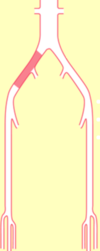
Briefly describe the role of perforating veins in the lower limb

- When the muscles relax, blood is “sucked” into the deep veins via the perforating veins from the superficial veins
- The valves in the perforating veins only allow unidirectional blood flow

Describe the pathology of the perforating veins of the lower limb
Varicosities result when the valves in the perforating veins become incompetent or diseased
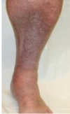
What are varicose veins?
Varicose veins are tortuous, twisted, or lengthened veins
ineffective blood flow
most common site are saphenous veins
walls of veins weaken = disrupt valves
pooling of blood and blood flows backwards (retrograde)

Describe the pathophysiology of varicose veins
The vein wall is inherently weak in varicose veins, which leads to dilatation and separation of valve cusps so that they become incompetent

What are the symptoms of varicose veins?
Occur along the vein itself (trunk varices):
- Heaviness
- Tension
- Aching
- Itching
What are the 5 complications of varicose veins resulting from venous hypertension due to calf muscle pump failure?
- Oedema
- Skin pigmentation
- Varicose eczema
- Lipodermatosclerosis
- Venous ulceration
What are the complications of varicose veins resulting from the vein itself?
- Haemorrhage
- Thrombophlebitis
What is thrombophlebitis?
Thrombophlebitis is an inflammatory process that causes a venous thrombosis to form, commonly in the legs:
- Superficial thrombophlebitis
- Deep vein thrombosis
What are the causes of calf muscle pump failure?
- Failure of calf muscle contraction – immobility, obesity, reduced movement
- Deep vein incompetence
- Superficial vein incompetence (volume overload)
What is the pathophysiology of thrombosis?
Virchow’s triad:
- Changes in the lining of the vessel wall
- Changes in the flow of blood
- Changes in the constituents of blood
In terms of Virchow’s triad, what is the most important factor in the pathophysiology of arterial thrombosis?
Changes in the lining of the vessel wall

In terms of Virchow’s triad, what is the most important factor in the pathophysiology of venous thrombosis?
Changes in the flow of blood
stasis
chemo
inflammatory conditions
platelet rich

Does stasis lead to thrombosis?
Stasis + another provocateur (trauma, oral contraceptive pill, dehydration, cancer)
Distinguish between arterial and venous thrombosis in response to bleeding
- Arterial thrombosis in response to bleeding involves vasoconstriction, platelets, extrinsic and then intrinsic pathways, hence arterial thrombi are platelet rich and caused by atheroma
- Venous thrombosis in response to bleeding involves venoconstriction, intrinsic then extrinsic pathways, hence, venous thrombi are fibrin rich
What is a deep vein thrombosis?
- A deep vein thrombosis is the formation of a thrombus within a deep vein, most commonly the deep calf veins
- It produces an inflammatory response - you will see lots of swelling (calor, dolor, rubor, tumor, functio laesa)
can be fatal if missed
What are the symptoms of DVT?
- Pain (cannot walk)
- Swelling
- Blue-red skin discolouration
What are the signs of DVT?
- Calf tenderness
- Skin warmth & discolouration
- Distended, warm superficial veins
- Oedema
- Pyrexia
What is the association between surgery and DVT?
- Immobility prior to surgery
- No calf muscle pump during surgery
- Immobility after surgery
- Surgery is trauma (body’s response → prothrombotic state)
What is the fatal consequence of DVT?
Pulmonary embolism
What is peripheral arterial disease?
Peripheral arterial disease is a common condition, in which a build-up of fatty deposits in the arteries restricts blood supply to leg muscles
alternative routes exist so blood can still pass and limit acute ischemia eg collateral circulation
What are the two types of peripheral arterial disease?
- Acute limb ischaemia
- Chronic limb ischaemia
Describe the pathophysiology of acute limb ischaemia
- The limb goes from a normal blood supply to greatly impaired blood supply over a period of minutes (sudden onset)
- No chance for collateral vessel development (weeks/months)
What are the commonest causes of acute limb ischaemia?
- Embolism (from heart / abdominal aortic aneurysm)
- Trauma