Regions of the neck Flashcards

1
Q

Describe the borders of the anterior and posterior neck?

A

Anterior: inferior border of mandible down to manubrium

Posterior: superior nuchal line down to C7/T1 disc

2
Q

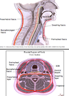
What are the four fascial layers of the neck?

A

Superficial fascia

Visceral fascia

Vascular fascia

Vertebral fascia

3
Q

Give a general description of the fascial layers of the neck?

A

4 longitudinal compartments each contained within layers of cervical fascia

4
Q

Describe the superificial fascial layer of the neck?

A

Contains a thin sheet of muscle called platysma

Blends with facial muscles and shares their nerve supply

Contains EJV

5
Q

Describe the superficial veins of the neck?

Include their course and structures that they drain.

A

EJV and AJV

EJV begins at angle of mandible, vertical descent on sternocleidomastoid

Together, drain superifical muscles of neck

6
Q

What is contained within the visceral fascial layer of the neck?

A

Thyroid, parathyroid and thymus glands

Also relates to trachea and oesophagus

7
Q

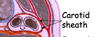
What is contained within the vascular fascial layer?

A

Cartoid sheath and contents

8
Q

What is contained within the vertebral fascial layer?

A

Cervical vertebrae and postural muscles

9
Q

Where is the investing fascia located?

What does it contain?

A

Deep to superificial fascia

Surrounds all layers

Splits to enclose sternocleidomastoid and trapezius

10
Q

Where is the prevertebral fascia loacted?

What does it contain?

A

Cylinder surrounding vertebral compartment

Contains vertebrae and postural muscles

11
Q

Where is the pretracheal fascia located?

What does it contain?

A

Comprises pretracheal and buccopharyngeal fascia

Preatracheal fascia extends up to hyoid bone

Together surround visceral compartment

12
Q

What is the significance of the pretracheal fascia connecting to the hyoid bone superiorly?

A

Hyoid bone moves up and down with swallowing > anything within pretracheal fascia will also move up and down with swallowing

13
Q

What is the significance of the fascial layers of the neck in terms of communication?

A

Spaces between fascial layers can communicate up as well as down to medistinum

Allows unrestricted spread of infection

14
Q

Describe the contents of the carotid sheath?

A

Common carotid artery/just afetr bifurcation

IJV

CN X

15
Q

Describe the borders of the anterior triangle of the neck?

A

In front of sternocleidomastoid

Below inferior border of mandible

To one side of midline of neck (two anterior triangles: right and left)

16
Q

Describe the borders of the posterior triangle of the neck?

A

Behind sternocleidomastoid

In front of trapezius

Above middle third of clavicle

17
Q

Describe the contents of the anterior triangle of the neck?

A

Anterior vertebral muscles

Carotid system

Veins of neck

Glossopharyngeal, vagus, accessory and hypoglossal nerves

Thyroid gland

18
Q

What do the anterior vertebral muscles consist of?

A

Suprahyoid and infrahyoid muscles

19
Q

Describe the suprahyoid and infrahyoid muscles?

A

Suprahyoid: connect hyoid bone to skull, constitute floor of mouth, elevate hyoid and larynx

Infrahyoid: anchor hyoid bone down to sternum/clavicle/scapula, depress hyoid or larynx

20
Q

In which fascial layer do the anterior verebral muscles lie?

A

Between investing fascia and pretracheal fascia

21
Q

Describe the actions of the anterior vertebral muscles?

A

Steady or move hyoid bone and larynx

Suprahyoid: elevates

Infrahyoid: depresses

22
Q

Describe the innervation of the anterior vertebral muscles?

A

Anterior rami of cervical nerves

23
Q

At which level in the neck does the common carotid artery bifurcate?

A

C3/4 (upper border of thyroid cartilage)

24
Q

Describe the braches of the internal carotid artery in the neck?

A

No branches in neck

25
Describe the branches of the external carotid artery in the neck?
Six branches Three off the front: superior thyroid artery, lingual artery, facial artery Two off the back: occipital artery, posterior auricular artery One deep: ascending pharyngeal artery
26
What do each of the branches of the external carotid artery in the neck supply?
Superior thyroid artery: turns downards to supply superior thyroid artery from above Lingual artery: tongue Facial artery: face Occipital and posterior auricular arteries: close off vascular supply to face and scalp Ascending pharyngeal artery: supplies deeper structures associated with pharynx and larynx
27
Which cranial nerves enter the anterior triangle?
9-12 Those that exit via jugular and hypoglossal formaminae
28
Describe the pathway of the glossopharyngeal, vagus and spinal accessory nerves into the anterior triangle?
CN IX: goes forwards to oropharynx CN X: goes downwards, descending in carotid sheath CN XI: goes backwards, through or beneath sternocleidomastoid into posterior triangle
29
Describe the pathway of the hypoglossal nerve in the anterior triangle of the neck?
Passes forwards between arterial and venous structures
30
Describe the structure and location of the thyroid gland?
2 lobes and isthmus, enclosed by pretracheal fascia Between upper border of thyroid cartilage and 6th tracheal ring
31
Which additional feature of the thyroid gland may be present in some individuals?
Pyramidal lobe In 50% people Often veers to left
32
Describe the location of the thyroid gland in relation to toher structures in the neck?
Deep to strap muscles Lateral to trachea and oesophagus as it extends Adjacent to and overlaps carotid sheaths
33
Which nervous strucutre is embedded in the thyroid fascia posteriorly?
Recurrent laryngeal nerve
34
How can enlargement of the thyroid gland present if it impinges on other structures?
Respiratory obstruction due to compression of trachea Dysphagia due to compression of oesophagus Hoarse voice due to compression of recurrent laryngeal nerve Venous distension due to compression of IJV
35
Describe the blood supply to the thyroid gland?
ARTERIES: Paried superior thyroid (first branch of external carotid) Paired inferior thyroid (branch of subclavian) Anastamoses across midline VEINS: Superior, middle and inferior thyroid
36
Describe the development of the thyroid gland? What is the significance of this?
Outgrowth from floor of embryonic pharynx Foramen caecum is origin Descends ventral to larynx before dividing into two lobes May end up with aberrant nodules of thyroid tissue anywhere along its descent
37
Describe the contents of the posterior triangle?
Deep musclesof neck Subclavian system CN XI Peripheral nerves
38
Where is the beginning of the brachial plexus situated?
In floor of posterior triangle, between scalenus median and anterior
39
What does the subclavian system supply?
Upper limb Almost 1/2 brain Most of spinal cord Part of thoracic wall
40
What are the main branches of the subclavian artery in the neck?
Vertebral (first branch) Thyrocervical trunk
41
Which important branch does the thyrocervical branch of the subclavian aretry give off?
Inferior thyroid artery
42
Describe the pathway of CNXI into the posterior trinagle?
Passes into top of anterior triangle from jugular foramen Turns backwards through sternocleidomastoid and drops onto floor of posterior triangle, crossing it obliquely, then disappears under cover of trapezius
43
Which peripheral nerves run in the posterior triangle of the neck? Which structures do they relate to?
Cervical plexus (under SCM, ventral rami of C1-4) Phrenic nerve (C3,4,5): descends on scalenus anterior Superficial (cutaneous) branches to skin: emerge from posterior border of SCM