Quiz 3- Lec 15-16 Flashcards

(57 cards)

1
Q

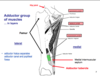
list the medial thigh muscles

A
  • pectineus
  • adductor brevis
  • a. longus
  • a. magnus
  • gracilis
  • obturator externus
2
Q

where does the saphenous nerve leave the adductor canal?

A

between sartorius and gracilis;

just above the knee on the medial side

3
Q

contents of the adductor canal

A

saphenous nerve

femoral artery

femoral vein

4
Q

pectineus:

origin/ insertion

A

o: superior pubic ramus & pectin pubis
i: prox. 1/2 of pectineal line of the femur
* crosses hip; “pecten” means tooth of comb*

5
Q

pectineus:

action/ innervation

A
  • act: hip flexion and weak hip adduction
  • inn: femoral nerve or obturator nerve (or both)
6
Q

adductor brevis:

origin, insertion

A

o: body and anterior/ventral surface of inferior pubic ramus
ins: distal part of pectineal line of femur & prox. 1/3 of medial lip of linea aspera
* crosses hip*

7
Q

adductor brevis:

action, innervation

A

act: ADDUCTion of hip, and flexion
inn: obturator nerve (anterior, posterior divisons, or both)

8
Q

adductor longus:

origin, insertion

A

o: ventral surface of body of pubic, adjacent to pubic tubercle
ins: middle 1/3 of medial lip of linea aspera

9
Q

adductor longus:

action, innervation

A

act: ADDUCTION of hip, flexion of hip joint
inn: anterior branch of obturator nerve

10
Q

gracilis

origin, insertion

A

o: ventral surface of body of pubis & inferior pubic ramus
ins: tibia (pes anserinus) - posterior to sartorius & superior to semitendinosus

11
Q

gracilis:

action, innervation

A

act: ADDUCTS hip, flexes the knee, medially rotates the leg while knee is flexed
inn: anterior division of obturator nerve

12
Q

adductor magnus:

origin, insertion

A
  • o:
    • hamstring part: inferolateral part of ischial tuberosity
    • adductor part: ventral surface of ischiopubic ramus
  • ins:
    • gluteal tuberosity & proximal ¼ of medial lip of linea aspera (minimus part of magnus) &
    • distal ¾ of medial lip of linea aspera (adductor part) adductor tubercle via tendon &
    • part of medial supracondylar ridge(hamstring part)
13
Q

adductor magnus:

action, innervation

A
  • act: ADDUCTION of hip, flexion of hip, (adductor part)
    • extension of hip w/ hamstrings (hamstring part)
  • inn:
    • hamstring: tibial nerve
    • adductor part: posterior branch of obturator nerve
14
Q

CC: groin pull

A

an injury to the muscle tendon unit that produces pain on palpation of the adductor tendons or its insertion on the pubic bone with or without pain during resisted adduction

15
Q

CC: riders’ bones

A

localized ossification sometimes seen on the inner aspect of the lower end of the tendon of the adductor muscle of the thigh in horseback riders.

16
Q

which adductor group muscles are found in the:

anterior layer

A
  1. pectineus
  2. adductor longus
17
Q

which adductor group muscles are found in:

middle layer

A
  • adductor brevis
  • gracilis
18
Q

which adductor group muscles are found in:

posterior layer

A

adductor magnus

19
Q

which adductor muscles are found in the

anterior, middle, and posterior layers

A
  • ANTERIOR
    • a. Longus
    • Pectineus
  • MIDDLE
    • a. Brevis
    • Gracilis
  • POSTERIOR
    • a. Magnus
20
Q

cc: which of the adductor muscles can be transplanted?

A

gracilis (part of all w/ neurovascular supply) can be transplanted to replace hand muscles &

to replace external anal sphincter muscle (free up distal attachment)

21
Q

which part of the adductor magnus is the “hamstring part”?

A

the ischiocondylar part

22
Q

identify the anterior and posterior divisions of the obturator nerve

A
  1. anterior division
  2. posterior division
23
Q

course of the internal iliac artery leaving pelvis

A

leaves pelvis through obturator canal;

terminates by dividing to anterior and posterior branches

24
Q

anterior branch of internal iliac artery supplies:

A

medial thigh muscles (adductor group)

25
**posterior branch of internal iliac artery** supplies:
* adductor magnus, hamstrings, acetabulum, including a small branch to the head of femur
26
which artery assists the **deep femoral artery** in supplying the **medial thigh muscles?**
the **obturator artery**
27
what is the major artery supplying blood to the muscles of the thigh?
deep femoral artery
28
which muscles are in the **posterior compartment** of the thigh?
* biceps femoris * semitendinosus * semimebranosus
29
another name for the posterior compartment muscles? why?
hamstrings; these are long skinny muscles; fuse w/ gluteus maximus
30
which 2 hamstring muscles have a common origin?
biceps femoris & semitendinosus
31
semitendinosus: origin/insertion
o: superomedial part of **ischial tuberosity** w/origin of long head of **biceps femoris** ins: pes anserinus * crosses hip and knee*
32
semitendinosus: action, innervation
act: extends hip and flexes knee; can medially rotate thigh and flexed leg inn: tibial nerve
33
semimembranosus (SM): origin, insertion
* o: as flat tendon from superolateral part of ischial tuberosity (also from sacrotuberous ligament & ischial attachment of adductor magnus) * ins: widespread, incl: * groove on medial condyle of tibia (posteriorly) * medial border of tibia * fascia covering politeus * knee joint capsule as oblique popliteal ligament
34
semimembranosus: act, inn
action: extends hip, flexes leg, medially rotates thigh and flexed leg inn: tibial nerve
35
biceps femoris: which head of muscle crosses both hip and knee?
long head crosses both hip and knee; short head ONLY crosses knee
36
biceps femoris: origin, insertion
* origin * long head: superomedial part of ischial tuberosity w/ semitendinosus & sacrotuberous ligament * short head: lateral lip of linea aspera, lateral supracondylar ridge, & lateral intermuscular septum * insert * 3 laminae/ layers of insertion: * **\*superficial laminae: head of fibula** * *deep & intermediate laminae are not important*
37
biceps femoris: action, innervation
* act: long head - extends & laterally rotates hip * both heads - flex knee and laterally rotate flexed knee * inn: * long head - tibial nerve * short head - common fibular nerve
38
lumbosacral trunk: assoc. rami
L4/L5
39
what contributes to SCIATIC NERVE?
**Common tibial/peroneal nerve** (dorsal/posterior division) & **Tibial** (ventral anterior division) w/in same sheath
40
which division innervates the **posterior cutaneous nerve of the thigh**?
both posterior and anterior division innervation
41
which ventral rami are involved in ## Footnote **Sacral Plexus**
L4- S4
42
nerves of the sacral plexus: list
1. superior gluteal 2. inferior gluteal 3. sciatic * common peroneal * tibial nerve 4. posterior femoral cutaneous 5. pudendal
43
mnemonic for sacral plexus
**Sacral innervation could totally prove preposterous** * S: superior gluteal nerve (L4, L5, S1) * I: inferior gluteal nerve (L5, S1, S2) * C: common fibular nerve (L4, L5, S1, S2) -- *sciatic* * T: tibial nerve (L4, L5, S1, S2, S3) -- *sciatic* * P: posterior femoral cutaneous nerve (S1, S2, S3) * P: pudendal nerve (S2, S3, S4)
44
which 2 nerves travel in same sheath to make sciatic nerve?
* C: common fibular nerve (L4, L5, S1, S2) * T: tibial nerve (L4, L5, S1, S2, S3)
45
superior gluteal nerve ventral rami
S: superior gluteal nerve (L4, L5, S1)
46
inferior gluteal nerve ventral rami
I: inferior gluteal nerve (L5, S1, S2)
47
posterior femoral cutaneous nerve ventral rami
P: posterior femoral cutaneous nerve (S1, S2, S3)
48
pudendal nerve ventral rami
P: pudendal nerve (S2, S3, S4)
49
posterior compartment muscle and its innervation
1. semitendinosus - TIBIAL 2. semimembranosus - TIBIAL 3. biceps femoris - * long head - TIBIAL * short head - COMMON FIBULAR
50
which nerves innervate the skin of the posterior thigh?
1. \*POSTERIOR FEMORAL CUTANEOUS 2. lateral femoral cutaneous 3. medial femoral cutaneous 4. obturator nerve
51
posterior femoral cutaneous nerve: ventral rami and fibers
* ventral rami of S1-S3 * dorsal division fibers from S2 * ventral division fibers from S2
52
major arteries supplying the thigh?
1. deep femoral artery 2. obturator artery 3. femoral artery 4. lateral circumflex femoral artery 5. medial circumflex femoral artery
53
relationship between **deep femoral artery** and **adductor longus / brevis/ magnus?**
deep femoral artery runs **DEEP to the adductor longus** muscle, and **SUPERFICIAL to adductor brevis and magnus**
54
Relationship of deep femoral artery and perforating arteries to femur and adductor magnus
* the external iliac artery --\> becomes the femoral artery when it **crosses under the inguinal ligament** and enters the femoral triangle. * **Lateral femoral circumflex artery** – Wraps round the anterior, lateral side of the femur, supplying _some of the muscles on the lateral aspect_ of the thigh. * **Medial femoral circumflex artery** – Wraps round the posterior side of the femur, _supplying its neck and head_. * **perforating arteries (3-4 arteries):** that perforate the adductor magnus, contributing to the supply of the muscles in the medial and posterior thigh
55
perforating branches of deep femoral artery: general characteristics
* 4 in number * all have descending & ascending branches that communicate * supply: adductors, hamstrings, vastus lateralis, and gluteus maximus
56
2nd perforating branch of deep femoral artery: function
provides nutrient artery to the femur
57
4th perforating branch of deep femoral artery: function
communicates with muscular branches of popliteal artery, providing **important collateral pathway** between thigh & leg (in femoral artery occlusion)