Physiology of the nervous system Flashcards
Functions of the nervous system
Directs immediate response to stimuli
Coordinates or moderates activities of other organ systems
Provides and interprets sensory information about external conditions
Major organs of the nervous system
Brain
Spinal cord
Periphal nerves
Sense organs
Central nervous system is made up of
Brain
Spinal cord
Periphal nervous system is made up of
All neurones outside of the brain and spinal cord
Functions of the CNS
Process and coordinate:
- Sensory data
- Motor comands
- Higher functions of the brain such as intelligence, memory, learning and emotion
Functions of PNS
Deliver sensory information to the CNS
Carry motor comands to peripheral tissues and effectors
2 cell types of neural tissue
Neurones
Neuroglia (glial cells)
2 types of neural tissue matter
Grey matter
White matter
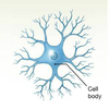
Neurones
Cells that send and recieve signals
Neuroglia
Glial cells
Cells that support and protect neurones
Types of neurones
Multipolar
Bipolar
Unipolar
Anaxonic
Examples of neuroglia
Ependymal cells
Astrocytes
Oligondendrocytes
Mircoglia
Schwann cells
Grey matter
Mainly cells bodies and unmyelinated neurones
White matter
Mainly axons of myenilated neurones
Functional classifications of neurones
Sensory neurones
Association neurones
Motor neurones
Sensory neurones
Afferent
From receptors to CNS
From lower to higher CNS levels
Interneurones
Association neurones
Link sensory to motor neurones
Motor neurones
Efferent
From CNS to muscles
From higher to lower CNS levels
Efferent autonomic nerve pathways
Have a 2 neurone arrangement
Pre- and post ganglionic nerve
Efferent somatic nerve pathway
Single neurone from CNS to effector
Which efferent pathway only has a single neurone
Somatic
Which efferent pathway has a pre and post-ganglionic nerve
Autonomic
Resting membrane potential
About -70mV
How resting membrane potential is achieved
Large, negatively charged proteins stuck in cell
More positive ions outside of cell
Na+/K+ pump
Membrane a lot more permeable to K+
Which ion is the cell membrane more permeable to
K+
The two types of force that influence the movement of ions across the plasma membrane
Chemical gradients
Electrical gradients
Chemical gradients in ion movement across membrane
Ions want to pass along concentration gradient - from high concentrations to low
Na+ wants to pass into cell
K+ wants to leave cell
Electrical gradients in the ion moevement across the cell membrane
Ions want to pass to areas of opposite charge
Inside of cell negatively charged so both Na+ and K+ want to enter
Electrochemical gradient effect on Na+ and K+ ions
Na+
- Both electrical and chemical gradients attract into cell
K+
- Oppose each other
- Chemical attracts out
- Electrical attracts in
Ion channels
Proteins spanning the lipid membrane
Determine the permeability to an ion

Types of ion channels
Passive
Gated
Passive ion channels
Also called leak channels
Always partially open
Gated ion channels
Open and close in response to specific stimuli
3 main types of gated channel
- Chemically regulated
- Voltage regulated
- Mehcanically regulated
3 possible states of gated ion channels
Activatable
- Closed but capable of opening
Activated
- Open
Refractory
- Closed and incapable of opening
Chemcially regulated ion channel
Channel opens when a chemical binds to it
Closes when the bound chemical is broken down
Voltage regulated ion channel
Reacts to changes in voltage
Mechanically regulated ion channel
Pressure causes gate to open
Closes when pressure disapears
1st step making a graded potential
Chemical neurotransmitter binds to receptor on chemically regulated Na+ channel
Channel opens
Na+ enters cell along it’s electrochemical gradient
What does the initial rush of Na+ ions entering the cell cause in producing a graded potential
Membrane becomes depolarised
This then also depolarises the adjacent mebrane
Stimulating and inhibiting influences on resting membrane potential
Stimulating
- Stimulating neurotransmitter
- Na+ influx
- Depolarisation
Inhibiting
- Inhibitory neurotransmitter
- K+ influx
- Hyperpolarisation
Are the influences on the resting membrane potential always trying to cause depolarisation?
No
Can also be sent an inhibitory neurotransmitter to inhibit
Graded potential
Tempory, localised change in resting potential
Caused by stimulus
Action potential
Electrical impulse and frequency signal
Produced by a graded potential that exceeds threshold
Propogates along the surface of axon to synapse
Size of action potential always the same
All or nothing principle
Difference between a graded potential and an action potential
Action potential is a result of a graded potential reaching threshold
Graded potential is localised, action potential propogates along axon
Anaxonic neuron
No axon, just dendrite
Small
Lots of dendrites
Found in brain and special sense organs
Bipolar neuron
One axon and one dendrite on opposite sides of the cell body
Occur in special sense organs

Unipolar neuron
Dendrite and axon fused and contineous
Cell body off to the side
Most neurones in the PNS and unipolar

Multipolar neuron
2 or more dendrites
Single axon
Most common type of neuron in CNS

Action potential sequence
Resting state
- Depolarisation to threshold
- Activation of Na+ channels and rapid depolarisation
- Innactivation of Na+ channels and activation of K+ channels
- Hyperpolarisation
- Return to normal permeability and resting state
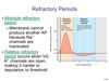
Absolute refractory period
No stimulus can cause an action potential to be generated
Na+ channels incapable of opening
Relative refractory period
Stronger than normal stimulus is required
When the refractory periods occur
Na+/K+ pump
Pumps Na+ out
Pumps K+ in
Has ATP binding site to provide energy needed
How is the current set up in neurones
One area of membrane is depolarises membrane
Membrane potential next to this graded potential is different
This sets up the current
Size of current depends on the size of the graded potential
What sets up an action potential
The current produced by the differce between a graded potential and the membrane potential next to it
All or nothing principle
Threshold for depolarisation must be met otherwise action potential will not be generated
What about an action potentail can the depolarising stimulas affect
If it happens or not
How often an action potential is generated
How an action potential is propogated along the axon
Depolarised membrane sets up a local current because of charge difference with neighbour
Neighbour’s voltage gated Na+ channels activated, causing it to also become depolarised
Current set up by differece between the neighbour and its neighbour
Process repeats along axon
Saltatory propagation
Speeds up propogation of action potentials
Axons myenlinated so only a few areas of cell membrane exposed
Ion exchange can only happen here
Rather than every part of axon being depolarised the local current causes impulse to jump from node to node
Node of Ranvier
Exposed area of myelin sheath
Where ion transfer is possible on myelinated axons
Schwann cells
Neuroglia cells
Produce myelin that wraps around axon covering it
Benifits of nerves being myelinated
Causes salvatory propogation which is quicker than normal propogation
Uses less energy as fewer ions need to cross the membrane
Post-synaptic cells of synapses could be…
Another nerve
Smooth muscle
Skeletal muscle
Glangular tissue
Types of synapse
Chemical
Electrical
Chemical synapse
Transfers from pre-synaptic cell to post-synpatic cell
Uses neurotransmitters
Electrical synapse
Gap junctions - pores in the membrane between cells
Pores allow passage of ions
Passage of ions means passage of their individual charge
Cholinergic synapses
Use aceytlcholine
Very common
Synapses that are cholinergic
Skeletal muscle neuromuscular junctions
Many synapses in CNS
All nerve-nerve synapses in ANS
All neuro-effector synapses in the parasymathetic nervous system
What synapse uses ACH
Cholinergic synapses
At what voltage do Na+ voltage gated channels open
-60mV
What causes more Na+ channels to open
Positive feedback
What does the increased movement of Na+ cause
As it enters the cell it causes the cell membrane to depolarise
At what voltage does the Na+ channels close and K+ channels open
+30mV
What does the opening of K+ channels in the propogation of an action potential cause
K+ ions flood out of cell
Lowers membrane charge
When the K+ ions leave the cell in propogating an action potential, with/against what gradient/s is it travelling
Electrical
- With
Chemcical
- Against
Ca2+ role at synapse
Enters synaptic knob
Stimulates excosytosis of ACh from synaptic vesicles and into synpatic cleft
Events at cholinergic synapse
- Ca2+ enters synaptic knob
- Causes excocytosis of ACh
- ACh diffuses across synaptic cleft
- ACh binds to receptors on post-synaptic membrane opening Na+ channels
- Post-synaptic membrane depolarised
- Acetylcholinesterase breaks down ACh
- Breakdown products recyled by pre-synaptic knob
AChE
Acetylcholinesterase
Breaks down ACh into choline and acetate
What recycles and produces new ACh at the synapse
In what part of synapse
Acetyl CoA
Synaptic knob
Inhibitory neurones
Release neurotransmitters that hyperpolarise the nerve cell membrane
Excitatory neurones
Release neurotransmitters that depolarise the nerve cell membrane
Summation of presynaptic inputs
Single EPSPs may not be enough to depolarise membrane to threshold
EPSPs can combine to acheive threshold
Propagation of action potentials
EPSP
Exitatory post-synaptic potential
Graded depolarisation caused by arival of neurotransmitter at post-synaptic membrane
Caused by opening of chemically gated Na+ channels
Propagation of action potentials
IPSP
Inhibitory post-synaptic potential
Graded depolarisation caused by arival of neurotransmitter at post-synaptic membrane
Maybe caused by opening of chemical gated K+ channels
Why would opening K+ channels decrease the likelihood of an action potential being propagated
K+ would leave the cell along it’s chemical gradient
Would cause the membrane to become hyperpolarised
Would take a larger than usual stimulus to reach threshold
Types of summation
Temporal
Spatial
Temporal summation
Multiple ESPSs in rapid succession from a single synapse
Spatial summation
Simultaneous mulitple EPSPs from different synapses
ANS effectors
Cardiac muscle
Smooth muscle
Glandular tissue
ANS
Autonomic nervous system
SNS
Somatic nervous system
SNS effectors
Skeletal muscle
SNS type of control
Voluntary
ANS type of control
Involuntary
SNS neural pathway
CNS direct to effector
ANS neural pathway
CNS
Pre-ganglionic fibre to synapse with post-ganglionic cell in ganglion
Effector
SNS action on effector
Always excitatory
ANS action on effector
Can be excitatory or inhibitory
Depends on ANS division and effector type
SNS neurotransmitters
ACh
ANS neurotransmitters
ACh
Noradrenaline
The different pathways of ANS
Sympathetic
Parasympathetic
Sympathetic pathway
Fight or flight
Long pre-ganglionic fibre
Short post-ganglionic fibre
Can stimulate adrenal medulla to produce hormones to travel in blood stream to affect target organs
Both pathways of ANS consist of
Pre-ganglionic cell and fibre
Post-ganglionic cell and fibre
Hormones used in sympathetic pathway
Adrenaline
Noradrenaline
Hormones used in parasympathetic pathway
Acetylcholine
Areas that the sympathetic pathway can affect
Eyes
Skin
Arteries
Heart
Adrenal gland
Pancreas
Lungs
GI tract
Liver
Adipose tissue
Areas that the parasympathetic pathway can affect
Eyes
Heart
Pancreas
Lungs
GI tract
Liver
Two types of receptors in ANS
Adrenergic
Cholinergic
Adrenergic receptors
α1
α2
β1
β2
Found where and does what
α1 recpetors
Part of ANS
Found in most tissues
Stimulates metaoblism
Activates enzymes and releases intracellular Ca2+
Found where and does what
α2 receptors
Sympathetic
- Found in sympathetic neuromuscular or neuroglandular junctions
- Inhibits effector cell
- Reduces cAMP concentrations
Parasympathetic
- Found in parasympathetic neuromuscular or neuroglandular junctions
- Inhibits neurotransmitter release
- Reduces cAMP concentrations
*
Found where and does what
β1 receptors
Found in heart, kindeys, liver and adipose tissue
Stimulates increased energy consumption by activating enzymes
Found where and does what
β2 receptors
Found in smooth muscle in vessels of heart and skeletal muscle and small muscle layers in intestines, lungs and bronchi
Causes muscles tissue to relax
Activates enzymes
Cholinergic receptors
Nicotinic
Muscarinic
Found where and does what
Nicotinic receptors
Found in all autonomic synapses between pre-ganglionic and ganglionic neurones
Also found in neuromusclular junctions of SNS
Causes muscular contraction
Opens chemically gated Na+ channels
Found where and does what
Muscarinic receptors
Found in all parasympathetic and cholinergic sympathetic neuromuscular or neuroglandular junctions
Activates enzymes that cause changes in membrane permeability to K+
Sympathetic effects on eye
Pupil dilation
Sympathetic effects on skin
Increased sweating
Sympathetic effects on ateries
Dilation in:
- Skin
- Heart
- Skeletal muscle
- Lungs
- Brain
Constriction of viscera and kidneys
Sympathetic effects on the heart
Increases heart rate
Increases force of contraction
Increases blood pressure
Sympathetic effects on the adrenal gland
Increased adrenaline and noradrenaline secretion
Sympathetic effects on the pancreas
Decreased insulin secretion
Sympathetic effects on the lungs
Increased airway diameter
Sympathetic effects on the GI tract
Activity decreased
Sympathetic effects on the liver
Glycogen breakdown
Glucose synthesis and release
Sympathetic effects on the andipose tissue
Lipolysis
Fatty acid release
Parasympathetic effects on the eye
Pupil constriction
Parasympathetic effects on the skin
No effect
Parasympathetic effects on the arteries
No effect
Parasympathetic effects on the heart
Decreased heart rate
Decreased blood pressure
Parasympathetic effects on the adrenal gland
No effect
Parasympathetic effects on the pancreas
Increased insulin secretion
Parasympathetic effects on the lungs
Decreased airway diameter
Parasympathetic effects on the GI tract
Increased activity
Parasympathetic effects on the liver
Glycogen synthesis
Parasympathetic effects on the adipose tissue
No effect
Process leading to contraction (neuromuscular junction)
Excitation contraction coupling
What are each muscle fibres controlled by
A single motor end plate
Location of CNS visceral motor neurones - sympathetic
Lateral grey horns of spinal segments
Location of CNS visceral motor neurones - parasympathetic
Brain stem and spinal segments
Location of PNS ganglia - sympathetic
Near vertebral column
Lengths of ganglionic fibres in sympathetic pathway
Short pre-ganglionic
Long post-ganglionic
Lengths of ganglionic fibres in parasympathetic pathway
Long pre-ganglionic
Short post-ganglionic
5 steps of relfex arc
- Receptor senses a stimulus
- Sensory neuron transmits signal up the PNS to the CNS
- Integration center decodes the signal
- Motor neurone sends directions back to the site of the stimulus
- Effector cells respond by contracting or secreting
