PBL 4 Flashcards
Peyer’s patches are large aggregations of what?
lymphoid cells
Where in the small intestine would you find Paneth cells?
base of crypts of lieberkuhn
The large intestine contains deep crypts - true or false?
True- colic crypts
What do the parietal cells of the stomach secrete?
Gastric acid (hydrochloric acid)
Which part of the small intestine produces the hormone cholecytoskinin pancrozymin (CCK)?
Duodenum
What are the large folds found in the small intestine called?
plica circulares
The duodenum contains specialised glands that secrete alkaline mucus. What are they called?
brunner’s glands
The space of Disse is found between ___ cells and hepatocytes in the liver
Endothelial

The liver receives blood from the hepatic artery, and also from which other vessel?
hepatic portal vein
what pH is the fluid secreted by the exocrine part of the pancreas - acid or alkaline?
Alkaline
Which cells in the pancreas produce the bicarbonate?
Centroacinar cells- they represent an extension of the intercalated duct into eahc pancreatic acinus
Also known as duct cells
The stomach has three layers of smooth muscle, circular, longitudinal and what?
oblique
What are the 4 regions of the large intestine?
- Caecum
- Colon
- Rectum
- Anal canal
What is the function of the caecum?
Receives soft, water-rich waste from the ileum.
Acts as a reservoir for ingesta received from the ileum
Contains a high number of aerobic microflora- function unknown
What are the subdivisions of the colon and what is its function?
Subdivisions:
- Ascending
- Transverse
- Descending
- Sigmoid
Function is to absorb water, salts, electrolytes and other essential vitamins and minerals back into the body
What is the blood supply to the caecum?
The ileocolic artery, which is a branch of the superior mesenteric artery.
What is the innervation to the caecum?
Innervated by the ileocolic branch of the superior mesenteric plexus
The subdivisions of the colon (Ascending, transverse, descending and sigmoid) form an arch with encircles what?
The small intestines
At which part of the colon is the colon is attached to the diaphragm? and what attaches it?
Located at the transverse colon
The phrenicocolic ligament attaches the transverse colon to the diaphragm.
Which parts of the colon are intraperitoneal?
Transverse colon
Sigmoid colon
Which parts of the colon are retroperitoneal?
Ascending colon
Descending colon
Which parts of the large intestine are intraperitoneal?
- Caecum
- Appendix
- Transverse colon
- Sigmoid colon
Which parts of the large intestine are retroperitoneal?
- Ascending colon
- Descending colon
- Rectum
- Anal canal
Which part of the colon is the least fixed and what is the potential consequence?
The transverse colon is the least fixed part
Consequence: it can vary in position (can dip into the pelvis in tall, thin individuals). No pathology associated with this.
Describe the Left colic flexure ( aka splenic flexure)?
The left colic flexure marks the transition from the transverse colon to the descending colon.
It is located at the spleen.
Describe the Right colic flexure (aka the hepatic flexure)?
The right colic flexure marks the transition from the ascending colon to the transverse colon.
It is located at the liver.
Describe the paracolic gutters?
The paracolic gutters are two spaces between the ascending/descending colon and the posterolateral abdominal wall.
Right paracolic gutter - between ascending colon and posterolateral abdominal wall
Left paracolic gutter - between descending colon and the poterolateral abdominal wall
What are the clinical importance of the paracolic gutters?
These structures are clinically important, as they allow material that has been released from inflamed or infected abdominal organs to accumulate elsewhere in the abdomen
What are the 3 anatomical structures that are unique to the large intestine?
- Appendices epiplociae
- Taeniae coli
- Haustrations

Describe the appendices epiploicae?
These are small pouches of peritoneum, filled with fat, attached to the surface of the large intestine.

Describe the taeniae coli?
The outer longitudinal layer of muscle in the muscularis externa is incomplete
There are 3 bands of thickened muscle, thicker longitudinal muscle layer.
These three bands are located on the outside of the ascending, transverse, descending and sigmoid colons

Describe the haustrations of the large intestine?
Sacculations found along the length of the large intestine, giving its appearance of segmented.
Describe how the haustration appearance was created in the large intestine
The sacculations are caused by the contraction of taenia coli. The taenia coli run the length of the large intestine.
Because the taenia coli are shorter than the large bowel itself, the colon becomes sacculated, forming the haustrations.
At what point does the unique features of the large intestine (taeniae coli, haustrations and appendices epiplociae) cease?
At the rectosigmoid junction

What is the function of the rectum?
Role as a temporary store of faeces until defecation
How is the rectum distinct from the colon?
Absence of taenia coli, haustra, and appendices epiploicae
What are the five flexures of the rectum?
Two antero-posterior flexures:
- Sacral flexure
- Anorectal flexure
Three lateral flexures:
- Upper flexure
- Middle flexure
- Lower flexire

What are the two major flexures of the large intestine?
- Left colic flexure (splenic flexure)
- Right colic flexure (hepatic flexure)
Describe the ampulla?
This is the final stage of the rectum
Relaxes to accumulate and temporary stores faeces.

Describe the rectovesical pouch?
A sac between the rectum and the urinary bladder in males that is formed by a folding of the peritoneum
only in males

Describe the rectouterine pouch?
Also known as the pouch of Douglas.
Only in females
An extension of the peritoneal cavity between the rectum and back wall of the uterus

Describe the peritoneal covering of the rectum
Superior third- the anterior surface and lateral sides are covered by peritoneum.
Middle third - an anterior peritoneal covering only.
Lower third - no peritoneum associated with it.
What are the arterial supply to the rectum?
Three main arteries:
- Superior rectal artery (terminal continuation of the IMA)
- Middle rectal artery (branch of the internal iliac artery)
- Inferior rectal artery (Branch of the internal pudendal artery)
What artery is the terminal continuation of the inferior mesenteric artery?
Superior rectal artery
What is the venous drainage of the rectum?
Superior, middle and inferior rectal veins.
The superior rectal vein: empties into the portal venous system.
Inferior and middle rectal vein: empties into the systemic venous system.
Describe the portocaval anastomosis?
Anastomoses between the portal and systemic veins are located in the wall of anal canal.
What is the function of the anal canal?
Role in defecation and maintains feacal continence.
It is located within the anal triangle of the perineum between the right and left ischioanal fossae.
Describe how the anal sphincters prevent the passage of faecal material?
The internal and external anal sphincetse collapse the anal canal preventing the passage of faecal material
Describe the internal anal sphincter?
Surrounds the upper 2/3 of the anal canal.
It is formed from a thickening of the involuntary circular smooth muscle in the bowel wall
Describe the external anal sphincter?
Surrounds the lower 2/3 of the anal canal.
Blends superiorly with the puborectalis muscle
Describe the layout of the anal sphincters?
Upper third: surrounded by the internal anal sphincter muscle.
Middle third: surrounded by the internal + external anal sphincter muscle.
Lower third: surrounded by the external anal sphincter muscle
Describe the anorectal ring?
Muscular ring at the junction of the rectum and anal canal.
Formed by the fusion of the internal anal sphincter, external anal sphincter and puborectalis muscle.
Able to palpate.

What are the 3 major functions of the large intestine?
- Recovery of water and electrolytes from ingesta
- Formation and storage of faece
- Microbial fermentation
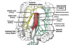
Name these parts of the large intestine?


Name these parts of the colon?


There are more than 400 different species of bacteria in the colon. Which type of the bacteria are of vast majority in colon?
Strict anaerobic
Which important vitamin is made and absorbed in the large intestine?
Vitamin K
The superior rectal artery originates from which artery?
Inferior mesenteric artery
The internal anal sphincter is supplied by which nerve?
Hypogastric nerve
Approximately 9 litres of water that go through the GI tract daily, roughly how much makes it out in the faeces?
100ml
When seeing a patient with diarrhoea, the most important investigation is?
A clear history
When treating someone with gastroenteritis, the most important management is?
Rehydration
Name these parts of the anal canal?


The external anal sphincter is supplied by which nerve?
pudendal nerve
Fill in the table in regards to the following volume of fluid?


Approximately 9 litres of water that go through the GI tract daily, roughly how much is absorbed in the large intestine?
1.4 litres
The majority of water absorption occurs where?
In the small intestine particularly the duodenum and jejunum
What are the major differences histologically between the large and small intestine?
LI is similar in feature to the SI except:
- No villi present
- Goblet cells are more abundant
- No Paneth cells
- Crypts are deeper and thicker
- Diameter of the lumen is 3x bigger
- Intestinal wall is much thinner
Name the 3 prominent patterns of motility in the large intestine?
Segmentation contractions
Mass movements
Gastrocolic reflex
What is the difference in the function of the proximal and distal portions of the colon?
Absorbing colon – proximal half of the colon’s function is absorption.
Storage colon – Distal half of the colon, function is for faecal storage.
What is the maximum capacity of the large intestine to absorb water and electrolytes?
Can absorb a maximum of 5 to 8 litres of fluid and electrolytes per day.
Describe the absorption of sodium in the large intestine?
The sodium gradient provides energy for active transport of other electrolytes.
The sodium is continuously excreted from the epithelial cells into the plasma via the sodium potassium pump (3 sodium out, 2 potassium in). This pump allows for the gradient changes of sodium but no electrical changes in the cell.
Hence, it creates a gradient allowing sodium to be absorbed in the lumen side of the cell.
I.e. constant gradient for sodium transport.
The absorption of water is dependent on which electrolyte?
Sodium
Describe the mechanism of water absorption into the large intestine?
In the intestine sodium drives the passive absorption of water. Water moves through the epithelium via osmosis.
Sodium comes into the enterocyte of the large intestine, resulting in the increase in concentration of the water in the lumen of the large intestine (i.e. higher osmolality – the total number of solutes particles per litre is higher). Results in a water gradient from the lumen into the plasma.
SGLT1 transporter is the contransporter of which electrolytes?
Sodium and glucose
Both into the enterocytes
Describe the transporting mechanism for sodium and water?
Sodium entry drives the entry of water.
Water enters via osmosis, down its gradient
Describe the transporting mechanism for sodium and hydrogen?
Exchanger
Sodium from the lumen into the enterocytes
Hydrogen out to the lumen from the enterocytes

Describe the absorption/secretion mechanism for chloride and bicarbonate?
The movement of sodium into the plasma produces an electrochemical gradient to allow absorption of chloride. Chloride ions are exchanged for bicarbonate ions (causing net bicarbonate secretion).

Describe the absorption/secretion process for potassium in the large intestine?
Absorption of water along the length of the bowel concentrates potassium in the lumen. This provides an electrochemical gradient for the movement of potassium into the plasma.
In the colon potassium may be absorbed or secreted depending on the remaining concentration in the lumen and the electrochemical gradient created by the active absorption of sodium.
What is the role of short chain fatty acids in the absorption mechanisms in the large intestine?
The main three SCFAs (acetate, propionate and butyrate) stimulates colonic sodium and fluid absorption.
SCFA absorption is occurs more readily in the proximal colon (‘leaky’ epithelium in the proximal colon and ‘tight’ and aldosterone dependent epithelium in the distal colon).
SCFA absorption is coupled with sodium, probably by the sodium/hydrogen exchanger.
How are short chain fatty acids produced?
Microbial digestion of indigested carbohydrates, particularly cellulose, results in short-chain fatty acids. The short-chain fatty acids are either used by the bacteria, or absorbed by the cells in the large intestine
Name two endocrine factors that can influence the absorption in the large intestine?
Aldosterone
Somatostatin
Both act to increase the net absorption of water and electrolytes by stimulating the basolateral sodium-potassium ATPase. This increases the electrochemical gradient and driving force for sodium absorption.
Aldosterone is produced by ___ glands in response to ____ blood levels.
Adrenal glands
Low blood levels
Describe the composition of the gut microbiota?
The gut microbiota contains a densely-populated microbial ecosystem with up to 1012 cells per gram of intestinal content.
The bacteria represent between 300-1,000 different species.
However, approx. 99% come from the about 30-40 species.
The bacteria from the gut microbiota make up which percentage of the dry mass of faeces?
60% of the dry mass of faeces
The majority of the bacteria are anaerobic or aerobic?
Anaerobic
Are the majority of the bacteria strict anaerobes or facultatve anaerobes?
A facultative anaerobe is an organism that makes ATP by aerobic respiration if oxygen is present, but is capable of switching to fermentation if oxygen is absent.
Majority are strict anaerobic
Name the 4 main domains of bacteria in the human gut are?
- Firmicutes
- Bacteroidetes
- Actinobacteria
- Proteobacteria
In the stomach and small intestine there are ____ species of bacteria present.
relatively few
The gut microbiota is mainly located in the what?
Large intestine
What organisms make up the microbiota of the gut?
Majority is bacteria
Also fungi, viruses and protists
What are the three most important roles of the gut microbiota?
- To maintain the integrity of the mucosal barrier
- Aid metabolism and provide nutrients
- Protect against pathogens.
Describe how the gut microbiota does not get destroyed by the immune system?
Both the gut microbiota and intestines develop at the same time. This results in the mucosal barrier tolerating the microbiota
The gut-associated lymphoid tissue can detect and react to pathogens. It develops tolerance to microbiota species but not to other microorganisms.
Describe how the gut microbiota maintains the integrity of the mucosal barrier?
The intestine is highly density populated with microbiota, therefore it provides a physical barrier against other microorganisms.
Provides competition for invading pathogens
Furthermore, the microbiota influences the quantity and properties of intestinal mucus.
Describe the gut micobiota’s mechanisms to protect against pathogens?
- Certain bacteria that compose the microbiota can produce and secrete molecules with bacteriostatic and bactericidal activities, which prevents pathogen growth.
- Some bacteria can interfere with pathogen gene expression. Overall, preventing the pathogens from colonising the gut.
- The bacteria are able to metabolise host derived molecules and convert them into secondary metabolites which can inhibit microorganisms from growing, e.g. C.difficile growth is inhibited by secondary bile acids, which is a metabolite of bile metabolism, via microbiota.
Describe how the gut microbiota can out compete with the invading pathogens for nutrient supply?
Competition from nutrient supply is a major way in which the microbiota protects against pathogens. The invading microorganisms and the already established microbiota compete for the same nutrient sources. However, the bacterial species already established are highly adapted to the specific gut environment, as no two peoples gut environment is the same.
Describe the role of gut microbiota in the metabolism process
The microbiota are important in metabolising undigested material, e.g. cellulose. The microbiota bacteria express carbohydrate-active enzymes, which breaks down undigested carbohydrates into short-chain fatty acids.
The SCFAs are absorbed by the enterocyte cells, where they are involved in the regulation of cellular processes e.g. gene expression. SCFAs are also important in stimulating the absorption of sodium, indirectly effecting the absorption of water.
The 3 predominant short-chain fatty acids are?
Butyrate, propionat and acetate (found in proportion of 3: 1: 1)
Microbiota bacteria has a role in the synthesis of which vitamins?
Vitamins B and K
Name a few factors that can affect the composition of the gut microbiota?
Composition of the microbiota are influenced by:
- Environmental factors such as diet and host genetics.
- Chemical, nutritional and immunological factors.
- Broad spectrum antibiotics
Describe the process of vitamin B12 absorption?
Occurs in the small intestine (in the ileum)
First, hydrochloric acid in the stomach separates vitamin B12 from the protein to which vitamin B12 is attached in food. After this, vitamin B12 combines with a protein, intrinsic factor, that is produced by the stomach. After which, B12 can be absorbed by the body in the ileum
Why are antibiotics not advised for diarrhoea?
Because its a self limiting condition i.e. gets better on its own.
The antibiotics will only reduce the time of the diarrhoea by a day or so
Why are broad spectrum antibiotics used in high risk patients with diarrhoea?
broad spectrum antibiotics are commonly used due to multiple reasons including:
- The length of time a culture would take
- Cultures have a high risk of a negative result (as the risk of the culture growing multiple species is high).
When potassium and bicarbonate are lost together what does not usually develop? and why?
Hypokalaemia
This is because the metabolic acidosis (a result of loss of bicarbonate) causes the potassium to move from the intracellular fluid to extracellular fluid in exchange for hydrogen ions. Therefore, the serum potassium levels are normal.
Define metabolic acidosis?
When the body produces excessive amounts of acids due to the kidneys being unable to remove enough acid or if the body loses too much bicarbonate.
A large amount of the bicarbonate is lost in the stool as it is not being reabsorbed. If the kidneys are functioning, the loss of bicarbonate is replaced by the kidneys.
Therefore metabolic acidosis ___ develop
Does not
Name these parts of the anal canal?


Name these parts of the anal canal?


Name these major arteries that supply the colon?

Name these veins that make up the venous drainage for the colon?


