Lab 3: Histology Flashcards
Histologically, how can you tell the difference between a vein and an artery?
Vein: thinner walled as there is no thick layer of smooth muscle
In what direction is the flow of fluid in the lobule for the following vessels:
A) Hepatic artery
B) portal vein
C) bile duct
A and B: towards the central vein
C: from the central vein
Describe the route for bile?
The hepatocytes secrete bile acids, bile pigments and cholesterol into canaliculi, which lie between adjacent cells

Would be at the center of the lobule if you wanted an anatomical view of it?
Center would be the central vein
Would be at the center of the lobule if you wanted an physiological view of it?
Center would be the portal tract
Describe the hepatic acinus zone theory?
The hepatic acinus can be described in terms of metabolic “zones”.
These three zones are based upon oxygen supply
Runs between two portal triads.
Zone 1: encircles the portal tracts where the oxygenated blood from hepatic arteries enters.
Zone 3: is located around central veins, where oxygenation is poor.
Zone 2 is located in between.

Which zone in the lobule does the paracetamol and alcohol have most effect?
Most effect in zone 3 because the metabolism of these drugs require oxygen
Describe the zone 1 hepatocytes?
Receive blood rich in oxygen and have a high level of metabolic activity.
The oxidative functions of the liver tend to be performed mainly by hepatocytes in zone 1

Describe zone 3 hepatocytes?
Function
Receive blood low in oxygen
Metabolic processes that can operate at lower oxygen tensions occurring mainly in zone 3
Important for glycolysis and cytochrome P450 drug detoxification

Which zone of the lobule is bile production and secretion mainly occuring in?
Mainly zone 1
Which zone is closest to the portal triad?
Zone 1 (high perfused)

Which zone is closest to the central vein?
Zone 3

Which zone of the lobule does glycolysis and cytochrome P450 drug detoxification occur in?
Mainly zone 3
Which zone is the site of most drug and alcohol detoxification?
zone 3
Which zone of the lobule does paracetamol overdose and alcohol effect?
Zone 3

Which zone of the lobule does phosphorous poisoning effect?
Zone 1
What is the recommended number of units of alcohol a week?
14 units per week spaced out
For both men and women
What is one unit of alcohol?
10ml of pure alcohol
Therefore approx 1 shot of spirit (as a shot of around 25ml is 40%)
Describe the pathogenesis of cirrhosis?
When liver parenchymal cells are destroyed, they are replaced with fibrous tissue that eventually contracts around the blood vessel.
This greatly impedes the flow of portal blood through the liver.
What are some common causes of cirrhosis of the liver?
- Chronic alcoholism
- Nonalcoholic steatohepatitis i.e. liver inflammation and damage caused by a buildup of fat in the liver
- Nonalcoholic fatty liver disease i.e. a term for a range of conditions caused by a build-up of fat in the liver.
- Ingestion of poisons e.g. carbon tetrachloride
- Viral disease e.g. hepatitis B
Name these parts of the liver?

- Coronary ligament
- Fissure for ligamentum venosum
- Porta hepatis
- Gallbladder
- Quadrate lobe
- Bare area
- Round ligament (ligamentum teres) of liver (obliterated umbilical vein) forming free border of falciform ligament
- Falciform ligament

Describe the fissure for ligamentum venosum
Fibrous remnant of the ductus venosus of the fetal circulation
Bile is ___ produced
Continously produced
Bile is only needed during which times?
During or after meals but it is continously produced.
Stored in the gallbladder when not required
Define the biliary tree?
A series of ducts which transport bile from the liver to the gallbladder and duodenum
Name these parts of the lobule of the liver?


Name these parts of the biliary tree?


Define the term “hepatic recesses”
What are their clinical importance?
Anatomical spaces between the liver and surrounding structures
Clinical importance as infection may collect in these areas
Name the hepatic recesses?
- Right subphrenic recess
- Left subphrenic recess
- Hepatorenal recess
Describe the left and right subphrenic recess?
Located between the diaphragm and the anterior and superior aspects of the liver.
They are divided into a right and left by the falciform ligament.
If infection is collected in this area, there will be a right sided diaphragmatic eventration (i.e. the diaphragm will be raised on an X-ray on the right side).
Describe the hepatorenal recess?
Also known as Morison’s pouch.
A potential space between the visceral surface of the liver and the right kidney
This is the deepest part of the peritoneal cavity when lying flat. Therefore, pathological abdominal fluid is most likely to collect in this region in a bedridden patient
Which ligament is the remnents of the umbilical vein?
The round ligament of the liver (or ligamentum teres)
Describe the vascular of the lobules of the liver?
Each lobule consists of a central vein surrounded by 6 portal triads

The small caudate lobe extends from which side of the right lobe and wraps around what anatomical structure?
The posterior side of the right lobe
Wraps around the inferior vena cava.

The small quadrate lobe is ___ to the caudate lobe and extends from the ___ side of the right lobe and wraps around the ___?
Inferior to the caudate lobe
Extends from the posterior side of the right lobe
Wraps around the gallbladder.

Define the term “hepatic acinus”?
The hepatic acinus is the functional unit of the liver
Thought of in metabolic zones, therefore represents a unit that is of more relevance to hepatic function due to oxygenation supply

There is several options how to describe structural organization of liver and thus understand its function. Name two structural and functional units that describe the liver?
Liver Lobules
Liver acinus
Describe the liver lobules view of the liver?
A way to describe the structure and function of the liver.
It is the anatomical model of the liver.
The hepatocytes are oriented radially to periphery of lobule.

What is the portal circulation?
Blood is drained from the GI tract, specifically the small intestine, right half of the colon and the spleen, by the portal vein.
This blood contains nutrients and toxins extracted from digested contents
The portal vein drains into the liver where the blood gets detoxified before being drained into the IVC.
Describe the liver acinus view of the liver?
A way to describe the structure and function of the liver.
It is diamond shape demonstrating blood flow, in terms of zones.
The area between the two portal triads is the most perfused. The blood travels from the zone (from zone 1 to zone 3) down towards the central vein. At each section the oxygenation of the blood is reduced. Bile has similar mechanism except goes from the central vein to the bile duct.

Which part of the gallbladder is seen from the anterior?

Fundus of the gallbladder
What are the 3 vessels make up the portal triad?
Portal vein
Hepatic artery
Bile duct
Name these parts of the biliary tree and gallbladder?


Describe Steatorrhoea?
Indicate dysfunctional bile production.
Fat located in faeces as less fat emulsification occurs
Either due to less bile being produced or pancreatic lipase is not being secreted
The faeces appear pale, floating and foul smelling, which is known as steatorrhoea.
Liver damage related to which two variables regarding alcohol intake
Amount and duration of alcohol intake
Describe the effect of alcohol on the liver?
Alcohol is a hepatotoxin
The acetaldehyde in alcohol can cause damage to hepatocytes as the hepatocytes cannot keep up with the oxidation from acetaldhyde -> acetyl-coA
Alcoholic Liver Disease can appear as any of three morphological patterns which can also overlap.
What are the 3 most widely recognised patterns of alcoholic liver disease?
- Alcoholic fatty liver (steatosis)
- Acute alcoholic hepatitis
- Alcoholic cirrhosis
How is alcohol metabolised by the body?
After ingestion, approximately 80% of the ethanol is metabolized exclusively by the liver. The ethanol is metabolised to acetaldyhyde by alcohol dehydrogenases (ADH). Acetaldehyde is then oxidized to acetate by aldehyde dehydrogenase (ALDH) into acetyl-CoA.
Remaining 20% of alcohol is metabolised by microsomal ethanol-oxidizing system (MEOS) pathway. Cytochrome P450-2E1 (CYP2E1), an enzyme, oxidises ethanol to acetate.
Chronic ethanol exposure increases the production of reactive oxygen species (ROS).
ROS are highly toxic when they surpass the antioxidative capacity of the hepatocyte. Leading to alcoholic liver disease
The vast majority (>90%) of those with alcoholic liver disease display as which type?
Alcoholic hepatic steatosis which can appear even after a single episode binge drinking
Describe Alcoholic Hepatic Steatosis?
- aka fatty liver
- Alcohol intake has altered the hepatocyte fat metabolism and thus there is an accumulation of cholesterol esters & fatty acids
- Over time, the lipid droplets grow to the point where it involves the entire hepatic lobule.
- Grossly, the liver may look enlarged, yellow, and fatty.
- Alcoholic hepatic steatosis is completely reversible and will resolve with cessation of drinking
Describe Alcoholic Hepatitis?
- Characterized by ballooning (swelling) of hepatocytes which can also undergo spotty necrosis.
- Inflammation is primarily characterized by the presence of neutrophils around injured hepatocytes.
- Fibrosis may be seen especially in the Space of Disse and around the terminal hepatic venule.
- Alcoholic hepatitis initiates the onset of irreversible hepatic injury and is considered an early stage in the development of alcoholic cirrhosis.
Describe alcohol cirrhosis?
- Represents the complete irreversible injury to the liver
- Fibrosis begins to develop between major liver anatomical landmarks e.g. portal triad.
- Nodules of regenerating hepatic tissue develop within these increasingly thick walls of fibrotic tissue
- Over time these nodules undergo necrosis due to ischaemia as the fibrosis further develops.
In alcoholic liver disease at which point is the damage irreversible?
Fibrosis marks the irreversible damage to the liver i.e. scarring of the liver
List these different parts of the pathway of alcoholic liver disease?

Note that not all patients will follow this pathway. Many will stay at one type and will not develop e.g. Alcoholic Hepatic Steatosis aka fatty liver

Alcohol is a major cause of fatty liver (a type of alcoholic liver disease). Name some of the other causes?
- Obesity
- Diabetes type 2
- Drugs e.g. methotrexate
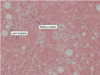
What would alcoholic steatosis look like microscopically?
- Will be able to see lipid droplets in the hepatocytes
- Remember alcoholic steatosis is rapidly reversed by abstinence (no alcohol intake)

Liver damage is more likely to occur in which group?
A) Continuous drinkers
B) Intermittent “binge” drinkers
A) Continuous drinkers
Why - because the “binge” pattern gives the liver the chance to recover
Name a few risk factors for alcoholic liver disease?
- Drinking pattern
- Continuous drinking pattern is more associated with ALD than “binge” drinking.
- Gender
- Incidence of ALD is increasing in women because women have a higher blood ethanol level after consuming the same amount of alcohol as men.
- Genetics
- Nutrition
- Obesity increases the incidence of liver-related mortality
How is alcoholic liver disease managed?
Cessation of alcohol consumption is the more important treatment and prognostic factor.
General health and life expectancy are improved- irrespective of the stage of ALD.
Fatty liver and alcoholic hepatitis (without fibrosis) is reversible therefore cessation of alcohol is a treatment.
For alcoholic hepatitis (with fibrosis) and cirrhosis- the damage is irreversible but cessation of alcohol would prevent further damage and improve life expectancy.
Describe the clinical features of fatty liver?
Usually asymptomatic
Rarely there is a slight tenderness due to the enlargement of the liver
Liver function tests are usually slightly elevated
What does the ALT and AST tests measure?
Measures enzymes that your liver releases in response to damage or disease
What would alcoholic hepatitis look like under the microscope?
- Lipid droplets in the liver (same as fatty liver)
- Ballooning of the hepatocytes (swollen)
- Areas of necrosis
- Mallory bodies in the cytoplasm
- Neutrophils may be present (multiple nuclei)
Function tests for liver.
If alcoholic hepatitis, if the AST:ALT ratio is greater than 2, what does this suggest?
Suggestive of alcohol being the cause
It is because alcohol induces the microsomal ethanol oxidising system
Describe how mallory bodies are formed in alcoholic hepatitis?
The hepatocytes are swollen which pushes the cytoskeleton.
Mallory bodies occur when the cytoskeleon eventully condenses under the pressure.
AST and ALT stand for what in the liver function test?
AST: Aspartate transaminase
ALT: Alanine transaminase
Only ___of alcoholic hepatitis cases develop cirrhosis
One third
Name some of the complications of alcoholic cirrhosis?
- Portal hypertension
- Liver failure
- Hepatocellular carcinoma in around 3-6% of cases
Name the 3 histological signs of alcoholic cirrhosis?
Characterized by fibrosis and conversion of the normal liver architecture into structurally abnormal nodules.
- Nodule formation
- Fibrosis
- Reduced liver diffusion i.e failure in the metabolic pathway in the liver
What is the most common cause of macronodular and micronodular cirrhosis
Macronodular cirrhosis: viral hepatitis (B and C)
Micronodular cirrhosis: Chronic alcoholism
What are transaminase values with alcoholic liver disease?
The aspartate aminotransferase is typically elevated more than alanine aminotransferase with alcohol-related liver disease
Define the term “Ascites”?
abnormal accumulation of fluid in the peritoneal cavity.
What is caput medusae?
Refers to the appearance of a network of painless, swollen veins around the abdomen.
A sign of an underlying condition, usually liver disease e.g. cirrhosis.

