Ostertagia 1, 2 and Control Flashcards
(20 cards)
What is the common name for ostertagia ostertagi?
Where in cattle is it found?
Brown stomach worm
Abomasum
Describe its morphology to explain how it is identified?
~1cm in length
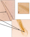
Slender, pinky-brown
Fine cervical papillae
Males have bursa and spicules

Describe the life cycle of Ostertagia ostertagi
What its the PPP?
Typical trichostrongyle- direct
- Egg passed in faeces
- L1-L3 in faecal pat, L3 infective stage (ensheathed)
- Cattle ingest L3
- L3 burrows into gastric glands of abomasum
- Develops to L4-L5
- L5 emerge into the lumen and matures to adults
- Mate and females lay eggs
PPP- 3 weeks
Describe ostertagia ostertagi eggs
Explain its appearance- bit of a weird Q but should make sense
Typical trichostrongyle egg
~90 x 45 um
barrel-shaped
undifferentiated content with thin-walled
Explanation- Undifferenentiated as development takes place on faecal pat, thin walls as hatched in the environment

L3 burrow and L5 emerge from gastric glands, what are their function?
Therefore what is the pathogenesis of O. ostergi?
Contain acid-producing parietal cells- pH, bacteriostatic, pepsin conversion
Pathogenesis-
- Disease associated with large numbers of developing larvae- 40,000
- Damage caused by developing larvae in gastric glands
- Damage to glands- leads to replacement by undifferentiated epithelial cells
- Loss of acid production
- Increase in abomasal pH
- Loss of bacteriostatic effect
- Increased permeability of mucosa
What are the clinical signs of ostertagia ostergi disease?
- Profuse watery diarrhoea
- Weight loss
- Loss of appetite
What nematodes are found in the abomasum of cattle?
HOT- yeh you are!
Haemonchus contortus- 3cm
Ostertagia ostergi- 1cm
Trichostrogylus axei -0.5cm
In order of size

What nematodes are found in the small intestine of cattle?
Ok bear with me- a bloke walks over and starts with ‘HOT’ (abomasum)
Aww bless but- ‘Not Thanks Clive’
Nematodirus spp- cotton wool
Trichostrongylus spp
Cooperia- watch spring
What nematodes are found in the large intestine of cattle?
Ok to ‘HOT’, ‘No thanks Clive’, a few beers are seen off you change your mind
‘Come On Then’- go on clive my boy
Chabertia spp
Oesophagostomum spp
Trichuris spp
What is the difference between type I and type II disease of ostertagia?
When do the different presentations occur?
Type 1-
Dairy replacement calves, end of the first grazing season
The disease occurs in summer- July to September
Calves ingest L3 in July
CS- green watery diarrhoea
Type 2-
Yearling calves
Disease in late winter, early spring
Acute- intermittent diarrhoea
CS- anaemia, thirst (high mortality)
What is hypobiosis?
How does it allow type II ostertagia disease?
Hypobiosis is the arrested development of L4 inside the host in response to a trigger received by the free-living L3
Type II disease-
L4 go into a state of hypobiosis in gastric glands due to temperature drop
Remains there over the winter and extends the PPP
At the end of winter, their development resumes
The simultaneous emergence of L5
Severe clinical disease
What factors affect the following:
development of pre-parasitic ostertagia stages?
What enables survival of L3?
Host factors for disease?
Development of pre-parasitic-
Temperature >10
Humidity
Dispersion from faecal pat (rainfall)
Survival of L3-
Ensheathed- wears L2 cuticle
Temperature- tolerates cold, hates heat
Moisture- desiccation is lethal
Limited food reserves
Host-
Age- 6-8 months
Immune status
Over dispersion- small population carry most of parasites
Describe what is happening in the Ostertagia life cycle in the following months:
May
June
July
August
Autumn- yes it’s not a month
Late winter-early spring
May-
first season dairy calves turned out, graze, ingest L3 (overwintered), no disease too few L3
June-
overwintered L3 die off- by the beginning of June, L3 mature, PPP 3 weeks, eggs onto pasture, the ambient temperature gradually start to increase, eggs start to develop
July-
eggs on pasture developing, development temperature-dependent, the peak of L3 in mid-July, ingested by calves
August-
large numbers of L3 develop in the abomasum of calves
type I disease 3 weeks later (august to Sep)
Autumn-
L3 released from pats with rainfall
Calves may be moved back onto contaminated pasture used to graze calves
L3 exposed to low temp- hypobiose L4
Late winter-early spring
Larval development resumes, the simultaneous emergence of L5, type II
How are beef herds affected?
Clinical disease is less common
Calves graze with immune mothers
Autumn born calves may be at risk from overwintered L3
How is ostertagia diagnosed?
Definitive?
Diagnosis-
Clinical signs
Grazing history
Definitive diagnosis- raised plasma pepsinogen levels
How would you treat Ostertagia infections?
What action would you take at a farm with yearlings suffering from type II ostertagiosis
Anthelmintics
Treat yearlings with an anthelmintic that targets L4, L4 and adults- MLs
What is the difference between treatment, metaphylaxis and prophylaxis?
What class of anthelmintics are:
White drenches
Yellow drenches
Clear drenches
Treatment- individual animal with disease
Metaphylaxis- timely mass medication of a group of animals to prevent or minimise an expected outbreak of disease
Prophylaxis- prevention of disease
White drench- 1-BZs
Yellow drench- 2-LVs- levamisole
Clear drench- 3-MLs- ivermectin
What is the dosing schedule for ivermectin used for turning out heifer calves?
Ivermectin kills L4/5 and adults
residual effects for 2 weeks
Explain you answer
Dose 3, 8 and 13 weeks after turnout
Calves turned out and ingest overwintered L3
Small numbers of worms in abomasum after 3 weeks (PPP)
Therefore stop eggs from being shed on pasture
Ivermectin lasts 2 weeks + 3 weeks PPP= 5
What does safe pasture and clean pasture mean?
Safe pasture- used the previous year but safe by the beginning of June
overwintered L3 die off
Clean pasture- not grazed by cattle for the previous 12 months


