Ortho Flashcards

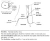
Ankle Xray Views & Metrics
Nomenclature of hip #’s

Distal phalanx #
- repair nailbed injury if present
- hairpin splint not involving PIP
- plastics within 2 weeks
Middle/Proximal Phalanx #
- correct any rotational deformity
- buddy tape (dynamic splint) if stable (transverse, non-displaced)
- radial or ulnar gutter splint if unstable
- plastics within 1 week
Bennett’s #
- intraarticular base of thumb MC # with dislocation/subluxation of CMC
- reduce, thumb spica
- plastics within 2-3 days
Rolando’s #
- comminuted # of base of thumb MC
- worse prognosis than Bennett’s
- thumb spica
- plastics within 2-3 days
DIP Dislocation
- reduce
- dorsal splint in full extension
- or buddy tape if stable post-reduction
- plastics within 1 week
PIP Dislocation
- reduce
- dorsal splint in 30 deg flexion at PIP
- or buddy tape if stable post-reduction
- plastics within 1 week (2-3 d if unstable)
Extensor zones of the hand
see Evernote “Hand Injuries”
Management of extensor tendon injuries of the hand
- Zone I, II open injuries: repair 5-0 sutures, splint in extension.
- Zone III injuries: modified Elson’s test to check for central slip damage. If open & have Boutonniere deformity, call plastics on call. If closed, place PIP in extension and f/u plastics (may leave DIP free).
- Zone IV injuries: primary repair with 5-0 sutures, splint MCP in 15 deg flexion.
- Zone V, VI injuries: primary repair with 4-0 sutures if clean laceration, splint.
- Zone VII, VIII injuries: splint, refer to plastics.
Management of Flexor Tendon Injuries of the Hand
- Splint in position of function
- Plastics within 1 week
Fingertip Amputation Zones
See Hand Injuries on Evernote
Describing Angulation in a #
- for midshaft #’s, angulation is direction of apex
- for distal fractures (e.g. Colles), angulation is direction of distal fragment
MCP Dislocation
- do not hyperextend during reduction
- reduce with wrist flexed to relax flexor tendon
- pressure and traction on base of prox phalanx
- splint in flexion
- volar dislocations usually need operative reduction
Scapholunate Ligament Injury
- FOOSH on thenar eminence
- clicking with wrist movement
- tender on dorsum of wrist just distal to Lister’s tubercle
- pain with ballottement of the scaphoid
-
scaphoid shift/Watson shift test
- wrist in ulnar deviation, thumb on scapohid prominence volarly –> move wrist into ulnar deviation
- test positive if scaphoid ‘clunks’ dorsally/gives or patient’s pain reproduced
- XRay
- 3 mm widening on PA view
- clenched fist view may help
- scaphoid shortening with dense ring (cortical ring sign)
- dorsal intercalated segment instability of lateral view (zig-zag pattern instead of 3 C’s)
- Tx: radial gutter splint
Triquetrolunate Ligament Injury
- ulnar equivalent to scapholunate injury
- FOOSH on hypothenar eminence
- volar intercalated segment instability on lateral xray
- ulnar gutter splint
Perilunate Dislocation
- FOOSH with great force
- posterior dislocation of carpal bones, lunate remains in place
- call ortho/plastics
Lunate Dislocation
- posterior dislocation of carpal bones with lunate facing anteriorly
- XRay
- piece of pie sign (lunate triangular on PA)
- spilled teacup sign (lunate displaced and angled palmar)
- if fracture associated, then add trans- to the name (e.g. transscaphoid lunate disclocation)
- call ortho/plastics
Scaphoid Fracture
- tender in snuffbox with ulnar deviation
- pain with resisted pronation/supination
- pain with axial load to thumb
- 10% initial xrays -ve
- may get dedicated scaphoid view
- thumb spica with mild wrist dorsiflexion and radial deviation (to compress # fragments)
Triquetrum Fracture
- often a dorsal avulsion # on lateral view
- sugartong splint
Lunate Fracture
- tender in dorsum wrist groove on flexion
- AVN possible (blood supply enters distally)
- xrays may be negatve
- thumb spica
Hamate Fracture
- interrupted bat/golf club swing
- carpal tunnel view
Colles’ Fracture
- reduction: > 20 deg angulation, intra-articular involvement, > 1 cm shortening, comminution
- criteria for adequate reduction
- At least 11 mm radial height
- At least 22 deg radial inclination
- At least 11 deg volar angulation
- practically, neutral is OK for age < 50 and 10 deg dorsal tilt is OK for age > 50
- Acceptable angulation in kids
- < 5 yrs = 30 deg
- 5-10 yrs = 20 deg
- 10-12 yrs = 10-15 deg
- +-2 mm ulnar variance
- < 3 mm impaction
- ulnar styloid often also fractured
Smith’s Fracture
- volar angulation of distal radius
Radial Styloid Fracture
- often with dislocation of the lunate
- major carpal ligaments insert at styloid so carpal instability
- short arm splint
Ulnar Styloid Fracture
- ulnar gutter splint
DRUJ Injuries
- ulnar deviation on lateral
- splint in supination for dorsal and pronation for volar dislocations
Compartment Syndrome
(Diagnosis, treatment)
- traditional, tissue pressure > 30-50 mm Hg
- better, delta pressure (diastolic - tissue pressure) > 30 mm Hg
- pain refractory to opioids, pain to passive stretch, firmness/fullness in compartment
- normal pulses/cap refill as tissue pressure less than arterial pressure
- Stryker kit
- pressures highest near injured area, obtain within 5 cm of # site
- 2 readings each compartment
- place limb at level of heart
- reverse anticoag/replace factors for hemophiliacs
Biceps Tendon Ruptures
-
proximal
- usually older, chronic tendonitis
- pain in anterior shoulder
- shoulder xray r/o avulsion #
- sling –> # clinic
-
distal
- usually younger, eccentric load
- pain in AC fossa
- Hook sign
- sling –> # clinic more urgently
Elbow Dislocation
- 90% are posterolateral
- assess (pre- and post-reduction):
- brachial artery (just medial to distal biceps tendon)
- ulnar, radial, median nerves
- Check for full ROM post-reduction, fragments often trapped
- call ortho if unstable on ROM or reduced ROM or NV compromise post-reduction
- splint in long-arm posterior splint in slightly less than 90 deg flexion and forearm in mild pronation
- NV f/u exam next-day

Supracondylar #
- common in 5-10 years of age
- common to injure anterior interosseous nerve
- motor only branch of median
- test OK sign
- common to injure anterior interosseous nerve
- extension-type (95%, posterior displacement)
- FOOSH in extension
- posterior fat pad or large anterior fat pad (sail sign), disruption of anterior humeral line
- long-arm posterior splint 90 deg, neutral rotation
- if only sign is fat pad then ortho f/u in 2-7 days
- if any angulation/break through cortex then fasting + ortho in ED
- flexion-type (5%, anterior displacement)
- rare, direct force, often open
Intercondylar #
- assume any supracondylar # in adult is intercondylar
- supracondylar + T or Y component separating condyles from each other and going intraarticular
- splint in long arm posterior splint at 90 deg in neutral position
Epicondyle #
- mostly medial, an apophyseal avulsion fracture
- pain, tenderness, swelling
- medial from repeat valgus stress such as throwing
- posterior splint in pronation
Condyle #
- mostly lateral
- a fracture through the condyle
- usually much larger/unstable than radiograph because mostly cartilaginous
Ossification Centres of Elbow
- all usually ossify by 12 years
- Capitellum
- Radial head
- Internal (medial) epicondyle
- Trochlear
- Olecranon
- External (lateral) epicondyle

Monteggia/Galeazzi #
- Fracture
- Ulna/radius
- Monteggia/galeazzi
- Elbow (radius)/wrist (ulna) dislocated
- may reduce, but often need operative management
Calcaneus #
- Boehler angle: line from highest part of anterior process of calcaneus and highest point of posterior articular surface of calcaneus + line between highest point of posterior articular surface of calcaneus and the most superior part of calcaneal tuberosity
- normal 25-40 deg
- < 25 deg suspect #
- posterior splint, NWB

Lisfranc Injury
- plantar flexion + axial load
- pain with torsion/dorsi/plantar flexion
- weight-bearing AP, lateral, 30 deg oblique
- 1 mm displacement base 1st/2nd MT
Base of 5th MT #
- Review Evernote “Foot Injuries”
Hip #
- Review Evernote
Ottawa Knee Rules
- age >= 2
- Xray if
- age >55
- tender at
- fibular head
- patella
- cannot flex > 90 deg
- cannot WB 4 steps immediately + in ED
Additional Knee Xray Views
- sunrise view
- patellar #/subluxation
- tunnel view
- intercondylar region/tibial spine #
- oblique view
- (internal for lateral, external for medial plateau #)
Treatment for Locked Knee
- usually meniscal tear
- sedation
- supine with knee 90 deg flexed hanging over edge
- longitudinal traction, internal + external rotation
- ortho if not successful
Knee Dislocation
- 50% self-reduce
- ++ injured & unstable multiple directions
- reduce, splint in 20 deg flexion
- if no vascular + ortho in house & NV intact, delay reduction for transfer
- CT angio post-reduction
Patellar Dislocation
- flex hip, hyperextend knee, posteromedial pressure on lateral border of patella
- 1st time dislocation: tensor, knee immobilizer, no flexion allowed, urgent ortho
- recurrent: less strict need for immobilization (ligaments already lax), semi-elective ortho
Tibial Shaft #, criteria for adequate reduction
- criteria for adequate reduction:
- <10 deg varus or valgus
- <10 deg anterior/posterior angulation - can accept more in the plane of joint motion
- <1 cm shortening
- min 50% apposition
- long leg splint, elevate
- ortho in ED
Pilon/Tibial Plafond #
- “mortal pestle” #
- axial load grinds tibia into talus
- look for L1 # & compartment syndrome
Gastrocnemius Tear
- sudden pop, swelling in calf
- RICE
- may splint in equinus
Ankle Syndesmosis Injury
- see Evernote

Grading Ankle Sprains
- Grade I: no tear, minimal functional loss, pain and ecchymosis
- Grade II: partial tear, some loss of function
- Grade III: complete tear, ++ swelling, bruising, usually NWB
- Any medial maleolar swelling/tenderness needs to be NWB and have close ortho f/u
- lateral mal # with medial mal swelling/tenderness needs posterior slab with medial molding
Weber Classification Distal Fibula #’s
- NWB with aircast unless avulsion #

Sternoclavicular Joint Dislocation
- CT imaging of choice
- US + aspiration if infectious/effusion (common in IVDU)
- anterior dislocation
- sling, ice, no need to reduce (won’t hold anyway)
- posterior dislocation
- ortho, open reduction
- closed reduction if mediastinal compromise
Clavicle #
- xray may miss #’s at extreme ends of bone
- 45 degree cephalad tilt view +- CT
-
distal
- displaced often operated on
- sling + early ortho f/u
-
middle third/distal #’s
- usually non-op unless athlete/cosmetic
- rule of 2’s for op mgmt
- 2 cm short
- 2 cm displaced
- 2 pieces
- sling, early ROM (in 3-5 days)
-
proximal third clavicle #’s
- rare, check with ortho
Scapular #
- dedicated views
- look for associated rib #’s/lung injury
- sling, ice
Anterior Shoulder Dislocation
- slight abduction + external rotation
- check deltoid sensation (axillary)
-
reduction
- 10-20 mL 1% lidocaine subacromial
-
Modified Hippocratic
- traction-countertraction
-
Snowbird
- belt looped over flexed elbow
- use foot to pull down on belt
-
Stimson
- prone, weights
-
Scapular Manipulation
- Stimson + rotate scapula (distal tip goes medial)
-
Kocher’s
- elbow 90 deg, slow external rotation
- may bring elbow anteriorly
-
Milch
- external rotation, arm straight
- arm abduction to 180 degrees
- push on humeral head upwards with R thumb
-
Cunningham
- sitting massage, shrug shoulders back
Posterior Shoulder Dislocation
- <1%
- usually held in internal rotation + adduction
- unable to external rotate + abduct
- reduce with longitudinal traction
Inferior Shoulder Dislocation (Luxatio erecta)
- hyperabduction force (levers neck of humerus against acromion)
- humerus fully abducted, elbow flexed, hand on or behind head
- traction upward + outward in line with humerus
Proximal Humerus #
- Neer classification of shoulder #’s
- a “part” is a fragment displaced > 1cm or angulated > 45 deg
- i.e. even if many fragments, if none angulated/displaced then it is a “one-part” #
- one part #
- sling + swathe, ice, early ROM
- more than one part # or #-dislocation
- ortho in ED

Humeral Shaft #
-
proximal
- accept up to 45 deg angulation, 1 cm displacement
- minimal displacement
- shoulder immobilizer, close f/u
- displacement/comminution
- d/w ortho
-
middle third #’s
- most common
- usually non-op
- radial nerve
- not comminuted
- sugar-tong, close follow-up
- comminuted
- call ortho
-
distal humerus #
- ED consultation re: NV structures
Jersey Finger
- FDP rupture from grabbing jersey
- splint in slight flexion
- hand clinic
Compression #’s
- Discuss all spine #’s with a surgeon
- if <40% loss of height, generally stable
- if >= 50% loss of height, or angle between damaged vertebra and spinal column is >25-30 deg usually unstable
- can often misdiagnose Chance (transverse) + burst #’s as compression #’s
- consider CT in all compression #’s found on plain films
- if truly stable, non-pathologic –> heat, massage, rest, f/u
Coccyx #
- pain meds, doughnut pillow
Prevertebral soft tissue spaces in cervical trauma
- 6 mm at C3
- 22 mm at C6
C-Spine Trauma Approach
- NEXUS
- CCSR
- Xray
- CT if inadequate
- If CT -ve but suspcious and MRI not available, may DC in firm foam collar and f/u in 3-5 days. If pain resolved, may DC collar
Neurogenic Shock vs. Spinal Shock
Neurogenic Shock
- loss of peripheral sympathetic innervation
- if T1-T4 then unopposed vagal to heart, bradycardia
Spinal Shock
- temporary loss of spinal reflex activity below injury that may recover
Thoracolumbar Spine Trauma
Xray vs. CT
- EAST recommends CT over xray (Level 1) although no studies in mildly injured patients
How Long to Immobilize a Shoulder Dislocation For?
- “8 minus decade of life”
- means if 75 years old then simple sling + ROM immediately
- max 3 weeks
- longer for first-time dislocators
Radial Head #
-
undisplaced, radial neck
- sling, f/u 1 week
-
undisplaced, intra-articular
- posterior slab, f/u 1 week
-
displaced
- call ortho to discuss
Coronoid Process #
- displaced, large fragment
- call ortho to discuss
- undisplaced
- posterior slab, 1 week
Olecranon #
- check triceps with arm horizontal (gravity eliminated)
- displaced
- call ortho to discuss
- undisplaced
- posterior slab, 1 week
Ulnar Collateral Ligament Injury
- 25% have Stener’s lesion (interposition of adductur pollicis between ends of ligament tear resulting in poor healing + chronic thumb pain)
-
Grade 1/2
- thumb spica
- plastics 1-2 weeks
-
Grade 3
- plastics within 2-3 days (operate within 1 week)
Toe Fracture
- Indications for referral (Great Toe)
- Fracture with dislocation
- Displaced intraarticular fractures
- Unstable, displaced fractures (ie, fractures initially reduced that immediately displace once traction is released unstable displaced fractures
- Indications for referral (lesser toes)
- Displaced intraarticular fractures
- Irreducible fractures
- Open fractures of non-distal phalanges
- Fractures that do not maintain acceptable position with buddy taping
Toddler’s #
- 9 mo to 5 yrs
- twisting of foot –> oblique tibial #
- often minor mechanism, subtle tenderness
- additional oblique views increase sensitivity
- above knee splint –> ortho 1 wk
Tillaux #
- girls age 11-13, boys age 12-15
- distal tibial growth plate closes from medial to lateral
- external rotation results in SH III # of the distal tibia
- ortho in ED
Patella #
- if has active knee extension: knee immobilizer, ortho 1 wk
- if no active knee extension: ortho in ED
Segond #
- vertically oriented avulsion # from lateral tibial plateau at the attachment of the lateral capsular ligament
- 75% association with ACL tear
- tensor, crutches, WBAT
- early ROM as tolerated
- early sports med f/u
Tibial Plateu #
- suspected/undisplaced
- long-leg splint, NWB, urgent ortho
- displaced
- ortho in ED
How to examine extensor tendons of fingers
- Test extension PIP/DIP with MCP in extension to remove lumbricals
- Modified Elson’s Test for zone III injuries
How to test SLR in knee exam
- test SLR seated to remove IT band
Mandible Dislocation
See Evernote “Dental”
How to Apply a Thomas Splint
See Evernote “Procedures”
Snowboarder’s #
See evernote “Leg + Ankle Injuries”
