Neuroradiology Flashcards
What was newly approved in 2011 regarding MRI studies?
- MRI-safe pacemakers.
- Safe for patient; cannot scan over the pacemaker region
- Good records, serial no. of pacemaker, etc. are required.
What is the significance of the origin of the opthalmic artery?
- It is the marker that the Internal Carotid Artery (ICA) is in the subarachnoid space, so the artery at this point is completely encased in the dura.
- Thus, at this point and beyond, an aneurysm that leaks or bursts would cause a subarachnoid hemorrhage.
Describe the complications caused by subarachnoid hemorrhage.
- Arachnoid granulations are blocked,
- CSF is not resorbed, ventricles expand (hydrocephalus), which leaves less room for cerebral blood, causing ischemia.
Identify the tagged artery in the following lateral carotid angiogram.
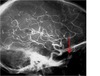
Ophthalmic artery
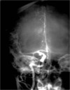
Identify the tagged artery in the AP carotid angiogram.

Posterior communicating artery
Identify the tagged artery.

Pericallosal artery (branch of the anterior cerebral artery)
What is the clinical significance of the pericallosal artery?
It courses above the corpus callosum, which is above the ventricular system, so the position of the pericallosal artery provides information about the ventricular system.
Identify the tagged artery.

Middle cerebral artery
What is tagged in this image?

The Sylvian point
What is the Sylvian point?
The most posterior and the most medial point of the MCA. The “end” of the MCA.
Where is the Sylvian triangle in the following image?

Sylvian triangle.

Describe the Sylvian triangle.
- Identifies all the branches as they come out of the Sylvian fissure (top line of triangle; coming “toward you”).
- Bottom line shows origin of the vessels.
- Must be in the normal location and shape. If the top line is “bowed up” there is a mass elevating it, and vice versa.
Identify the tagged artery.
Posterior communicating artery.
What artery is the origin of PICA?
Vertebral a. (not Basilar!)
Identify the tagged artery.

Posterior inferior cerebral artery (PICA)
What is wrong with the Sylvian triangle in this image?

- There is elevation of the triangle. The shape of the top portion bows up.
- Thus, there is a lesion below. There is a mass in the temporal lobe elevating the Sylvian triangle.
- (A frontal lobe lesion would cause the opposite, a depressed “bowed down” triangle).
Knowing that there is a mass in the temporal lobe, what symptoms will the patient present with?
Seizures.
What does water look like on a T1 weighted image?
LOW signal (black)
Edema that does not involve all of the gray matter is termed?
Vasogenic edema
Small collections of contrast that arise from an artery are called?
Aneurysms
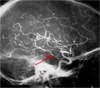
Describe the pathology of the following image.
- Posterior communicating artery aneurysm (actual aneurysm is on the ICA - PComm is fine)
- Sylvian point is laterally displaced. Large hypovascular area (enlarged ventricle).
What symptoms might a patient with a Posterior communicating artyer aneurysm experience?
“Worst headache of my life”
What is the pathology of this vertebral angiogram?

Large aneurysm of the basilar artery. There is a not-sharp, fuzzy border on one side. This is due to thrombus.
The lesion of the following image is in what lobe?
What symptoms will the patient present with?

- Parietal lobe
- Progressive hemiparesis
Is the lesion an infarct, a tumor, or an abscess?

Tumor
What lobe is the following lesion?
What symptoms will the patient present with?

- Temporal lobe
- Uncinate fits
What type of lesion is this?

Either a tumor or and abscess
Where is the lesion in the following CT?
What symptoms will the patient present with?

- Pineal gland
- Parinaud’s Syndrome: Paralysis of upward gaze by compression of the superior colliculus
