Neuropathology 1 Flashcards
1) The Fasciculus Gracilis exist at _______of spinal cord and DOES contain ______ ____ from the ___
vs.
Fasciculus Cuneatus exist in {__-__} = ____ and ______ and contains _____ ____ from the ___
1B) Which Pathway are these Fasciculus columns associated with?
2) What happens to 2nd order neurons of this Pathway?
2B) Where are these 2nd order neurons located?
3) What happens to 3rd Order neurons of this Pathway?
4) How does [Proprioreception/Vibration/2-point discrimination] get to 2nd order neurons? [5]
**Position/Vibration/2-point discrimination travels from periphery in the ______ ______—–> passes ______—>passes ______ ______ Gray mater–>ultimately travels upwards using ______ ______ or ______ ______
—>travel up into [______ MEDULLA] to synapse 1st time in ______ ______ or ______ ______
1) The Fasciculus Gracilis exist at ALL LEVELS of spinal cord and DOES contain [ascending sensory fibers] from the LE
vs.
Fasciculus Cuneatus only exist in {C1-T6} = Thoracic segments (T1-T6) and Cervical segments (C1-C8) and contains [ascending sensory fibers] from UE
**DORSAL COLUMN PATHWAY**
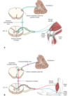
2) 2nd order neurons in [nucleus gracilis] AND [nucleus cuneatus] decussate as [internal arcuate fibers] in LOWER MEDULLA—>cross midline to form [Medial Lemniscus]—>this travels to VPL nucleus of thalamus!
3) 3rd order neurons in [VPL nucleus of thalamus] travel in internal capsule and terminate in [Area 312 POSTcentral gyrus]
4) Position/Vibration/2-point discrimination travels from periphery in [Dorsal Root]—> passes DRG—>passes Dorsal Horn Gray mater–>bends in {Fasciculus cuneatus or gracilis}—>travel up into [LOWER MEDULLA] to synapse 1st time in {nucleus cuneatus or gracilis}
A) [Lateral Dorsal Root] Synapses in ____ _____ of ____ Horn and then becomes the _____ ______ . What sensations is this transmitting? [2]
B) SPINOTHALAMIC TRACT DECUSATES in _____ _______ and BEGINS ASCENT in the __________
—->It then travels ultimately travel up to [VPL nucleus of Thalamus] via the _____ _____ of spinal cord
C) Cutting STT in CORD will affect Pain on ____[same/Opposite] side of Body
A) [Lateral Dorsal Root] Synapses in Nucleus Proprius of [Gray Dorsal Lateral Horn] to become SPINOTHALAMIC TRACT= USED FOR PAIN and TEMPERATURE OF THE BODY
B) SPINOTHALAMIC TRACT DECUSATES in [ANT Commissure of Spinal Cord] and begins ascent in the [white Anterolateral Fasciculi] —->STT then ultimately travels up to [VPL nucleus of Thalamus] via [LATERAL FUNICULUS] of spinal cord
C) Cutting STT in CORD will affect Pain on OPPOSITE of Body

A: [Dorsal Spinocerebellar tracts] travel in ____ ______ and synapse either on _____(below C8) OR _____(Above C8). This tract is used for ____ ______ ______
B: Muscle Spindles use Sensory Affarent fibers (which bend in the [Fasciculus ______]) and transmit __ ______ ______ to the [____ _____ _____ nucleus] located in ___ ______
C: What is the [cuneocerebellar tract] ?
**A tract that receives ______ info from ______ nucleus & then ______[contralaterally/Ipsilaterally] sends this to the __ ___ ____ which relays info to ______ ______
D: [Dorsal Spinocerebellar tracts] AND [cuneocerebellar tract] use the__ ___ ____ to enter Cerebellum Vermis
A: [Dorsal Spinocerebellar tracts] travel in LATERAL FUNICULUS and synapse either on [Dorsalis Nucleus of Clarke] (below C8) OR lateral accessory cuneate nucleus for UNCONSCIOUS IPSILATERAL PROPRIORECEPTION
B: Muscle Spindles use Sensory Affarent fibers to bend in the [Fasciculus CUNEATUS] and transmit UE unconscious positioning to the LACN [lateral accessory cuneate nucleus] located in Lower Medulla.

C: [cuneocerebellar tract] = tract that receives UNCONSCIOUS positioning info from LACN [lateral accessory cuneate nucleus] & then ipsilaterally sends this to the ICP—> Cerebellum VERMIS
D: [Dorsal Spinocerebellar tracts] use the [inferior cerebellar Peduncle] to enter Cerebellum Vermis
A: How are the Bipolar Cells, Internueonrs & Ganglion cell bodies arranged near the Fovea? What about blood vessels?
B: How does Fovea and surrounding Macula receive metabolic supply?
C: Fovea is an _____[vascular/Avascular] retinal area with the ___[least/most] visual acuity. It has a __ ___ ____layer to prevent light barrier and is EXCLUSIVELY _____. There is a 1:1 ratio with [__:___] -which means ___ are more specific
D: Fovea is only __% of Retina and transmits __[least/most] of visual information to ________
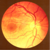
A: Near Fovea, Bipolar Cells, Interneurons & Ganglion cell bodies are pushed LATERALLY–>makes a clear path for light = High visual acuity.
**Fovea is also Devoid of any blood vessels for same reason
B: Since Fovea is Devoid of any blood vessels, Fovea and surrounding Macula depend on diffusion from underlying choroid vessels for metabolic needs
C: The Fovea is an AVASCULAR retinal area with the MOST visual acuity. It has a laterally flat inner layer to prevent light barriers and is EXCLUSIVELY CONES. There is a 1:1 ratio with
[ganglion cell body: Cone receptor] –>Cones are more specific!
D: Fovea is only 5% of Retina but transmits MOST of visual information to [Lateral geniculate nucleus of thalamus]

A: What are the 7 Steps of Visual Pathway starting with light entering eye?
B: Light Entering Eye:
[TEMPORAL Field axons] pass _____ [crossed/uncrossed]
vs.
[nasal field axons] pass ____ [crossed/uncrossed], which means what for optic chiasm lesions?
A: Pathway of Visual Info from Retina [RN CT L DV]
“RN (use) CT (to) Learn Direct Vision”
1st: Light enters —> hits Retina
2nd: Travels in Optic N.
3rd: Optic Chiasm [Temporal Field Axons CROSS HERE]
4th: Optic Tract
5th: [Lateral Geniculate Nucleus of Thalamus]
6th: Optic RaDiations [Meyer’s Temporal Loop vs. Parietal direct path]
7th: [Area 17 -Calcarine Primary Visual cortex]
B: Light Entering Eye:
TEMPORAL Retina Fields pass CROSSED = [Temporal fields] decussaTe at Optic Chiasm
vs.
nasal retina fields pass UnCrossed @ Optic Chiasm = NOT AFFECTED BY Central OPTIC CHIASM LESIONS

A: [P-type] ganglion cell bodies are used by _____. [M-type ganglion cell bodies are used by ______.
B: Describe each of these
[2 each]
A: There are 2 MAJOR classes of [innermost ganglion cell bodies]
1. P-Type [USED BY CONES] “Pretty Colors”
ºsmall receptive fields –> BEST with [color/fine detail/high acuity]
ºtons of it near Fovea
- M-type[used by rods]
ºLARGE Bodies and many axons share 1 cell body –> LARGE Receptive fields
ºBEST with [rapidly transient adaptation] &response to mvmnt or large objects
A: Although Visual Perception begins in ______, collateral visual info enters ____ via Pretectal Area= for ___ _____ and _____ _____ for ________
B: Pretectal Area uses ___ nuclei of _____ for ___ _____ and projects both ipsilateral & contralateral via ____ _____
A: Although Perception of vision begins in [Area 17 CPVC-Calcarine Primary Visual Cortex] collateral visual info enters
- Brainstem via Pretectal Area=FOR PUPILLARY REFLEX and
- [SUP Colliculus]=For [head&eye movement]
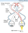
B: Pretectal Area uses [Edinger-Westphal nuclei] of midbrain for PUPILLARY REFLEX and projects both ipsilateral & contralateral via POSTERIOR commissure
What would occur if there was a Lesion in …
1. R Optic N. —->
- *Optic Chiasm —–>
- R Optic Tract—>
- R [lower radiation meyer’s loop] –>
- R [Lateral Geniculate nucleus of thalamus]–>
- R [upper radiation fibers]
- R [Area 17 CPVC]
B: Which 2 lesions present the same Visual sx?
C: Which Visual Field Decussates at Optic Chiasm?
Lesion in … (using R side damage as example)
- R Optic N. —-> BLIND RIGHT EYE
- ——————————————————————————- - *Optic Chiasm —–> [Bitemproal Heteronymous hemianopsia] (both temporal fields knocked out) since Temporal fields decussaTe at Optic Chiasm
- ——————————————————————————- - R Optic Tract—>[CTL homonymous hemianopsia]
- ——————————————————————————- - R [lower radiation meyer’s loop] –> “Pie in the Sky Lesion’ = [CTL homonymous upper quadrantanopsia]
- ——————————————————————————- - R [Lateral Geniculate nucleus of thalamus]–>[CTL homonymous hemianopsia]
- ——————————————————————————- - R [upper radiation fibers]–>[CTL homonymous LOWER quadrantanopsia]
- ——————————————————————————- - R [Area 17 CPVC]–> [CTL homonymous hemianopsia with macular/fovea sparing]
- ——————————————————————————-

**Lesions of Optic Tract {4} and [Lateral Geniculate Nucleus of thalamus] {6} - PRESENT SAME VISUAL SX!
C: [Temporal fields] decussaTe at Optic Chiasm
CTL = Contralateral or (Left side in this case)
A: The Pupillary Light Reflex involves a ___ reflex and ____ reflex. Describe each?
B: After light is shone thru 1 eye, BOTH pupils constrict becuz ___ connections to [_____ nucleus] from ____ fibers traveling thru [___ ____]–> activates [Oculomotor CN3] fibers to synapse in the ___ganglion and constrict ____ _____ m.
C: What part of the brain does this test?
D: [Optic Nerve damage] presents how? Why is this?
vs.
E: [Oculomotor Nerve damage] presents how? Why is this?
A: DIRECT reflex = light is shone thru L pupil and the L pupil constricts
CONSENSUAL reflex= light is shone thru L pupil BUT R pupil constricts also
B: both pupils constrict becuz bilateral connections to [Edinger-Westphal nucleus] from optic nerve fibers traveling thru [SUP colliculus] activates [Oculomotor CN3] which travels to Ciliary ganglion ipsilaterally—>constrict [sphincter pupillae m].
C: Rostral Midbrain
D: Optic Nerve Damage—-> EQUAL PUPILS becuz signal is NEVER sent to [Edinger-Westphal nucleus]= NO REFLEX on either side
vs.
E: [Oculomotor CN3] damage—>[constricted pupil on 1 side and Dilated pupil on other]. Signal is sent from Optic N. but only 1 [Oculomotor CN3] is working = 1 sided constriction

A: Explain how the [Pupillary Dark Reflex] is transmitted?
B: Explain what causes Horner’s Syndrome and the 4 manifestations?
A: Darkness activates [Optic Tract] which –> terminates at hypOthalamus –> hypOthalamus sends its axons to descend in spinal cord and terminate at the [sympathetic preganglionic neurons of the T1-T3 lateral horn]–>sends its axons to [SUP cervical ganglion]–> Postganglionic axons in the [ciliary n.] –> Activate [iris Dilator pupillae m.]
B: Horner’s Syndrome= Damaged [SUP cervical ganglion]
1) loss of [Face Vasculature] –> flushing
2) loss of [Sweat/Lacrimal glands] –> (anhidrosis)
3) loss of [Eyelid Tarsal Muscles]—> (Ptosis)
4) loss of [iris Dilator pupillae m.] –> (miosis)

In order to change gaze & focus on very close objects you need the ____ REFLEX, which involves what 3 things?
In order to change gaze & focus on very close object you need the ACCOMMODATION REFLEX which involves..
1. eye convergence via [medial recti m.]
- Ciliary m. constriction —>Lens thicken
- Constriction of both pupils–>DEC light entering due to greater reflectance from close object

A: What are the 2 ways to Detect Motion?
B: *Motion Perpendicular to orientation axis can be detected by____ ___ ____ but___ ____is needed for more complex movements **
C: What test is used to test for Color Blindness?
2 ways to detect Motion
1. Image moves temporarily across retina while eye remains still = “temporal association”
- [Head & Eyes] move to fix the [object image] onto the fovea
B: *Motion Perpendicular to orientation axis can be detected by [Area 17 CPVC] but [V5 Temporal area] is needed for complex movements **
C: Ishihara Test
**DIFFERENCES BETWEEN RODS & CONES **
1. ____ are better for DAY Vision with ___[higher/lower] sensitivity to light. They Saturate only in _____ Light
- ____ Capture MORE light and are great for _____ Vision and ______ light. ___ have ___[FAST/slow] response time and long integration.
- Cones have Less photopigment AND Less amplification per cell
- ___ are AChromatic and ____ are Chromatic ! Explain this
- Which photoreceptor CAN’T do Bright Light?
- CONES are better for DAY Vision with Lower sensitivity to light. They Saturate only in INTENSE Light
- RODS Capture MORE light and are great for NIGHT Vision and Scattered light. Rods have slooow response time and long integration.
- Cones have Less photopigment AND Less amplification per cell
- Rods are AChromatic and Cones are Chromatic!
**Chromatic Cones = 3 types of photopigment in Cones (each sensitive to diff visible light spectrums –> Red/Blue/Green
**AChromatic Rods= ONLY 1 TYPE of photopigment in Rods
- RODS CAN’T DO BRIGHT LIGHT (since they capture more light)
A: In the OUTER ear the ___ & ______ direct sound/vibrations to tympanic membrane
B: Once in The MIDDLE ear _______ conduct sound from the tympanic membrane to the ___ _____
C: What does the EUSTACHIAN Tube do and where is it located?
A: In the OUTER ear the Pinna & [EAM Ear Canal] direct sound to the tympanic membrane
B: The MIDDLE ear [MIS ossicle bones]–>{Malleus/Incus/Stapes} conduct sound from tympanic membrane to the [Oval vestibular window]

EAM = External Auditory Meatus
A: [Bony Labyrinth] is the __-SHAPED CAVE of the ____ ear that surrounds a bony core called the _______. It’s housed in the ____ _______ bone and has a [_____ Snail Apex]. The [Bony Labyrinth] is divided into 3 canals.
B: These 3 Canals are separated by the ____ ___ _____ & ____ ___.
C: Describe These 3 Canals
A: [Bony Labyrinth] is the SNAIL-SHAPED CAVE of the INNER ear that surrounds a bony core called the [MODIOLUS]. It’s housed in the Petrous temporal bone and has a [Helicotrema Snail Apex]. [Bony Labyrinth] is divided into 3 canals.
B: 3 Canals: [Scala are separated by [Reissner’s Vestibular Membrane] & basilar membrane]
1. Scala Vestibuli (Contains perilymph & is continuous with bony labyrinth of vestibular apparatus)
- Membranous Labyrinth (contains _endo_lymph and is inbetween 2 Scala)
- Scala Tympani (Contains perilymph and separated from Cochlear Duct by Basilar membrane of the round window)

A: [Membranous Labyrinth] is filled with ______ & forms the ___ ____ (or ___ ____) which contains [________]. This is where __________
B: How far does the Cochlear Duct extend throughout the cochlea in the [Membranous Labyrinth]?
A: [Membranous Labyrinth] is filled with ENDOLYMPH & forms the COCHLEAR DUCT (or scala media) which contains [Organ of Corti]. This is where sound wave transduction—>nerve impulses occur.
B: Cochlear Duct extends throughout the cochlea, EXCEPT FOR HELICOTREMA

1) The [Organ of Corti] contains __inner row and __ OUTER rose of hair cells. Describe each of these rows (OUTER vs. inner row)
2) What are Stereocilia
3) Is hearing an Active or passive process?
[Organ of Corti] contains 1 inner row and [3 OUTER ROWS] of [hair cell cilia]
*OUTER ROW= contain Stereocilia that insert into tectorial membrane & can receive inhibitory inputs via olivocochlear n. fibers= amplifies/sharpens/suppresses responsiveness of inner row
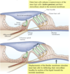
*inner row= When cochlear fluid displaces Basilar membrane–>displaces Tectorial membrane—-> deflects [Stereocilia of inner row to move in endolymph]–> TRANSDUCTS MOST OF SENSORY AFFERENT VIA CN8
2) Stereocilia= [Tethered Hair cell cilia] arranged in a “V” and when 1 is bent –> causes neighboring Stereocilia to bend
3) Hearing is an ACTIVE PROCESS

•Cochlear nerve fibers enter brainstem at ___ _____ and terminate in ___________
B: Ventral cochlear nucleus is ___[smaller/Larger] and it projects to to the _______ nuclei in the _____. What is the TRAPEZOID BODY?
C: dorsal cochlear nucleus terminates in the ___ and ____ nuclei
D: [inferior Colliculus] relays ____ info (via ____) to_____ nucleus in the Thalamus —->sends ______ projections to _______ where it is interpreted
E: [T or F] Auditory Cortex is sensitive to lesions and can easily —> sound discrimination problems
•Cochlear nerve fibers enter brainstem at PONTOMEDULLARY JUNCTION and terminate in [Dorsal/Ventral cochlear nuclei]
B: Ventral cochlear nucleus is LARGER and it projects to ipsilateral & contralateral [SUP olivary nuclei] in Pons. **Midline crossing of the [Ventral cochlear nuclei] axons = TRAPEZOID BODY
C: dorsal cochlear nucleus terminates in the [inferior Colliculus] and [lateral lemniscus nuclei]
D: [inferior Colliculus] relays auditory info (via brachium) to [Medial Geniculate nucleus] in Thalamus —->sends auditory projections to [Area 41 Heschl’s Gyri]
E: FALSE! Auditory Cortex Lesions have to be EXTENSIVE in order to affect sound discrimination

C: Where is the Tensor Tympani m. & what is it innervated by?
D: Where is the Stapedius m. & what is it innervated by?
D2: What is the OVERALL PURPOSE of these 2 Middle ear muscles?
C: Tensor Tympani m.= Fast Striated m. (middle ear wall fibers) near eustachian tube that attaches to Malleus
{{Innervated by CN5B3}}
D: Stapedius m.= Fast Striated m. (middle ear wall fibers) tht attaches to [Stapes] near its connection with [Incus]
{{{Innervated by Facial CN7}}}
D2: Purpose of these MIDDLE EAR m. is to DEC amplification of sound oscillation—> protect us from LOUD SOUNDS & adjust loudness of voice b4 we speak[using Facial CN7]

A: Describe the Semicircular Canals
C: Utricle and Saccule are Dilations of the __ ___ that contain _____ which allows them to ______ . Why can they do this? How are they related to the Maculae?
D: What’s the difference between Utricle and Saccule in regards to Planes?
E: ºThe ______ [Utricle/Saccule] connects the [3 semicircular canals]
ºThe ______[Utricle/Saccule] is continuous with Cochlea
ºBOTH Utricle & Saccule have an ______ ______ for [______, ______ & ______ detection]
A: 3 interconnected tubes positioned at right angles to one another in 3 planes of space (1 horizontal, 2 perpendicular)

C: Utricle and Saccule are Dilations of the [Membranous Labyrinth] that contain [Ca+carbonate Otolithic granules]—>allows them to respond to gravity/acceleration because it Adds weight. They also EACH have a Maculae which contains the [Vestibular sensory Hair cells]
——————————————————————————
1. Utricle = sensory hair cells oriented in HORIZONTAL plane = sensitive to linear acceleration/deceleration in HORIZONTAL DIRECTION
-
SACcule =sensory hair cells oriented in vertical plane = sensitive to linear/acceleration/deceleration in VERTICAL DIRECTION. “We SAC those dudes Vertically”
- ——————————————————————————
E:
ºThe Utricle connects the [3 semicircular canals]
ºThe Saccule is continuous with Cochlea
ºBOTH have an Otolithic Membrane for [Gravity, Acceleration & position detection]
[PPRF] Paramedian pontine reticular formation
1. Where is this located?
- What happens when this is stimulated?
- Describe its projection
- ——————————————————————————- - Explain what the [mesencephalic reticular formation] does and is located?
- A: Vestibuloocular Reflexes (VORs) reflexly move eyes in Direction ____ of Head mvmnt. It uses ____ to get from ____ and ____ to the ____
B: Absent of (VORs) means there is ___ damage
[PPRF] Paramedian pontine reticular formation
1. Near abducens nucleus (so sometimes called parabducens nucleus)
- Stimulation = Horizontal Eye movement
- Receives multiple afferents and projects to Ocular nuclei 3, 4 and 6 using the [Ascending MLF]
- ——————————————————————————- - [mesencephalic reticular formation] = includes a [Cajal interstitial nucleus] and controls VERTICAL eye mvmnt . Is located in the ROSTRAL [PPRF]
- A: Vestibuloocular Reflexes (VORs) reflexly move eyes in Direction OPPOSITE of Head mvmnt. It uses [Ascending MLF] to get from [medial vestibular nuclei] and [SUP vestibular nuclei] —->PPRF—>[CN nuclei 3, 4 & 6]
B: Absent of (VORs) = brain STEM damage

What are the 3 ways we can get “Dizzy”?
- Vestibular input without vision (i.e. spinning in a chair with eyes closed–> constant motion that eventually results in cupola membrane returning to its baseline)
- [Motion detection from Visual system] but WITHOUT [Vestibular confirmation] (looking out car window when an adjacent car moves away= false sense of motion)
- [Motion detection from Vestibular system] but WITHOUT [Visual confirmation] (sitting in cabin of a boat during a storm= MOTION SICKNESS)
Nystagmus are __ ___ ___ that have both _ and __ components (but during test refers to ___ component only) *There are 3 types*
B1. SPONTANEOUS Nystagmus is ____[sometimes/always] pathological and is caused by ___ ____ 2º to ______
B2. What are the 3 sx?
Nystagmus are OSCILLATING EYE MOVEMENTS that have both slow and Fast components (but test refers to Fast component only) .
B1. SPONTANEOUS Nystagmus is ALWAYS PATHOLOGICAL and is caused by [Vestibular/ Brainstem/ Cerebellum] imbalance 2º to irritating or destructive lesions.
B2: Sx= [N/V] + [bp DEC] + Tachycardia
A: Tracking/Smooth Pursuit involves _____ using ______. Is Tracking/Smooth Pursuit voluntary and can it be done without Visual Stimuli?
B: What is the [Fixation Reflex]? Give example
C: [Optokinetic Railway Nystagmus]? Give example
A: Tracking/Smooth Pursuit involves “Locking” eyes onto perceived moving object using [Occipital Eye Fields]. Although These mvmnts are voluntary, smooth sweep of eyes CAN’T be done in ABSENCE of visual stimuli
B: Fixation Reflex = same as smooth pursuit mvmnt but allows us to fix on an object when BOTH person AND object are moving [Ex: Reading road sign while driving on bumpy road]
C: [Optokinetic Railway Nystagmus] = (to-and-from) oscillating eye mvmnts made when ur fixating on moving objects [Ex: Looking @ telephone pole out window of moving train]
A: Frontal eye fields are located in the __ __ ___ and are considered the _______. Frontal eye fields influence ____ nuclei using ___ and ____
B: Lesioned [Frontal Eye Fields] will lead to what?
A: Frontal eye fields are located in the MIDDLE FRONTAL GYRUS and are considered the [center for Voluntary eye Saccades]. FEF influence Ocular nuclei using [SUP colliculus] and PPRF
B: Lesioned FEF —->inability to look contralaterally (with reflex eye movements intact)
ASCENDING Medial Longitudinal Fasciculus (MLF)
1. Arises from what 2 nuclei?
- Describe where it projects to?
- what is its purpose? (2)
- Explain how CROSSED Ascending MLF Fibers are different than UnCrossed Ascending MLF Fibers?
**[ASCENDING Medial Longitudinal Fasciculus] (MLF)**
1. Arises from [medial vestibular nuclei] and [SUP vestibular nuclei]
- Projects BILATERALLY to CN3, 4 and 6’s nucleus!
- Coordinates
- [conjugate Eye mvmnts] (via reticular formation)
+/- [Head mvmnt = i.e. vestibuloocular reflex]
- ***CROSSED Ascending MLF = EXCITES XtraOcular m.
vs.
*UNCrossed Ascending MLF= inhibits xtraocular m.

Describe the 3 Vestibulospinal Tracts
- Lateral vestibulospinal tract Arises from [_____ nucleus] & descends ___ to ___ spinal cord levels to activate _____
- MEDial vestibulospinal tract is the MAJOR component of ___ __ and comes from [_____ nucleus] . Projects to ___ spinal cord levels to innervate ______
- Spinovestibular tract
- Lateral vestibulospinal tract
Arises from [lateral vestibular nucleus] & descends ipsilaterally to ALL spinal cord levels to activate EXTENSOR motor neurons - MEDial vestibulospinal tract
is the MAJOR component of DESCENDING MLF and comes from [medial vestibular nucleus] . Projects to CERVICAL spinal cord levels to innervate neck muscles - Spinovestibular tract = collaterals of spinoCEREbellar projections
A: _____ hair cells of the [Crista ampullaris] produce a gelatinous [_____] to cover and protect them.
B: How are the Semicircular Canals paired up?
A: Stereocilia hair cells of the [Crista ampullaris] produce a gelatinous [CUPULA] to cover and protect them.
B: ANT canal is parallel to POST canal from other side. Horizontal canals from both sides form functional pair together
A: Equilibrium is Maintained and Controlled by what 3 sensory inputs? How many is needed to maintain Balance
B: What are the Sx (3) of Vertigo?
B2: Causes of Vertigo (2)?
A: Equilibrium is Maintained and Controlled by
1. Vestibular apparatus of internal ear
2. Vision
3. Proprioreception
[It only requires 2 out of the 3! to maintain Balance]
——————————————————————————
B: Vertigo Sx = DNP = Dizziness, NV and Pallor
B2: Causes: [Degenerated Ca+ Crystal Otoliths] vs. [Acute Labyrinthitis]
What are the 6 Sensory Receptors that utilize the [Dorsal Spinocerebellar Tract] Pathway for ______ ______ ______.
5 Sensory Receptors that utilize the [Dorsal Spinocerebellar Tract] Pathway for UNCONSCIOUS IPSILATERAL PROPRIORECEPTION
MMM JRP
- Meissner’s Corpuscle
- Pacinian Corpuscle
- Merkels Disk
- Ruffini
- Joint Receptors
- Muscle Spindles

Describe This Receptor:
[Free Nerve Ending]
- Is it Encapsulated?
- Function (2)
- Distribution (2)
[Free Nerve Ending] Receptor:
- NOT Encapsulated (“Free Hairy Merkel”)
- Pain/Temp
- Deep skin & viscera

Describe This Receptor:
[Merkel’s Disk]
- Is it Encapsulated?
- Function
- Distribution [4]
[Merkel’s Disk] Receptor:
- NOT Encapsulated (“Free Hairy Merkel”)
- Touch
- Feet/hands/genitalia / lips

Describe This Receptor:
[Hair Follicles]
- Is it Encapsulated?
- Function
- Distribution
[Hair Follicles] Receptor:
- NOT Encapsulated (“Free Hairy Merkel”)
- Touch
- Anything with Hair

Describe This Receptor:
[Meissner’s Corpuscle]
- Is it Encapsulated?
- Function
- Distribution [4]
[Meissner’s Corpuscle] Receptor:
- Encapsulated !!
- 2 Point Discrimination
- [hairLESS Skin] / joints / ligaments / fingertips

Describe This Receptor:
[Pacinian Corpuscle]
- Is it Encapsulated? ]
- Function
- Distribution [4]
[Pacinian Corpuscle] Receptor:
- Encapsulated !!
- Vibration
- Fingers & Toes / mesenteries / peritoneum

Describe This Receptor:
[Ruffini Ending]
- Is it Encapsulated?
- Function
- Distribution
[Ruffini Ending] Receptor:
- Encapsulated !!
- Stretch / pressure
- Dermis

Describe This Receptor:
[Joint Receptor]
- Is it Encapsulated?
- Function
- Distribution [2]
[Joint] Receptor:
- Encapsulated !!
- Joint Position
- [Joint Capsules] & Ligaments

Describe This Receptor:
[Neuromuscular Spindle]
- Is it Encapsulated?
- Function
- Distribution
[Neuromuscular Spindle] Receptor:
- Encapsulated !!
- Limb muscle Stretch
- Muscles

Describe This Receptor:
[Golgi Tendon Organs]
- Is it Encapsulated?
- Function
- Distribution
[Golgi Tendon Organs] Receptor:
- Encapsulated !!
- MUSCLE Tension
- Muscle tendon Junctions

[Dorsal Posterior Horn] contains the [Substantia gelatinosa] and [Nucleus Proprius]
Describe the [Substantia gelatinosa] (3)
**[Substantia gelatinosa]**
ºIs “Pain Gate keeper” & filters sensory information by synapsing on dendrites in [Nucleus Proprius]
ºHomologous to [spinal trigeminal nucleus]
ºAxons ascend & descend 1 to 4 segments in [Dorsolateral Fasciculus /Zone of Lissauer]
- [Pain, temperature, position sense, vibration from skin/body wall and pressure] come from the ___ component
- [Motor projections to Viscera, Glands & blood vessels] = ____ component
- Pain, sensations of FULLNESS/STRETCH come from the ____ component
*Alar Plates derive into the ____/____ root
Basal Plates derive into the ___/_____ root
- [Pain, temperature, position sense, vibration from skin/body wall and pressure] come from the GSA component
- [Motor projections to Viscera, Glands & blood vessels] = GVE component
- Pain, sensations of FULLNESS/STRETCH from viscera come from the GVA component
*Alar Plates Derive into SENSORY/DORSAL ROOT
{Afferent and Alar = Sensory}
*Basal Plates derive into MOTOR/VENTRAL Root
A: What’s special about the CLOSED medulla?
B: What are the Corpora Quadragemini? How is it related to “SLOW AIM”
C: Describe the 3 Cerebellar Peduncles and what their attached to
D: Which Cerebellar Peduncle DECUSSATES in the Caudal Midbrain?
A: The CLOSED Medulla is the part of Medulla NOT UNDER 4th Ventricle
B: [Corpora Quadragemini] are 4 Colliculi tht sit on DORSUM of Midbrain and is AKA [TECUM OF MIDBRAIN].
**SLOW = SUP colliculi talk with [Lateral geniculate] for Optic “WVision”
**AIM = Auditory system uses Inferior Colliculi which talks with [MEDIAL geniculate]
————————————————————————————–
C:3 Cerebellar Peduncles
1- **SUP Cerebellar Peduncle = MOSTLY EFFERENT except [Ventral Spinocerebellar tract] and is in midbrain
2-MIDDLE cerebellar Peduncle= LARGEST, is afferent and attaches to Pons
D: **SUP Cerebellar Peduncle DECUSSATES inbetween [substantia nigra] of caudal MIDBRAIN …on its way to [Red Nucleus of midbrain]
A: [Crus Cerebri Cerebral Peduncles] are on the ______[Dorsal/Ventral] Midbrain and responsible ……
B: Where does the Infundibulum Stalk sit in relation to the [Crus Cerebri Cerebral Peduncles]? What does it suspend?
C: Cerebellar Peduncles would stain ____ with myelin stain. Why?
A: [Crus Cerebri Cerebral Peduncles] are on the VENTRAL Midbrain and responsible for connecting Cerebrum with brainstem/spinal cord
B: Infundibulum Stalk hangs Between [Crus Cerebri Cerebral Peduncles] of midbrain and suspends PITUITARY GLAND ventrally
C: Cerebellar Peduncles would stain BLACK with myelin stain because they’re white fibers lol
C: Describe the Pathway of [Spinal CN11]
————————————————————————————–
D: What happens when the _____ ______ ____ squishes [Oculomotor CN3] and why?
D: When the Temporal Lobe UNCUS squishes [Oculomotor CN3] —> LATERAL GAZE! (because CN6 Abducens just takes over)
- Which Artery perfuses the [LATERAL Medulla] and what Parent Artery does it come from?
- What’s the Name of Dz that occurs when this Artery becomes occluded?
- Name the Sx of this dz and what syndrome is causes? (5)
- The PICA (Daughter of Vertebral a.) perfuses Lateral Medulla
- Can Cause [Lateral Medullary syndrome of Wallenberg]
- causes ischmia and —> Lucy Has 2 VPN
Limb Dysmetria ipsilateral- from [inf cereberllar peduncle]
Horner’s Syndrome ipsilateral- from [descending sympathetic fibers]
2TVP loss Contralaterally-from [medial lemniscus involvement]
[Vocal Cord/Palatal Weakness] ipsilateral- from [nc. ambiguous]
[Pain & Temp] impairment ipsilateral face/Contralateral body (STT)- from [descending CN5, n & t] & STT
Nystagmus- from vestibular nc.

1) Where is the [Nucleus of Solitary Tract] [NST] located?
2) What is it responsible for? (2)
- [NST] Nucleus of Solitary Tract are in the [Medulla]
- Taste [upper NST] &; GVA Sensation [Lower NST]
B: What are these Structures PERFUSED By?
- Olive (2)
- Basal Bons
- [Middle cerebellar peduncle] (2)
- [Crus Cerebri Cerebral Peduncle]
- .[inf cerebellar peduncle] AKA ___ ____
PERFUSIONS!
6. Olive perfused by [PICA or actual Parent Vertebral a.]
- Basal Pons perfused by Basilar a.
- [Middle cerebellar peduncle] perfused by [AICA and Basilar a.]
- [Crus Cerebri Cerebral Peduncle] perfused by PCA {POST cerebral a.}
10 .[inf cerebellar peduncle] (AKA RESTIFORM BODY) perfused by PICA

A: [Lower Motor Neurons] are Motor neurons of the ___ & _____ They are arranged into 4 columns and release ____ onto _____ receptors of ____ _____ Lower Motor Neurons are recruited based on __ & ____
B: What are the 4 Column Arrangements and which muscles do they innervate?
A: Lower Motor Neurons are Motor neurons of the Brainstem & Spinal Cord.
They are arranged into 4 columns and release ACETYLCHOLINE onto nicotinic receptors of target m.
Lower Motor Neurons are recruited based on size & Force
B: 4 Column Arrangement:
- medial LMN–>axial trunk m.
- Lateral LMN—>Distal Limb m. (extremities)
- Dorsal LMN—>FLEXORS
- venTral LMN——->exTensors
Describe the Descending pathways for Lower Motor Neurons. These all act as 1 of the ___ ______ in the Spinal Cord
A: Corticospinal tract (lateral)
B: Vestibulospinal tract [2]
C: Reticulospinal tract [2]
D: Tectospinal tract
E: Rubrospinal tract
Descending pathways = CONTROL SYSTEM in spinal cord LMNs
A: Lateral CST=ALL Excitatory (Glutamate is the transmitter)
B: Vestibulospinal= Head mvmnt & postural adjustments
C: Reticulospinal= locomotion & postural control
D: Tectospinal = reflex of turning head in response to visual/auditory stimuli
E: Rubrospinal= no significance in humans
A: CorticoBULBar tract (AKA ___ tract) is an ____ MOTOR NEURON tht descends ANterior to _____ tract. It usually ends on ____ of the ___ _____ but sometimes ends on _____inside the ____.
B: Name the 3 CN nucleus that do NOT receive DIRECT CorticoBULBar innervation.
A: CorticoBULBar tract (AKA Corticonuclear tract) is an UPPER MOTOR NEURON that descends ANterior to Corticospinal tract. It usually ends on interneurons of [Reticular formation] but sometimes ends on [motor neurons of CN] inside the brainstem.
B: Oculomotor, Trochlear & Abducens nucleus does NOT receive DIRECT CorticoBULBar innervation.
Describe The Cortex Origin of Upper Motor Neuron for CN nuclei involving:
- Chewing ex. Upper Motor Neurons for Chewing (CN__) comes from ____[R vs. BOTH] side(s) of the Cortex before reaching the Nucleus
- Facial Droop vs. Bell’s Palsy
- [speaking & swallowing]
- tongue mvmnt
Cortex Origin of Upper Motor Neuron for CN nuclei involving:
- Chewing ex. UMN for Chewing (CN5) comes from [Both BUT More of 1 side] before reaching Nucleus
-
Facial Droop vs. Bell’s Palsy: UMN come from BOTH Cortex before reaching CN7 Nucleus –> ONLY 1 LMN Innervates Entire Face ipsilaterally
* Facial Droop= Lesion of Facial UMN*
* BeLLs Palsy= Lesion of Facial LMN = complete face droop* - tongue mvmnt: UMN comes from [Both BUT More of 1 side Cortex] before reaching CN12 Nucleus

What are Betz cells?
Make up 3% of [CST-Corticospinal Tract] and are concentrated there
Primary Motor Cortex makes up 50% of CST and the rest comes from [Adjacent Frontal motor and Parietal areas]
Area 44 = ______
Area 22 = ______
Area 41 = ______
Hearing is ______[unilateral/Bilateral] above the cochlear nucleus
**99% of hearing comes from ______[Outer/inner] row of hair cells
What is the Other row for?
Area 44 = Broca’s
Area 22 = Wernicke’s
Area 41 = Heschel’s Gyrus / Primary Auditory Cortex
Hearing is BILATERAL above the cochlear nucleus = why it’s hard to knock out
**99% of hearing comes from inner row
Outer row = displacement sensitive so controls tectorial membrane
A: The _____ Artery (_____ circulation) perfuses MOST of the CEREBRUM.
It bifurcates into the ______ and ______ artery
B: What are the 3 daughter branches of this Artery?
The INTERNAL CAROTID Artery (ANTERIOR circulation) perfuses MOST of Cerebrum (70%). (Vertebral a. perfuse 30%)
It bifurcates into….. [Anterior Cerebral a.]—->medial cortex and [middle cerebral a.]–>lateral cortex
B: Daughter branches: 1) ophthalmic artery 2) ANT Choroidal a. 3) POST communicating a.

A: The _____ system (______circulation) perfuses Brainstem, Cerebellum & Spinal Cord. It Bifurcates into ____ cerebral arteries.
B: Describe this system
C: What are the daughter branches for each of these Arteries?
The VERTEBROBASILAR system (POSTERIOR circulation) perfuses Brainstem, Cerebellum & Spinal Cord.
B: 2 Vertebral a. Join—> 1 Basilar ARtery–>Bifurcates into PCA [POST cerebral a.]
C:
*Basilar branches = [(AICA) ANT inf. cerebellar a.] & [(SCA)SUP cerebellar a.]
**Vertebral branches = PAP! (, ASA PSA, PICA) -ANT Spinal a. -POST Spinal a. -POST inferior cerebellar a.

A: 80% of Strokes arise from occlusion in the ____ a. which is a bifurcated branch of the ______ Artery. What part of the brain is perfused by this bifurcated branch?
B: What part of the Brain does the [POST cerebral a.] perfuse? [2]
80% of strokes<—–[middle cerebral a.] which is a bifurcated branch of INTERNAL CAROTID ARTERY.
[middle cerebral a.] perfuses lateral cortex
B: [POST cerebral a.] perfuses Occipital lobe & Temporal Lobes (memory lost)

A: The Circle of Willis Interconnects the ____ and ____ circulations. How does it do this exactly?
B: How is it related to perfusion of deep cerebral structures? (2)
C: What are the Anterior/Posterior Perforated Substance?
{Circle of Willis} Interconnects ANTERIOR and POSTERIOR circulations. It forms 1. [ANT communicating a.] between the two [ANT cerebral a.] and [POST communicating a.] between [Internal carotid] and [POST cerebral a.]
B: [Circle of Willis] surrounds brain base it gives rise to small perforating ganglion arteries (via MCA) called [lenticulostriate a.] –perfuse—> [deep cerebral structures (internal capsule/basal ganglia/thalamus)]
C: entry points of perforating a. on Brain Base

Blood Brain Barriers are formed by _____ ___ _____which use ___ junctions between ____ cells to filter blood coming from _______.
B: What are Pericyte? Where is it located (2)?
Blood Brain Barriers are formed by [ASTROCYTE GLIAL CELLS] which use TIGHT junctions between ENDOTHELIAL cells and [ENDOTHELIAL CELL LAMINA] to “gatekeep” blood coming from from CAPILLARIES(which never make direct contact with brain tissue)
B: Pleuripotent cell that gives rise to other blood vessels and regulates endothelial cells. Located [under basal lamina] but [ON TOP OF ENDOTHELIUM]

The Brain is ___% of Body weight BUT uses ___% Oxygen!
A: metabolic: INC neuronal activity—>________ released —>[______ _______ _______ activation]—>________ ________released at feet —> applied to vessels to ________ ________ in that area
B: How is Control of Blood Flow AUTOregulated?
C: How is Control of Blood Flow regulated neuronally?
D: What is the normal Flow of Blood and what happens when that number is low? [2]
Brain is 2% Body Weight BUT uses 25% Oxygen!
-Control of Blood Flow:
A: metabolic: INC neuronal activity—>Glutamate released —>[astrocyte feet receptor activation]—>VasoDIOLATES factors released at feet —> applied to vessels to DILATE VESSELS IN THAT AREA
B: AUTOREGULATION: Arterial & Smooth muscle cell mediated
C: neuronally: autonomic fibers innervate Cerebral vessels
D: Normally= [55 ml Blood/100 g in 1 minute]
20 ml = neurons stop electrically firing
10 ml = NECROSIS OF BRAIN!
Valveless Cerebral Veins —(drain into)—>___________—–(drain into)—–> _____ ______ ______ + [Basilar Venous Plexus]
B: The [Basilar Venous Plexus] drains mostly ____ _____ and communicates with +_____________
C: Where does the [Cerebellum and Brainstem] draiiin their veiiiinnns?
Valveless Cerebral Veins—(drain into)—>[DURA VENOUS SINUSES]—-(drain into)—-> [INTERNAL JUGULAR VEIN] + [Basilar Venous Plexus]
B: [Basilar Venous Plexus] mostly drains BASE OF BRAIN and communicates with [SPINAL CORD EPIDURAL VENOUS PLEXUS]
C: [Cerebellum and BrainSTEM] drain their veins into the [GREAT VEIN OF GALEN]! (along w/Deep Veins)
Describe the 2 Major Divisions of Cranial Venous Drainage -Superficial Veins (5) vs. Deep Veins (6)
****Superficial Veins*** 1. Superficial group = Dumps INto Superior/inferior Sagittal Sinuses 2. Inferior group= empties with transverse AND cavernous sinus —->[SUP/inf sagittal sinus]—-> **[SINUS CONFLUENCE]**—->Transverse sinuses—>IJV
vs.
Deep Veins = empty into [Internal cerebral Veins] —>[GREAT VEIN OF GALEN]—–>Straight Sinus—->**[SINUS CONFLUENCE]**—–>Transverse sinuses ——>IJV

What does the CT scan delineate?

SubArachnoid Hemorrhage going into the Cisterns

A: What type of Nystagmus does Drugs/Metabolic produce?
B: What type of Nystagmus does Anatomical lesions produce?
C: Give an example of Physiological Nystagmus
A: Symmetrical Nystagmus
B: Asymmetrical nystagmus
C: Rotating in a swivel chair and then suddenly stopping
A: MOD for Bell’s Palsy
B: Clinical Manifestation (3)
C: Cause
D: Short-term tx
A: BeLLs Palsy= Sudden and Non-traumatic Lesion of Facial LMN = complete face droop
+
[possible taste loss over ipsilateral ANT Tongue (chorda tympani branch)]
+
[possible ipsilateral hyperacusis (stapedius branch)]
C: [Facial CN7 inflammation in petrous bone (possibly viral)]
D: Steroids

List Clinical Manifestation for these Facial CN7 lesions
- [Facial CN7 LMN]
2a. [Petrous Bone: Chorda tympani fibers]
2B. [Petrous Bone: Stapedius fiber]
- [CAAN- Cerebellopontine Angle Acoustic Neuroma]
- [Pontine Lesion]
FTSDL

[Facial Paralysis ipsilateral / Taste loss / Sound sensitivity INC / Deafness&Tinnitus / Lateral Gaze impairement]
- [Facial CN7 LMN] = F
2a. [Petrous Bone: Chorda tympani fibers] = FT
2B. [Petrous Bone: Stapedius fiber] = FTS
- [CAAN- Cerebellopontine Angle Acoustic Neuroma] = FTSD
- [Pontine Lesion] = FTSDL
Brain Stem Synromes
Describe:
A: Brain stem lesions (2)
B: [R Pontine Infarct] (2)
A. Brain stem lesion –>
- ipsilateral CN deficit
- Contralateral UMN limb weakness
B: R Pontine Infarct–>
- [R LMN Facial Weakness from CN7 deficit]
- [L Hemiparesis from (Corticospinal Pyramidal tract) deficit]

Stroke - Brain Stem Syndromes
Describe:
[Medial Midbrain of Weber syndrome]
A. [Medial Midbrain of Weber syndrome]
**Occlusion of [POST Cerebral a.] –>
- ipsilateral CN3 lesion
- Contralateral hemiplegia (Cerebral Peduncle)

A: Explain the *Rinne Test* [3]
B: What two Auditory Defects does it differentiate between?
1st: Place vibrating tuning fork on mastoid process of suspected side. Pt should hear vibrations in that ear = Bone conduction route intact
2nd: While tuning fork is still vibrating move prongs of fork to [outside ear pinna]–> where *Airconductionis 100x more sensitive than bone*
3rd: Pt should still hear sound for 15 more seconds. If pt can’t hear sound for that long = air conduction deafness on that side
B: Differentiates between [Air and SensoriNeural: 2nd step] defects

A: Explain **WEBER’S TEST** [4]
B: What two Auditory Defects does it differentiate between?
A: WEBER’S TEST
1st: Tuning Fork placed on Skull midline
2nd: Normally sound is conducted simultaneously by both [ossicular air] and [bone] routes which are out of phase w/each other (1vibration up and 1vibration dwn)
3rd: Being out of phase—>Cancel each other out on both sides of head–>sound perceived as coming from midline
4th: If [ossicular air] route loses conduction, cancellation effect on tested ear is DEC and net vibration will be GREATER on the AFFECTED SIDE
* can also be caused by [CTL Ear has SensoriNeural defect]*
B: Differentiates between [Air and SensoriNeural defect: 1st step]

A: Clinical Course of Carbon Monoxide Poisoning
B: Tx
C: Which drugs cause Drug-related Stroke Syndrome (4)
1st: HA / vomiting / blurred vision
2nd: Coma / Seizures / Cardiopulm arrest (Amnesia / Parkinsonism)
B: [Hyperbaric O2 chamber]
C: [Cocaine (most common) / Amphetamine / PCP / LSD]–> vasoconstriction or abrupt HTN–>cerebral infarct/hemorrhage
A: Describe the 2 stages of EtOH Withdrawal
B: What factors can be Fatal during EtOH Withdrawal (2)
C: When is a Brain Scan during EtOH Withdrawal warranted?
- [Early Hypersympathetic Stage] = tremors + sweaty + tachycardia + [limited convulsive Tonic–>Clonic seizures] tht occur 12 hrs-3 days post drinking]
- [Later (3-4 days post drinking) Delirium Tremen Stage] = [Fluctuating motor/autonomic activity] + confusion + hallucinations(visual and auditory)
B: Coexisting infectio vs. Trauma
C: If pt has [Partial / Focal Seizure] this suggest focal lesion

Chronic Alcoholism Syndrome
A: Causes (3)
B: What type of Head Trauma can it cause? (2)
C: Clinical Manifestations (4)
A:
- EtOH itself
- Malnutrition from EtOH
- Vitamin (B1) Deficiencies from EtOh
B: [Subdural vs. Cerebral Hemorrhage]
C:
- [Wernicke Encephalopathy] —> [Korsakoff Psychosis]
- [Alcoholic (ANT SUP Cerebellar vermis) Degeneration]
- Peripheral Neuropathy
- Dementia
Chronic Alcoholism Syndrome
A: [Wernicke Encephalopathy]
- Sx (4)
- Tx
B: [Korsakoff Psychosis]
-Sx of [Korsakoff Psychosis]
A: [Wernicke Encephalopathy]
- [Nystagmus vs. Ophthalmoplegia vs. (Gait ataxia) vs. Confusion]
- Tx = Thiamine B1 supplement
B: [Korsakoff Psychosis] = CHRONIC PHASE!
-Amnesia compensated with Comnfabulation

A: Brain uses _____ metabolism intaking [___% Cardiac Output] and [____% O2 consumption]
B: Describe pathogenic course once Cerebral ischemia occurs (2)
C: Which brain cells are most ischemia sensitive? (4)
D: Which brain cells have variable ischemia sensititvity (3)
A: Brain uses Aerobic metabolism intaking [20% Cardiac Output] and [15% O2 consumption].

Brain has NO O2 reserve = SENSITIVE TO ISCHEMIA
B: Post cerebral ischemia –> [Normal for 8-10 seconds] –> [Irreversible Damage AFTER 6-8 minutes!]
C: Neurons > Oligodendrocytes > [Endothelial Cells] > Astrocytes
D:
- Cerebellar Purkinje
- [(SHC-Sommer’s Hippocampus CA1) Pyramidal neurons (long term memory)]
- [Cortex Watershed Layers 3 & 5] Pyramidal neurons (–>Laminar Necrosis if pt survives Global ischemia longer than 3 days)
A: What type of pathology makes up the lesser porition of Stroke cause? Describe its etiologies (2)
A: Intracranial Hemorrhage makes up 15% of causes of Stroke (Cerebral Ischemia is most common)
- Intraparenchymal Hemorrhage (HTN vs. amyloid)
- SubArachnoid Hemorrhage (Saccular Aneurysms vs. AVM)
A: Brain Histology post Global Cerebral Ischemia (3)
B: When do these Histological changes occur?
A:
- [Red Dead Neurons] (neuronal cytoplasm appears red because necrosis attracts eosinophilia)
- DEC Nissl substance
- Dark Pyknotic nuclei
B: [6-12 hours post insult]


