Neurology: Embryology Flashcards

1
Q

What does the neural plate give rise to?

A
  • Neural tube
  • Neural crest cells
2
Q

What does the notochord become?

A

Nucleus pulpous of intervertebral disc in adult

3
Q

What induces overlying ectoderm to differentiate into neuroectoderm and form neural plate?

A

Notochord

4
Q

Is the alar plate (dorsal) sensory or motor?

A

Sensory

5
Q

Is the basal plate (ventral) sensory or motor?

A

Motor

6
Q

What are the three primary vesicles in the developing brain (superior to inferior)?

A
  1. Forebrain (prosencephalon)
  2. Midbrain (mesencephalon)
  3. Hindbrain (rhombencephalon)
7
Q

What are the five secondary vesicles in the developing brain (superior to inferior)?

A
  1. Telencephalon (from forebrain)
  2. Diencephalon (from forebrain)
  3. Mesencephalon (from midbrain)
  4. Metencephalon (from hindbrain)
  5. Myelencephalon (from hindbrain)
8
Q

What are the adult derivatives of the walls and cavities of the telencephalon?

A

Walls

  • Cerebral hemispheres
  • Basal ganglia

Cavities

  • Lateral ventricles
9
Q

What are the adult derivatives of the walls and cavities of the diencephalon?

A

Walls

  • Thalamus
  • Hypothalamus

Cavities

  • Third ventricles
10
Q

What are the adult derivatives of the walls and cavities of the mesencephalon?

A

Walls

  • Midbrain

Cavities

  • Cerebral Aqueduct
11
Q

What are the adult derivatives of the walls and cavities of the metencephalon?

A

Walls

  • Pons
  • Cerebellum

Cavities

  • Upper part of fourth ventricle
12
Q

What are the adult derivatives of the walls and cavities of the myelencephalon?

A

Walls

  • Medulla

Cavities

  • Lower part of fourth ventricle
13
Q

What develops from the neuroepithelia in the neural tube?

A
  • CNS neurons
  • Ependymal cells (inner lining of venticles, make CSF)
  • Oligodendrocytes
  • Astrocytes
14
Q

What develops from the neural crest?

A
  • PNS neurons
  • Schwann cells
15
Q

What develops from the mesoderm?

A
  • Microglia (like Macrophages)
16
Q
  • Failure of caudal neuropore to close, but with no herniation
  • Usually seen at lower vertebral levels
  • Dura is intact
  • Associated with tuft of hair or skin dimple at level of bony defect
A

Spina bifida occulta

17
Q

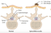
Meninges (but no neural tissue) herniate through bony defect

A

Meningocele

18
Q

Meninges and neural tissue (eg. cauda equina) herniate through bony defect

A

Myelomeningocele

19
Q
  • Increased alpha-fetoprotein (AFP) in amniotic fluid and matternal serum
  • Increased acetylcholinesterase (AChE) in amniotic fluid
A

Neural tube defects (except spina bifida occulta = normal AFP)

20
Q
  • Neuropores fail to fuse (4th week)
  • Persistant connection between amniotic cavity and spinal canal
A

Neural tube defects

21
Q

What deficiencies are neural tube defects associated with?

A
  • Maternal diabetes
  • Folate deficiency
22
Q

Exposed, unfused neural tissue without skin/meningeal covering

A
  • Myeloschisis
  • Also known as rachischisis
23
Q
  • Failure of rostral neuropore to close → No forebrain, open calvarium
  • Clinical findings: polyhydramnios (no swallowing centre in brain)
A

Anencephaly

24
Q

MRI:

  • Monoventricle
  • Fusion of basal ganglia (star)
A

Holoprosencephaly

25
Failure of the embryonic forebrain (prosencephalon) to seperate into 2 cerebral hemispheres (usually occures durning weeks 5-6)
Holoprosencephaly
26
What signalling pathway may be related to holoprosencephaly
Mutations in sonic hedgehog signaling pathway
27
What conditions are associated with holoprosencephaly
* Trisomy 13 * Fetal alcohol syndrome
28
Signs: * Moderate form has cleft lip/palate * Severe from results in cyclopia
Holoprosencephaly
29
* Failure of neuronal migration resulting in a "smooth brain" that lacks sulci and gyri * May be associated with microcephaly and ventriculomegaly
Lissencephaly
30
* **Ectopia of cerebellar tonsils** inferior to foramen magnum * Congenital, usually asymptomatic in childhood * Manifests in adulthood with headaches and cerebellar symptoms * Associated with spinal cavitation (eg. **syringomyelia**)
Chiari **I** malformation | (ONE structure)
31
* **Herniation of cerebellar vermis and tonsils** through foramen magnum with aqueductal stenosis → non-communicating hydrocephalus * Usually associated with **lumbosacral myelomenigocele** * May present as paralysis/sensory loss at and below the level of the lesion
Chiari **II** malformation | (TWO structures)
32
* **Agenesis of cerebellar vermis** leads to cystic enlargement of 4th ventricle that fills the enlarged posterior fossa * Associated with non-communicating hydrocephalus, **spina bifida**
Dandy-Walker malformation
33
* Cystic cavity (syrinx) within central canal of spinal cord (yellow arrows) * Associated with Chiari I malformation (red arrows show low-lying cerebellar tonsils) * Aquired causes include trauma and tumours
Syringomyelia
34
Which fibres are typically damaged first in syringomyelia?
Fibres crossing in anterior white commissure (spinothalamic tract)
35
Symptoms of syringomyelia
* Cape-like, bilateral, symmetrical loss of pain and temperature sensation in upper extremities * Fine touch sensation is preserved
36
Most common location of syringomyelia
Cervical \> thoracic \>\> lumbar
37
What pharyngeal arches form the **anterior 2/3 of the tongue**?
1st and 2nd pharyngeal arches
38
What nerves supply sensation and taste for the **anterior 2/3 of tongue**?
* Sensation: CN V3 * Taste: CN VII
39
What pharyngeal arches form the **posterior 1/3 of tongue**?
3rd and 4th pharyngeal arches
40
What nerves supply sensation and taste for the **posterior 1/3 tongue**?
**CN IX** and extreme posterior via CN X
41
Which muscle and motor supply is responsible for **retracting and depressing the tongue**?
**Hyoglossus** via **Hypoglossal** nerve (CN XII)
42
Which muscle and nerve supply are responsible for **protruding the tongue**?
Genioglossus via CN XII
43
Which muscle and nerve supply are responsible for **drawing the sides the tongue upwards** to create a trough for **swallowing**?
Styloglossus via CN XII
44
Which muscle and nerve supply are responsible for **elevating the posterior tongue** during **swallowing**?
Palattoglossus via CN X
45
What cranial nerves are involved with **taste**?
* CN VII: anterior 2/3 * CN IX: posterior 1/3 * CN X: extreme posterior
46
What cranial nerves are involved in **tongue sensation**?
* CN V3: anterior 2/3 * CN IX: posterior 1/3 * CN X: extreme posterior
47
What cranial nerves are involved with **motor control of the tongue**?
* CN X: palatoglossus (swallowing) * CN XII: hyoglossus (retracts, depresses), **geni**oglossus (**protrudes**), **styl**oglossus (**swallowing**) *"The **geni**e comes **out** of the lamp **in styl**e"*