Muscle Physiology (Sept 19) Flashcards
What are the three types of muscle?
-skeletal -cardiac -smooth
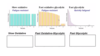
What type of muscle cell is this? What are some of this muscle type’s functions?

- smooth muscle cell
- storing and moving substances
What type of muscle cell is this? What are some of this muscle type’s functions?

-cardiac muscle
What type of muscle cell is this? What are some of this muscle type’s functions?

- skeletal muscle
- movement, stabilization
- thermoregulation
- contractions account for all body movements
Fill in the chart below


What are the layers of mysium and their functions?
- endomysium: surrounds each individual muscle fibre
- perimysium: surrounds a bundle of muscle fibres
(fascicle: bundle of muscle fibres surrounded by perimysium) - epimysium: surrounds the entire muscle
- all 3 layers contribute to the muscle sheath and become the tendon which connects 2 bones together
- tendon becomes continuous with the periosteum (wrapping around bone)
Myocyte/myofibre structure
- muscle fibre is composed of myofibrils: long tube-like structures, sarcomeres in series (collection of sarcomeres which allow muscles to contract)
- sarcomeres: basic contractile unit of muscle
- made up of a thick filament (myosin) and thin filament (actin)
Label the diagram

- Tendon
- Muscle
- Epimysium
- Perimysium
- Blood vessel
- Endomysium
- Muscle fibre
Label the diagram

- Myofibril
- Muscle fibre
- I band
- A band
- Sarcomere
- Z line
- thick filament (myosin)
- Thin filament (actin)

What happens when stressor is put on muscles?
- hypertrophy!
- muscles enlarge with use
- exercise stimulates production of actin and myosin filaments
- increased number of myofilaments expands the fibre causing muscle enlargement and definition
- want more sarcomeres to handle that stress
What happens if you remove stress from muscle?
- atrophy
- have lots of metabolically active tissue but it’s not being used so it takes proteins and sarcomeres away to use for something else
- aging, spinal cord injury, space flight all cause atrophy
What is the sliding filament theory?
- contraction of sarcomeres happens through sliding of filaments
- thin filament slides over thick filament (thick doesn’t move)
- bring Z lines closer together
- when sarcomere shortens, fill H zone and space between 1/2 I bands is being removed (distance between thick filament, A band, and Z line)
Label the diagram

- A band
- 1/2 I band
- H zone
- Relaxed
- Contraction
What is outlined in yellow? What are represented by dark black dots?

- muscle fibre
- thick filament (composed of myosin)
Label the diagram. What function does this structure serve?

- it is a thick filament and the projections are cross bridges
- the cross bridges are what makes the thick filament interact in 3D
- each thick filament can interact with 6 thin filaments
- each thin filament (actin) can interact with 3 thick filaments (myosin)

What is thick filament composed of?
- several myosin molecules with their head sticking up
- tail is lying down in parallel
- wants to grab on to thin filament

What is thin filament composed of? What is the role of each component? Where does Ca2+ come in to play?
- three proteins: actin, tropomyosin, troponin
- actin: form 2 coiled chains (which is the “thin filament”)
- tropomyosin: run along actin blocking cross bridge binding site (tropo: to turn or react)
- troponin: holds tropomyosin in place
- Ca2+ binds troponin and changes its conformation which pulls tropomyosin away from cross bridge binding site which allows for myosin-actin interaction to occur
- when Ca2+ is removed, tropomyosin moves back to block cross bridge binding site (no actin-myosin interaction)

What is this diagram depicting?

- cross bridge cycling
- Ca2+ must be present
- binds and releases
Complete the diagram

-if no ATP is present, can’t release cross bridge so there is tension in the muscle (rigor mortis)

What is excitation?
- motor neuron attaches to every muscle fibre
- action potential comes, ACh comes across NMJ, hits muscle fibre
- muscle fibre propogates action potential
- action potential spreads over muscle fibre’s membrane
- muscle fibres are large and have a large diameter
What are T tubules? Terminal cisternae?
- invaginations of the membrane
- tunnels that go deep into muscle fibre and allow for synchronous release of calcium along the muscle fibre
- T tubule wraps around myofilaments as it goes deep into muscle fibre
- sarcoplasmic reticulum contains calcium in terminal cisternae
- terminal cisternae is up against t tubule
- triad consists of one t tubule with a terminal cisternae on either side

What is the function of the DHP receptor?
- located on t tubule membrane
- reaches out and grabs “plug” on ryanodine receptor and pulls it open with arrival of action potential
- electrically sensitive and undergoes confirmational change
- ryanodine receptor located on terminal cisternae
- calcium rushes out and follows concentration gradient (lots inside terminal cisternae so it rushes out)
What are calcium ATPases?
- bring calcium against its concentration gradient to pull it back into terminal cisternae
- so it’s ready for next action potential and try to stop contraction
- release is fast but this uptake is slower which allows for the contraction to last a certain amount of time
Label the diagram


What is occurring during latent period?

-time between action potential and muscle starting to shorten is the time taken that calcium is being released so there is enough pouring out to start contraction
How do you adjust amount of force?
- muscle length (easier to lift it when bicep is at optimal length rather than when it’s stretched)
- action potential frequency (how many signals being sent to make calcium release)
- number of fibres per motor unit and the cross sectional area of those muscle fibres (motor unit: one motor neuron and its branches and how many muscle fibres that innervates- if one branches 5 times it can active 5 muscle fibres)
How does muscle length affect force?
- optimal number of cross bridges that can form
- L knot is in range where number of cross bridges that can attach to thin filament are at optimal length to produce maximal contraction
- if muscle is too contracted, thin filaments start to overlap so you can’t get them to go more
- if muscle is too stretched, thin filaments get pulled away from centre then have myosin heads that can’t reach thin filament and create cross bridge
How does action potential frequency affect force?
- one single action potential is a twitch
- most movements require multiple action potentials to generate a sustained amount of calcium release
- send multiple APs and it releases a certain amt of calcium
- before calcium ATPase can take it all up, more calcium gets released with another AP
Describe the graph

a) single AP
b) AP followed by another AP- summation of force
c) APs were spaced far enough apart that with each release of Ca, some Ca was taken up so force drops a bit then goes back up (unfused tetanus)
d) APs so closely spaced that Ca keeps pouring out (force is 3-5x greater than single AP)
How do motor units affect force?
- 3 types of motor units exist based on muscle fibre types
- muscle fibre types differ in size and ease of activation
- size determines the amount of force produced
Slow versus fast muscle fibres
- slow allow you to run marathons whereas fast give you power
- differ on speed of contraction and what kind of energy they use
- speed of contraction: dependent on rate of myosin ATPase action- slow fibre types contract more slowly but are more fatigue resistant, fast fibre types generate more power but fatigue rapidly
- aerobic metabolism versus anaerobic metabolism
Oxidative and glycolytic fibres
- oxidative: primarly use O2 for ATP production (aerobic)
- glycolytic: high capacity for ATP production without O2 (anaerobic)
What is an isometric contraction?
- muscle force=load
- no movement of load
Fill in characteristics of each fibre type and describe the size principle
- motor units are recruited according to size
- first to be recruited are slow oxidative, then fast oxidative glycolytic, then fast glycolytic
- this way, fatigue resistant fibres are always first
- the number of motor units recruited depends on the force required to perform the task

What is an isotonic contraction?
- muscle force does not equal load
- force > load gives a concentric contraction
- force < load gives eccentric contraction

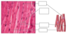
Identify the specimen and label the parts. Where is the specimen found?

- smooth muscle
- smooth muscle fibre
- nucleus of smooth muscle fibre
- lines the “hollow tubes of the body” (arteries, digestive system, respiratory, urinary and reproductive tracts)
- can be electrically coupled by gap junctions
Does smooth muscle contain thick and thin filaments? If so, do they differ from skeletal muscle?
Yes but they are less organized than skeletal muscle
- no striations
- filaments are anchored to dense bodies on the intermediate filament network
Label the diagram


What kind of contractions does smooth muscle make? What are the contractions regulated by?
- very slow to contract
- can maintain prolonged tension
- contraction can be regulated by: ANS (NTs), hormones, stretching, other local factors (Nitric oxide), pacemaker cells
What are some functions of smooth muscle?
- storing and propelling substances (peristalsis)
- mixing and churning
Identify the specimen and label the diagram. What are some key features of the specimen?
- cardiac muscle
- cardiomyocytes generally have 1-2 nuclei and are branched

How does cardiac muscle contract?
- involuntary contraction
- intrinsic conduction system (pacemaker cells)
- regulated by ANS
What are intercalated discs?
- specialized areas where cardiomyocytes connect
- contain desmosomes and gap junctions
- desmosomes allow for interaction between cells (stabilize cell to cell contacts)
- gap junctions allow electrical signal to pass (direct communication between adjacent cells)
Label the diagram


What is sarcolemma?
- muscle cell membrane
- t tubule is invagination of sarcolemma
What is sarcoplasm?
-muscle cell cytoplasm (contains myoglobin and glycogen)
What is sarcoplasmic reticulum?
-endoplasmic reticulum in muscle cell
