Morphologic Diagnosis Flashcards
What identifiers should be included in morphologic diagnosis?
- ) Organ +/- anatomic modifiers
- ) Exudate (Nature of the lesion)
- ) Distribution
- ) Duration
- ) Extent/ severity
What are the modifiers for exudate?
- Serous, similar to mucoid and/or catarrhal
- Fibrinous
- Suppurative or purulent
- Granulomatous
- Necrotizing
- Hemorrhagic
- Lymphoplasmacytic
- Eosinophilic
What are the different types of distributions of lesions?
- Focal
- Focally extensive
- Multifocal
- Diffuse
- Other
What is the different durations of lesions?
- Peracute
- acute
- subacute
- chronic
- chronic-active
What are the different extents of lesions?
- minimal
- mild
- moderate
- marked
- severe
What is the anatomic modifier for inflammation in the skin? Liver? Gallbladder? Lymph node?
Skin- Dermatitis
Liver- Hepatitis
Gallbladder- Cholecystitis
Lymph node- Lymphadentits
What is the anatomic modifier for inflammation in the lungs? Large intestines? Eyelid? Heart? Muscle? Cecum?
Lungs- pneumonia
Large intestines- colitis
Eyelid- blepharitis
Heart: myocarditis
Muscle- myositis
Cecum- Typhlitis
What is inflammation of the adipose tissue called? Epididymus? Air sac? Gland (any)? Salivary gland? Sinus?
- Steatitis/ Panniculitis
- Epididymitis
- Air sacculitis
- Adenitis
- Sialoadenitis
- Sinusitis
What does the ending itis mean? What about osis? Pathy?
Itis indicates inflammation
Osis indicates non inflammatory lesion
Pathy should not really be used, it means unknown.
How would you reference kidney inflammation with the anatomic modifiers? Glomerular inflammation? Medulla and calix ( with tubules) inflammation? Non inflammatory lesion of the kidney? an unknown kidney lesion?
- Kidney inflammation: Nephritis
- Glomerular inflammation : Glomerulonephritis
- Medulla and calix ( with tubules) inflammation: Pyelonephritis
- Non inflammatory lesion of the kidney: Nephrosis
- an non inflemmatory unknown kidney lesion? Nephropathy
What is a cause of bronchopneumonia? How about interstitial pneumonia? Aspiration pneumonia?
Bronchopneumoni usually comes from something that is inhaled
Interstitial pneumonia is typically something that comes from the blood that reaches the lungs.
Aspiration pneumonia- From aspirating contents into the lungs.
What kind of necrosis can be associated with purulent exudate? What about granulomatous?
Purulent: Liquifactive necrosis
Granulomatous: Caceous necrosis
How is exudate classified?
- Predominant type of inflammatory cells
- Plasma protein content
- Amount of fluid
What is serous exudate? Timeframe associated Causes? Gross appearence?
-Definition :Fluid rich in protein, few cells. On body surface or mucosa
- Time: Usually acute
Causes:
- May be a dominant pattern of exudation for a wide variety of mild injuries.
- Examples: trauma, cold, blisters, sunburn
- Gross Appearance: Straw-yellow or clear fluid
What is fibrinous exudate? What is its cause?
Can be in tissue or body cavities
- Increased vascular permeability (inflammatory edema)
- Leakage of fibrinogen
- Fibrinogen turns into fibrin
- Fibrin clots
What is the exudate seen here?

fibrinous exudate ( yellow egg color, no fibrin strands)
What is the timeframe for fibrinous exudate? What will you see on histology? What are the outcomes of this type of exudate?
- Acute - can form in seconds
- Histo: Thread-like eosinophilic meshwork or solid amorphous material, few neutrophils
- Outcome: Provides the support for fibroblasts and new capillaries (organization)

What is these arrows pointing at?

Fiberous adhesions
What are fiberous adhesisons?
Fibrous adhesions: common sequelae of fibrinous exudate. It is a scar, cannot remove -> chronic lesion.
What is suppurative exudate? What is its synonym? What cells can be seen?
- Synonym: Purulent Exudate
- Many neutrophils, necrotic cells and debris
- Pus due to proteolytic enzymes in neutrophilic granules (i.e) Myeloperoxidase
What is the kind of exudate seen in this image?

SUPPURATIVE EXUDATE
or
Purulent Exudate

What is the kind of exudate seen in this image?

SUPPURATIVE EXUDATE
or
Purulent Exudate
Do birds get suppurative exudate? Why or why not?
No they do not.
- Avian species, amphibians, fish, reptiles and some mammals have heterophils or granulocytes, instead of neutrophils. These will lack myeloperoxidase which is needed for suppurative exudate formation
Whart is caceous exudate?
Hard pus essentially ( looks like cheese) it occurs in birds due to lack of myeloperoxidase in heterophils.

What is occuring in this image?

Caceous exudate ( particularly in the air sac of a bird (aspirgillosis)
What are abcesses?
ABSCESS:
Localized form of suppurative inflammation, walled off by a connective tissue capsule. Suppurative lesions are often of bacterial origin!

What is a pyothorax? What kind of exudate will be found?
Pyothorax (pleural empyema): pus in the thoracic cavity
Suppurative exudate/ purulent
What is seen in this image?

Pyothorax
Note the neutrophils & fibrin strands
What is occuring in this image?

FIBRINOSUPPURATIVE EXUDATE
Pus + fibrinous exudate
Pus is in the bronchioles
What can be seen in this image?


What is granulomatous exudate? How is it characterized? What should be present to make this characterization?
Characterized by the presence of macrophages, lymphocytes and plasma cells. Macrophages can be clustered around the foreign material and can be “multinucleated” or “epithelioid”.
Some pathologist will not call it granulomatous unless there are multinucleated giant cells present.
Whatis circled in this image?

Multinucleated giant cells.
What can granulomatous inflammation tell us about the lesion?
Time: almost always chronic
Etiology: Non-digestible organism/ particle, a chronic inflammatory stimulus.
E.g.:
- Fungus
- Mycobacteria
- Vegetable material
- Mineral crystals
- Suture material
- Sperm
- Keratin, etc.
- Delayed-type hyper-sensitivity is often required
What is a classical clinical example of granulomatous inflammation? What can be seen grossly? Histologically?
Example:
Johne’s disease*
Morphologic dx: Colitis, granulomatous
Etiology: Mycobacterium avium spp. paratuberculosis
Etiologic dx: Mycobacterial colitis
Name of disease: Paratuberculosis*
You typically will see hystiocytic inflammation
Grossly: Mucosa gets very thick and makes it hard for patients to absorb nutrients. Mycobacteria proliferate within the lamina mucosa.
Histologically: yon acid fast you will see mycobacteria within macrophages.

What is pyogranulomatous inflammation?
Definition: neutrophilic + granulomatous inflammation.
e.g.: Morphologic dx: Lymphadenitis, pyogranulomatous
What is etiological diagnosis?
Reason causing the disease.
What is occuring in this image? What kind of exudate is seen? Why?

FIBRINONECROTIC EXUDATE
Necrosis of well vascularized tissue = necrosis + fibrin exudation
E.g.: Morphologic diagnosis: Enteritis, fibrinonecrotizing
What occurs when you have fibronecrotic exudate in a tubular organ?
- Casts of friable material, often yellow, fill the lumen
- Composed of fibrin and necrotic debris
•Easily broken apart and from the tissue
- Pseudomembrane or diphtheric membrane
What are the black arrows pointing to in this image?

Fibronecrotic area
What is hemorrhagic inflammation? What is its typical causes? What is its duration typically? What can be present with it?
- Hemorrhage predominates
- Due to severe injury to blood vessels from:
- Thromobosis/vascular obstruction
- Bacterial toxins
- Proteolytic enzymes
- Most often acute or peracute
- Often accompanied by necrosis (necrohemorrhagic inflammation)
What is occuring in this image?

Necrohemorrhagic inflammation
What do you call blood in the thorax? Air in the thorax?
Hemothorax
pneumothorax
What is mucoid exudate? Mucopurulent? Catarrhal?
- Mucus predominates, with few inflammatory cells
- Mucopurulent exudate: Combination of mucus and neutrophilic (suppurative) exudate
- Catarrhal: inflammation of mucous membrane w/marked increased flow
- Mucous or mucopurulent
What is occuring in this image?

Mucoid exudate

What is eosinophillic inflammation? What color can it be grossly? What is it usually associated with?
- Eosinophils predominate
- May be green, grossly
- Usually associated with:
- Parasites ** most common
- Allergies
- Sometimes with foreign bodies
What kind of inflammation can be seen in this image?

eosinophillic inflammation
What is non suppirative inflammation? What can you see microscopically? What are some examples of this?
- Microscopic Dx
- Lymphocytes & plasma cells predominate
- Used mostly in encephalitis (viral)
Examples: - Lymphocytic - lymphocytes
- Plasmacytic - plasma cells
- Histiocytic- macrophages
- Lymphoplasmacytic
- Lymphohistiocytic
What kind of inflammation is occuring in this image?

Non suppurative inflammation ( lymphocytic)
What is occuring in this image? What else can it be called? What does it normally occur as?

LYMPHOCYTIC INFLAMMATION
- Lymphocytes predominate
- Sometimes called subacute
- Often lymphoplasmacytic
What can the different types of exudate tell you?
Fibrinous
• Blood vessel damage
• Bacteria, virus - ACUTE
Suppurative
• Bacteria
Granulomatous/caseous
• Bacteria, fungus – CHRONIC**
Hemorrhagic
• Toxin (bacterial or otherwise)
• Ischemia
Eosinophilic
• Parasites
- allergens
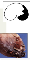
What is the distribution of this lesion?

Focal.
Can be focally extensive.
What is the distribution seen in this image? What does it mean? What are the possible origins?
Focally extensive
Definition
- Involvement of considerable area within an organ
- AKA: Locally extensive
Possible origins:
- Severe local reactions that spread into adjacent tissue
- Coalescence of foci in a multifocal reaction
What is this kind of distribution seen in this image? What is the definition of this kind? What is the size?

Multifocal
Definition Arising from or pertaining to many foci
Size: Variable
- Each focus of inflammation is separated from other inflamed foci by an intervening zone of relatively normal tissue.
- In some cases, foci coalesce, (multifocal to coalescing).
What is occuring in this image?

Multifocal to coalescing
- Multifocal to coalescing lesions start as multiple foci, but as they grow, merge into fewer larger nodules.
What is the distribution seen in this image? What is its typical etiology?

Diffuse
- Involves entire organ or tissue
- Can vary in severity
- Not definitely limited or localized
- Etiology often viral or toxic
What is peracute inflammation? What is its cause? What can you see grossly/ histologically? What is an example?
Definition: Very Acute ¡ Usually due to a potent stimulus
Little/no time to respond to stimulus
Grossly or histologically – no/minimal lesions
Less common than acute
- Usually occurs over a few hours
Ex: Fatal muocardial infarction
What are the important features of peracute inflammation? i.e (timeframe, vascular involvment, presence of inflammatory cells, clinical signs)
Definition: Very Acute
Features
0-4 hours – time
Minimal vascular involvement
Few inflammatory cells
Clinical signs
- None – Sudden
- Shock
What likely caused the lesion on this bird and was the cause of death? What will you likely see histologically? What kind of lesion is it?

Gross: electrocution
Histological: Epidermal and dermal coagulative necrosis with nuclear
streaming
Peracute lesion

What is acute inflammation? ( i.e: timeframe, vascular involvement, inflammatory cells seen, other features?
- Time: 4-6 hours to 3-5 days
Vascular Involvement
- Active Hyperemia: Edema, Occasional Fibrin thrombi.
- Inflammatory Cells: Neutrophils
- Cardinal Signs seen
- Lymphatics: Role to remove exudate
What is occuring in this image?


What is subacute inflammation? What is the timeframe, cells seen and vascular involvement?
- Transition period between acute and chronic
- Time: Varies from a few days to a week or so.
- Cells: mostly lymphocytes and plasma cells, fewer macrophages or neutrophils
- Vascular involvement: Less hemorrhage, hyperemia and edem
What are the 5 cardinal signs of inflammation?
- Redness
- Heat
- Swelling
- Pain
- Inability to function/ loss of function
What is the duration used to name a primary lymphocytic inflammation of viral origin? What other term could be used to describe this?
- Some viral infections may result in a primarily lymphocytic accumulation.
- These are often referred to as subacute.
- Non-suppurative is often used instead of lymphocytic or lymphoplasmacytic, particularly in viral disease of the brain.
What is the morphological diagnosis of this lesion? If it is of viral origin and in the brain, what is the etiological diagnosis?

Morph. Dx: Encephalitis, non-suppurative, focal, subacute Etiol. Dx: Viral disease (several)
What is chronic inflammation? What are its features (i.e. - causes, timeframe, ect)
- Inflammation that persists over a long period of time
Features
- Result of a persistent inflammatory stimulus
- Not completely resolved
- Accompanied by an immune response
- Host tissue response in terms of repair
- Time: weeks to years
What is the likely duration of the lesion seen here?

Chronic
Will chronic issues always have signs? What is an example?
May be an insidious, low-grade, subclinical process with no evidence of an acute episode
Johnes Disease
What can you see histologically for chronic inflammation?
- Mononuclear inflammatory cells – MQ, LQ, PL, MGC
- Fibroblasts and collagen
- Proliferating vasculature - angiogenesis
What can be seen when their is repair from granulation tissue?
Scar
Granulation tissue (REPAIR): Organized neovascularization and fibrous tissue proliferation
What is seen in this image?

Scar
What is a lesion that is mild? What would you see in terms of inflammatory cells, tissue cells, vascular involvement ect?
- Mild - not too bad
- Tissue damage: Absent or minimal
- Inflammatory cells: Few inflammatory cells
- Vascular involvement: A small amount of edema and/or congestion
What extent would you say the injury is to the tissue?

Mild
What would you see with moderate tissue inflammation? What would you see in terms of tissue damage, inflammatory cells, and vascular involvement?
- Moderate - fairly bad
- Tissue damage: Some obvious damage present
- Inflammatory cells: Yes - there are some inflammatory cells - Vascular involvement: Edema and hemorrhage likely
To what extent would you consider this tissue damaged?

Moderate
What would you see with severe tissue inflammation? What would you see in terms of tissue damage, inflammatory cells, and vascular involvement?
- Severe - really bad
- Tissue damage: Lots of tissue damage
- Inflammatory cells: Lots of inflammatory cells
- Vascular involvement: May see lots of edema and hemorrhage
To What extent would you consider the extent of the damage to this tissue?

marked - severe
