Chronic Inflammation Flashcards
When will you have chronic inflammation?
When cause of issue is hard to neutralize.
What is the temporal appearance of cells and exudates of inflammation in each of these categories (at their highest point)? (Acute, Subacute, Chronic)
Initial: Macrophages ( macrophages will be present throughout all stages)
Acute: Fluid, Fibrin, Neutrophils
Subacute: Lymphocytes, plasma cells.
Chronic: Neovascularization, fibroblasts, collagen
What are fibroblasts? Neovascularization?
Fibroblasts: Create collagen
Neovascularization: Creation of new blood vessels
What is seen in this image? Is this acute or chronic inflammation?

Acute inflammation
- Neutrophils present which are smaller than macrophages.
What is occuring in this image? Is it acute or chronic inflammation?

Chronic: Large numbers of macrophages surrounding this blood vessel, larger cells, non segmented nuclei.
What are the causes of chronic inflammation?
• Resistant/persistent agents
- Bacteria, Fungi, Protozoa/Parasites
• Isolation of pathogens within the pus
• Foreign bodies
• Autoimmune diseases
• Unidentified mechanisms
(I.e: Canine granulomatous meningoencephalitis (GME)
Why would bacteria cause chronic inflammation? Why may the body not clear bacteria?
Some bacteria live inside macrophages and will eventually rupture the macrophage. Another macrophage will come and will phagocytize bacteria. Bacteria will continue to proliferate and cycle will continue.
Why may some fungi cause chronic inflammation?
Some fungi are large and have antiphagocytic properties so they cannot be destroyed by macrophages. This is one reason why blastomyces / histoplasma
What is an example of a parasite that causes chronic inflammation and why? What about a virus?
Parasite: Leismania ( too large)
Virus: Some virus targets macrophage. FIP is an example and it targets macrophages.
What is chronic inflammation? What is the duration/ time frame for classification? What is it characterized by?
Continuous/ long inflammatory process.
Severe injury with necrosis, neutrophils (acute) occurs first ( mononuclear cells with macrophages, plasma cells, lymphocytes ect.
Abcesses can form and they are characterized by macrophages - will secrete alot of cytokines to induce neovascularization ( granulation tissue formation) and fibrosis.
What is each one of these labeled cells?

A. Macrophage
B. Lymphocyte:
C.) Plasma cell: Basophillic in periphery of cell.
- Perinuclear halo -> golgi complex, produces immunoglobulins.

What is each letter signifying in this image?

A.) Macrophage
B.) Agent ( in this case budding yeast cryptococcis neoformans) (Capsule does not stain with H + E)
C.) Lymphocyte
D.) Plasma cell

What does the type of inflammatory cell within the exudate indicate? What kind of exudate would be indicated if you see dead neutrophils surrounded by capsule of fiberous connective tissue? What about the presence of epithelioid macrophages? What about the presence of lymphocytes and plasma cells?
- ) Type of exudate.
- ) Presence of dead neutrophils surrounded by a capsule of fibrous connective tissue = ABSCESS - purulent exudate
- ) Presence of epithelioid macrophages = GRANULOMATOUS
- ) Presence of lymphocytes and plasma cells = LYMPHOPLASMACYTIC
In this picture, what cells should be seen in it based on the type of exudate?

Large numbers of lymphocutes, plasmacells, macrophages + fibrosis.
In this picture, what cells should be seen in it based on the type of exudate/ tissue present?

Alot tof lymphocytes
In this picture, what cells should be seen in it based on the type of exudate?

Dead neutrophils
What is the difference between an abcess and granuloma?
Abcess: Liquifactive necrosis, purulent discharge, alot of dead neutrophils.
Granuloma: epitheliod macrophages. Multinucleated giant cells.
What is the image of A? What is the image of B? How can you tell?

A.) Abcess - purulent exudate, with liquifactive necrosis present. Dead neutrophils should be present.
B.) Granuloma: due to granulomas presence and lack of purulent exudate. Would be better to confirm via .
What is a multinucleated giant cell? How can you describe it:?
Irt is a bunch of macrophages that joined together. It has nucleii in periphery.

How can you tell apart fibrin and fibrosis? What would it tell you about the duration of the injury?
Fibrin: Acute inflammation, scrabled eggs, from vascular leakage, yellow/ friable.
Fibrosis: Chronic inflammation, white/ grey, over time fibrin -> collagen, firmly adhered.

What is occuring in this image? What causes the lesion seen? What is the result of seeing lesions such as this.

This is milk spot liver in pigs ( ascaris suum larval migration)
- Due to ascaris larval migration ( caused fibrosis)
Usually non clinical in animla but meat will be condemed at slaughter.
What are the different types of macrophages and what do they do? Where do they come from?
Macrophage Types:
Osteoclast- Bone
Microglial -CNS
Alveolar Macrophage - Lung
Kupffer cell- Liver
Histiocyte - Connective tissue
White- pulp, red- pulp, marginal- zone, and metalophillic macrophage. - Spleen
These are already in tissues but their jobs depend on location.
- Bone marrow produces monocytes -> migrate during times of inflammation to tissues and are then called macrophages

What is involved in inflammation? How does each cell know what it needs to do? What is the communication between the cells called? What is the macrophage in broad terms? What is its role?
- Inflammation = complex communication system • Cells “tell” each other what to do
- Communication between cells = inflammatory mediators
- Cytokines
- Chemokines
- Macrophage – MAESTRO ( it will prepare tissue for repair)
How do macrophages kill their enemies?
- ) Engulfment and recognition of bacteria. Bacteria will be held within phagosome
- ) Diffusion between lysozomes and phagyosome = Phagolysozomes.
- ) Enxymes will be released to kill bacteria. Main method of destruction is via ROS.

What activates macrophages? What occurs once they are activated? How can you identify these cells? What is the 2 types of cells that can result from activation, and what determines what each one will become?
- Macrophage activated via cytokines,
- Once activated they change into epitheliod macrophage.
- Epitheliod macrophages have more eosinophillic cytoplasm, more cytoplasm, ect. There are 2 kinds of macrophages that can occur when activated, but which kind it becomes is dependent on the cytokines/ t lyphocytes.
1. ) M1 : Causes inflammation, kills bacteria, ect.
M2: Heals tissues

How are macrophages activated?
• Microbes and their products (such as endotoxins)
- Leads to macrophage release of ROS, NO
• Cytokines (from activated T lymphocytes):
– Classical macrophage activation (M1)
– Alternative macrophage activation (M2)
What is the classical activation of macrophages? What is the alternative? What is the role of each? What will cause each cells function to occur?
M1 is classical activation: it results in increased ROS, NO, and lysosomal enzymes and has microbicidal actions due to these factors. When influenced by IL-1, IL-12, IL-23 and chemokines it will lead to inflammation.
M2 is alternative activation: it results in promotion of angiogenesis, activation of
fibroblasts, and stimulation of collagen synthesis → Wound healing, fibrosis. If M2 is influenced by IL-10 or TGF- B you will see antiinflammatory effects of M2. If M2 is influenced by arginase, proline polyaminasesm or TGF-B then you will see wound repair / fibrosis.
What dierects the macrophage to form each specific type of granuloma?
- IFN-γ and IL-12 = Th1 Granuloma (classic activation, M1)
- IL-4, IL-5, & IL-10 = Th2 Granuloma (alternative activation, M2)
What is the granuloma composition?
- Central area (with or without necrosis) containing the AGENT
- Surrounded by EPITHELIOID MACROPHAGES
- FUSED MACROPHAGES = GIANT CELLS
- Langhans-type
- Foreign body-type
- Lymphocytes and Plasma cells
• Fibrosis

What does the multinucleated conformation help macrophages do? How may the conformation change based on the cause/ agent?
Multinucleated giant cells working against microbacterium in langerhans will have multinucleated giant cells with nucleus in the periphery.
Multinucleated giant cells working against foreign material (i.e suture material) has nuclei placed at random within the multinucleated cell.

What is the difference between nodular granulomas and diffuse granulomas?
•Th1 reaction = Nodular granuloma (tuberculoid)
Histologically: Looks nodular.
•Th2 reaction = Diffuse granuloma (lepromatous)
Histologically: Dont form nodules, forms sheet of epithelioid macrophages in the tissues that will replace normal tissues.
In this histological image are you seeing a diffuse or nodular granuloma?

Nodular
In this histological image, are you seeing nodular or diffuse granulomas?

Diffuse

What kind of granuloma is seen for A? B?

B is nodular
A is diffuse ( Johnes disease, showing markedly thickened folds due to lamina propria of intestinal epithelium being replaced by epithelioid macrophages.
What is a classic example of nodular granuloma as seen in this image ? Where is it seen? What animals? What kind of reaction causes it? Can M bovis be seen in other animals?

Mycobacterium bovis, M tuberculosis
• Bovine, small ruminants, wild animals
• Respiratory tract → Lung
It is a Th1 reaction
M bovis can be affect many species

What other animals can get mycobacterium tuberculosis? Where does it affect?
- Humans (trainers) → Elephants
- Monkeys
- Affects Lungs
What characteristics are seen of a classic granuloma?
- Center of necrosis (caseous) - Epithelioid macrophages - Multinucleated giant cells, Langhans type - Lymphocytes and Plasma cells - Fibrosis
What is seen in this image and why?

Areas of caceous necrosis in center, fibrosis also present, multinucleated giant cells are seen ( langhans type) and lymphocytes. plasma cells can be seen.
What do you see in this image and what do you think you are seeing?

- Epithelioid macrophages
- Multinucleated giant cells, Langhans type
- Lymphocytes and Plasma cells
- Fibrosis
this is coccidioides immitus: another fungal infection that can cause systemic disease in companion animals. It is also a classic granuloma.
What is the cause of Johnes disease? Where is it found? What can you see grossly? What kind of granuloma is it? What is its origin T lymphocyte?
- Mycobacterium avium subspecies paratuberculosis
• Paratuberculosis or Johne’s disease
• Intestine of ruminants
Y0ou will see markedly thickend mucosa.

What is seen in this histological segment of a patient with Johnes disease?
- Lamina propria is expanded by a large numbere if epithelioid macrophages. It has loss of villi = no nutrient absorption which causes diarrhea + weight loss. Crypts are widely seperated from one anouter.

What are the clinical signs of Johnes disease?
Diarrhea and weight loss

What occurs in visceral leishmaniasis in dogs? What causes the enlargement of the spleen? What kind of granuloma is it? What is its Tlymphocyte origin?
Visceral leishmaniasis in dogs -> causes
splenomegaly due to infiltration of large numbers of epithelioid macrophages
Diffuse granuloma: you can see epithelioid macrophages with amastigotes of leishmania ( protozoa) within.
What is occuring in this image?

Epithelioid macrophages with amastigotes of leismania ( protozoa) within.
What can cause chroniginicity?
- Bacteria, fungi, protozoa, ect that live within a macrophage.
What is more efficient at destroying mycobacterium?
Nodular ( this is because it walls it off and can attack it vs making a large area full of them.
How does mycobacterium persist within the cell?
• Inhibition of the formation of phagolysosome
- They inhibit this and can proliferate within the macrophage
What are other types of granulomatous reactions? What do they contain? What is an example of them ?
- Pyogranulomatous (neutrophils + epithelioid macrophages)
- Feline coronavirus (Feline Infections Peritonitis)
- Fungi
what kind of granulomatous reaction is seen in this image?

Pyogranulomatous (due to presence of both neutrophils and epitheliod macrophages.

What kind of granulomatous reaction is occuring in this image?

• Eosinophilic and Granulomatous (Parasites and Fungi)
Larva of parasite can be seen within the center of multinucleated cells. Eosinophils attack parasites.

What kind of granulomatous reaction is occuring in this image?

• Eosinophilic and Granulomatous (Parasites and Fungi)
Nasal turbinates are completely destroyed from inflammation, and multinucleated giant cells can be seen phagocytizing yeast.

What is occuring in this image? What is the cause? What is the toxin/ agent and what does it cause?

Hairy vetch poisoning (Vicia villosa)
• Citrus pulp toxicosis
• Type IV hypersensitivity
- Pathogenesis not well understood.
Toxin enduces systemic granulomatous inflammation.
What is the timeframe for healing ?
Hemostasis/ acute inflammation occurs: day 0-3
Proliferation occurs day 0.3 to day 10 (granulation tissue formation)
Remodeling occurs from day 3 to 30 ( wound maturation and contraction)
Collagen accumulation starts day 3 and continues on.

What quantity of collagen will you have with minor tissue damage? Moderate?
Mild: little collagen
Moderate: more collagen.
What are the 4 stages of tissue repair?
- Hemostatic
- Inflammatory
- Proliferative
- Remodeling
What occurs during each stage?
4 phases:
• Hemostatic:
• Inflammatory: clot formation, chemotaxis
• Proliferative: Re-epithelialization, angiogenesis and granulation tissue, provisional matrix
• Remodeling: collagen matrix, wound contraction.
How do macrophages ( and fibroblasts) effect proliferation of reparative cells?
• Macrophages (& fibroblasts) release growth factors that enhance proliferation of:
1. Endothelial cells (neovascularization)
- Fibroblasts (ECM deposition)
- Myofibroblasts (wound contraction)
- Parenchymal cells (tissue return to normal structure and function)
What are the types of tissue repair? What does each have that indicates one or the other?
First intention: Minimal granulation tissue. Edges are close together.
Second intention: Granulation tissue present: Scar with contraction. Reepithelialization, ect. Myofibroblasts will contract to decrease space between wound edges. Be careful with infection and open wounds.

Each image is healing by a different intention. What is each one healing by?

A. First intention
B. ) second intention, granulation tissue seen.

What is seen in this image labeled with the orange arrows? The red? How can you tell?

Orange arrow is fibroblasts ( which are running perpendicular to blood vessels)
Red arrows ( are blood vessels)
What is seen in each section of these segments showing reepithelialization?

The histologic slides show: 1, a skin ulcer with a large gap between the edges of the lesion; 2, a thin layer of epidermal re- epithelialization and extensive granulation tissue formation in the dermis; and 3, continuing re-epithelialization of the epidermis and wound contraction.
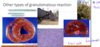
What are examples of exuberant granulation tissue?
- Hypertrophic scar – horses
- Keloid - humans

What can occur with hypertrophic scars in horses if not managed appropriately?
This area is always moving, If wound is not managed and this occurs than it can cause decreased mobility for the horse.
What is occuring in this final step ( step four) of remodeling ? What induces it to occur?

•Fibroblast maturation and collagen apposition (induced by TGF-β)
What is occuring in this image? What tissue is this? Is there regeneration occurng? Why or why not?

•Fibroblast maturation and collagen apposition (induced by TGF-β)
- Loss of cardiac myofibers can be seen. Deposition of collagen is occuring in the 2nd image. No regeneration in the heart ( you got what you got)

What occurs during repair of nervous tissues?
- Collapse of the parenchyma or cystic cavity
- Macrophages: Gitter Cells → malacia (Areas of necrosis for the CNS)
- Scar: astrocytes (gemistocytes)
What is seen in this image?

Malacia of spinal cord
What can be found in this image?

Glitter cells
What is seen in this image?

Gemistocytic astrocytes.

What are the most important take aways about chronic inflammation?
Chronic inflammation
• Macrophages = The most important cells
• Granuloma = Lesion (epithelioid macrophages + multinucleated giant cells)
- Tissue Repair
- Try to go back to what it was before
- Proliferation phase - granulation tissue
- Uncontrolled proliferation = Exuberant
- Fibrosis = fibrous tissue = has collagen
Intentions:
1st: wound edges close together
2nd: Wound edges further away
