Circulatory Disturbances - Part 1 Flashcards
What are parts of the circulatory system?
Consists of blood, central pump, blood distribution (arterial (O2+ nutrients) and collection (venous (metabolites, products ect)).
What are the lymphatics?
Lymphatics that parallel the veins contribute to circulation by draining fluid from the extravascular spaces into the blood vascular system.
What is the part of the circulatory system with the largest cross sectional area?
Capillaries (part of a whole)
What part of the circulatory system is the largest vessel?
What has the lowest pressure
Aorta
Vena cava
What are the thickest areas of veins? Arteries? Capillaries?
◦ Vein: tunica externa, veins also have valves.
◦ Artery: tunica media
◦ Capillary would just be basement membrane with endothelial tissue.
What is microcirculation? What occurs in this area?
- microcirculation is where metabolic exchange occurs due to slowing down of circulation.
In terms of vascular endothelium, What is hemostasis? What is its role in hemostasis?
- Provides anti thrombic and pro fibrinolytic in normal state and pro thrombotic and anti fibrinolytic during injury.
• Hemostasis is the arrest bleeding by physiologic properties of vasoconstriction and coagulation or by surgical means. Bleeding can be stopped.
What state do you want blood in when in normal conditions? What will help with that?
- Liquid state
- anti thrombic, pro thrombic, anti fibrinolytic, fibrinolytic factors help with keeping the blood in a liquid state.
The formation of this substance is very important for the formation of a clot.
◦ Formation of fibrin is very important for formation of clot.
What will injury cause to repair the vessel?
• Injury will cause prothrombotic, and antifibrinolytic which will help to repair the vessel.
What will occur if there is a leaking vessel after an injury?
• Leaking vessels after an injury, and endothelial cells will produce cytokines so WBC can go and repair the area. This makes the area more more able to absorb nutrients and bring cells to repair the area.
What modulates perfusion in vascular endothelium? What causes vasodilation? What causes vasoconstriction.
NO ( nitric oxide) relaxes and causes vasodilation.
Endothelin causes vasoconstriction.
What is the role in inflammation of vascular endothelium?
- Regulates traffic of inflammatory cells.
- Produces pro-inflammatory cytokines
- Control angiogenesis and tissue repair
What is the break down the total body water? What is the body percentage referencing water?
Total Body Water: 65 % total body weight
- Plasma 5%
- Interstitial Fluid 15%
- Intracellular Fluid 40%
- Transcellular Fluid 5%
What changes total fluid content in the body?
Age. Infant have increased water content in their blood. Fluid loss occurs with age, and that is why wrinkles occur.
What is the interstitium? What is ECM?
- Interstitium: Space between tissue compartments. It is the medium through which all metabolic products must pass and is made up of ECM and supporting cells.
- ECM: Collagen, reticulin, elastic fibers) ground substance (glycoproteins, ect)
What is the hydrostatic pressure? What is osmotic pressure? What is the role of both of these?
- Hydrostatic: Pressure of fluid within the vasculature
- Osmotic: Pressure based on presence of solutes, primarily proteins, albumin.
- These control the amount of fluids that will be within the vasculature vs. in the third space.
What does it mean if there is increased hydrostatic pressure and decreased osmotic pressure ? What can occur with fluid backup ?
Increased Filtration.
Edema.
What occurs on the arteriole side ? what occurs on the venous side? what happens to the remaining fluid.
• Arteriole side has increase of hydrostatic pressure( increased in fluid exchange), venous side has increase in oncotic pressure. remaining fluid is drained by lymphatic system
◦ Arteriole: Favors filtration
◦ Veins: Favor absorption.
What will cause extravascular fluid to accumulate?
• Increased hydrostatic pressure or diminished plasma osmotic pressure within capillaries will cause extravascular fluid to accumulate. If capacity of lymph drainage is exceeded tissue edema will result.
What are the categories of circulatory disturbances?
Edema
Hyperemia & Congestion
Hemostasis
Hemorrhage
Thrombosis, Embolism, DIC
Infarction
Shock
What is edema?
- abnormal accumulation of excess extracellular fluid.
- Fluid outside vascular compartment.
What are the pathomechanisms of edema?
• Increased hydrostatic pressure
◦ (Right sided CHF, Tightly bandaged limb)
◦ Impairing venous return will increase hydrostatic pressure.
• Decreased plasma colloidal osmotic pressure aka oncotic pressure ( decreased protein absorption, decreased protein synthesis, increased protein loss)
◦ (proteinuria can indicate protein loss via damage of glomeruli in kidneys. This can lead to generalized edema in an individual
◦ Starvation, hypoproteinemia, PLE, ect.
• Lymphatic obstruction: Damage/ obstruction of lymphatics
◦ Tumor, surgery, inflammation ect.
• Increased vascular permeability
◦ inflammation, cytokines will decrease vascular permeability, and cause localized edema.
What is occurring in this image? What is the cause? What other causes can cause similar effects?

- Diffusely swollen leg, this is due to lymphatic obstruction.
- Some tumors of dogs (i.e melanoma which this dog had), can spread along the lymphatics)

What is occuring in this image?

Local lymphatics are distended (lymphangiectasia) and filled with neoplastic cells.
What are the classifications of edema, and what are the associated signs of each?
• Edema can be classified as:
◦ Inflammatory: Increased vascular permeability, - Refers to “exudate” -> high protein, high specific gravity, higher cellularity, less than 7,000 cells/uL
◦ Non Inflammatory: Edema of CHF, Edema of Liver failure) Referred to as a “transudate” -> Low protein content, low specific gravity, low cellularity, less than 1500 cells/uL
Inflammatory may indicate peritonitis.
What is the gross appearence of edema in a patient post mortem?
Wet
Gelatinous and Heavy
Swollen organs
Fluid weeps from cut surfaces
May be yellow
What is occuring in this image?

Horse Gastric wall edema
What can you see on a histological slide of edematous tissue?
- Clear or pale eosinophilic staining depending on whether it is non-inflammatory or inflammatory edema
- Spaces are distended
- Blood vessels may be filled with RBC
- Lymphatics are dilated
- Collagen bundles are separated

What is pitting edema?
When pressure applied to an area depression or dent results as excessive interstitial fluid is forced to adjacent areas.
Takes a few seconds for area to return to normal.
What is occurring in the image in the circled part?

Pitting edema
What is a hydrothorax? When can you see it?
• Presence of fluid w/in the thoracic cavity - hydrothorax
◦ Can be seen with chronic pneumonia
◦ Something in the lungs that causes pulmonary hypertension.
◦ Resistance to blood being pumped into the lungs

What can cause resistance of blood being pumped into the lungs?
‣ Increase of hydrostatic pressure, increase amount of plasma within those blood vessels and this causes edema.
‣ Left sided CHF will eventually back up into the lungs and cause all of the same issues as you see above.
What is pericardial effusion? What is an example of a condition that causes pericardial eddusion from Vitamin E / Selenium deficiency?
• Pericardial effusion: yellowish in color, contains a bit of an exudate
• Mulberry heart disease - related to vitamin E selenium deficiency.
◦ Increased protein in fluid, fibrin strands and cloudy with high cellularity, kind of inflammatory conditions.
What is occuring in this image?

• Mulberry heart disease - related to vitamin E selenium deficiency.
◦ Increased protein in fluid, fibrin strands and cloudy with high cellularity, kind of inflammatory conditions.
What is occuring in this image?
• Hydroperitoneum/ Ascites: Fluid ( transudate) within peritoneal cavity.
What is hydroperitoneum/ ascites? What can cause more diffuse edema?
- Hydroperitoneum/ Ascites: Fluid ( transudate) within peritoneal cavity.
- R sided heart-failure can lead to more diffuse edema.
What is occuring in this image?

Ascites in a horse
What is occuring to the puppies in this image? When does this typically occur?

Anasarca
This can be seen typically in aborted fetus’ with severe congenital heart malformations .
What is anasarca?
Anasarca- Generalized edema with profuse accumulation of fluid within the subcutaneous tissue.
What is another term for submandibular edema? What can cause it? Why?
- Submandibular edema: bottle jaw is commonly associated with severe GI parasitism and hypoproteinemia in sheep.
- Specifically can be seen with infestations of Haemonchus contortus (barber pole worm) . Translucent fluid.
- Decreased oncotic pressure from hypoproteinemia from heavy parasitism.
What parasite is more commonly the cause of submandibular edema?
Haemonchus contortus (barber pole worm)
What is occuring in this image?

Bottle Jaw/ Submandibular edema
What other conditions can cause generalized edema?
Protein losses (i.e protein loosing enteropathy, protein loosing nephropathy)

What is the clinical significance of edema?
Dependent upon: extent, location and duration.
Tissue may become firm and distorted due to an increase in fibrous connective tissue after prolonged edema
What is pulmonary edema? What are the types?
Non-inflammatory edema: e.g.: Associated to left-sided
congestive heart failure (CHF).
Inflammatory edema: Damage to pulmonary capillary
endothelium -> e.g.: pneumonia
What is acute respiratory distress syndrome?
ARDS (Acute respiratory distress syndrome) Sudden, diffuse and direct- increase in vascular permeability: high fatality rate -> Followed by pneumonia if animal survives
What is occuring in this image?

• Pulmonary edema
◦ Lungs slightly enlarged, heavier, fluid coming out, you will see frothy material when you open sections of trachea, lungs, ect.
◦ Gelatinous material in the tissues.
What is chronic pulmonary edema? How is it seen in histological slides?
Most commonly associated with cardiac failure
Alveolar walls become thickened -> may lead to fibrosis
Congestion, micro-hemorrhages -> and accumulation of heart failure cells
What is being shown in this image?

• Macrophages with hemosiderin are indicators of micro hemorrhage/ pulmonary hemorrhage.
◦ these are considered heart failure cells or siderophages.
What is hyperemia?
Hyperemia indicates increase of arteriole-mediated engorgement of the vascular bed. Blood is oxygenated (red).
(i.e: exercise, inflammation)

What is congestion?
Congestion indicates passive, venous engorgement. Blood is not oxygenated (blue).
(i.e local obstruction, congestive heart failure)

What are examples of physiologic hyperemia?
Digestion: Inc blood to GI
Exercise: Inc blood to muscles
To dissipate heat: Inc blood to skin to cool down
Neurovascular: Involuntary inc blood in face as result of embarrassment/ emotional distress
What is pathologic hyperemia?
Caused by an underlying pathological process – usually inflammation.
Arteriolar dilatation->secondary to inflammatory stimuli (inflammatory mediators).
Redness (“rubor”) is one of the 5 cardinal signs of inflammation : tumor (swelling), calor (heat), rubor (redness), dolor (pain) and loss of function.
Swelling/ edema occurs
What are the 5 cardinal signs of inflammation?
Redness (“rubor”) is one of the 5 cardinal signs of inflammation : tumor (swelling), calor (heat), rubor (redness), dolor (pain) and loss of function.
What is occuring in this image? Is it pathologic hyperemia or physiologic?

Gingivitis (pathologic hyperemia)
What is occuring in this image? Is it pathologic hyperemia or physiologic?

Bulbar and palpebral conjunctivitis, human
What is congestion? What are its classifications?
Since the vascular beds are engorged with poorly oxygenated blood tissues are dark red to blue (cyanotic), depending on the degree of stagnation. Like other lesions it can be classified according to duration (acute or chronic) and its extend: localized (e.g. isolated area of venous obstruction); generalized: Systemic change like in CH
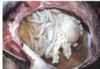
What is occuring in this image? Is this congestion? What is the pathophysiology behind the damage?

- GDV usually includes splenic involvement.
- signs -> abd pain, distention, ect.
- Obstruction of gastric veins which will cause hypoxic damage due to the lack of blood-flow. Sepsis can occur, peritonitis, ect.
- Can happen in swine as well when they get over excited.
- Necrosis in the walls of vessel will cause blood to leak into abdomen after death.
What is occuring in this image?

Intestinal volvulus, horse
What is occuring in this image?

colonic torsion, horse
What is occuring in this image? What is the cause of the discoloration?
- L side of heart can cause pulmonary congestion/ edema and this can cause brownish accumulation on lungs.
- Lungs will be enlarged, brown changes on lungs is deposition of heart failure cells and hypoxic damage (sign chronic pulmonary edema)
- Lungs also wet looking
What occurs with pulmonary hypertension and how does it lead to chronic hepatic congestion? What will the livers look like in these instances?
- Pulmonary Hypertension makes it hard for R side of heart to pump blood so R side will also start failure
- Increase hydrostatic pressure,
- Backup of poorly O2 blood will back up into liver and can show congestion in there.
- Liver will be round, and enlarged, and you will see fibrosis/ discoloration due to backup of blood.
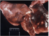
What is occuring in this picture? What is the likely cause? What are the differences from normal pathology?

Chronic hepatic congestion - > likely the result of Right sided CHF
Livers are enlarged and exhibit rounded edges
What is occuring in this image? What is this an indication of?

Chronic hepatic congestion: “Nutmeg liver”

What is the cause of nutmeg liver?
Chronic hepatic congestion -> nutmeg liver
• Poorly O2 blood/ increased around portal vein, and hypoxic damage due to poor oxygenation.
