Exam # 2 Flashcards
(224 cards)
What is hemorrhage?
Is defined as the escape of blood from the blood vessels (extravasation)
Can be external or internal (within tissues or body cavities)
What are the causes of hemorrhage?
Trauma
Sepsis, viremia, bacteremia or toxic conditions
Abdominal neoplasia may lead to hemoperitoneum
Coagulation abnormalities (platelet and coagulation factor defects or deficiencies)
What is the difference between hemorrhage and hyperemia & congestion?
Hemorrhage- blood is outside the vessel wall
Hyperemia & congestion blood is within the blood vessels
- eg: Photo shows congestion. This will lead to hypoxic damage.

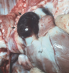
What is occuring in this image?

Hemorrhage
• Hemopericardium -> leads to fatal cardiac tamponade ( Blood cannot pump effectively due to pressure build up preventing it from being able to make full contractions. Can happen because rib punctured lung after HBC, ect)
What determines the clinical significance of hemorrhage?
Determined by the location and the severity
e.g.: Profuse blood loss is the most common cause of hypovolemic shock; Hemorrhage in the brain or heart can be fatal.
What can occur with boxers after a boxing match?

• Hemorrhage post boxing match-> fighter can be fine, then start feeling terrible and collapse. Meningeal arteries can cause alot of bleeding
What can occur with enough bloodloss?
• enough blood-loss can cause hypovolemic shock. Hemorrhage in brain can be fatal.
What can occur with Hemangiosarcoma located in the right atrium?
• Hemangiosarcoma -> sometimes in the right atrium. This will create cystic lesions filled with blood and can just collapse and die. Death is attributed to rupture of R atrium and blood build up in pericardial sac and then tamponade which lead to death.
What is hemorrhage by rhexis?
Due to a substantial rent or tear in the vascular wall (or heart).

What is occuring in this image? What is a potential cause of this in humans? What about in Pigs?
In humans: aortic dissection, dissecting hematoma: dissection of blood between and along the laminar planes of the media (blood- filled channel within the aortic wall)-> can result in rupture and fatal hemorrhage
Dissecting aneurysm, pig with Copper deficiency
What is an aneurysm?
• aneurysm - focal dilation of a blood vessel - > likely an artery. Usually due to weakening of vessel. Could be secondary, but if located in cerebral artery then rupture can lead to death.
What can copper deficiency cause in young male racing greyhounds?
• Copper deficiency can cause dissecting aneurysm. Can occur in coronary/ renal arteries of young male, racing greyhounds leading to arterial rupture and fatal hemorrhage.
What can be ruptured in turkeys by an aneurysm?
abdominal aorta
What is hemorrhage by diapedesis?
Hemorrhage due to a small defect in the vessel wall or rbc’s passing through the vessel wall in cases of
inflammation or congestion (like in the lungs of animals with left-sided CHF…)

What occurs in patients with hemorrhagic diathesis? In what conditions can you see hemorrhagic diathesis?
Increased tendency to hemorrhage from usually insignificant injuries (seen in a wide variety of clotting
disorders).
What would you call “ Blood in the thoracic cavity”?
Hemothorax
What is a hemoperitoneum?
blood in the peritoneum
What is hemoarthrosis?
blood within a joint space
What is occuring in this image?

Blood in a joint space
What is hemoptysis?
Coughing up of blood or blood-stained sputum from the lungs or airways.
What is occuring in this image?

Hemoptysis
What is epistaxis?
bleeding from the nose
What are the causes of epistaxis?
◦ Causes:
‣ Hypo coagulability (rat poison, anticoagulant)
‣ Trauma
‣ Presence of tumor in nasal cavity
‣ Severe inflammation (rhinitis)
‣ Exercise induced pulmonary hemorrhage in horses, mycotic infection in guttural pouches
What is occuring in this image?

Epistaxis