Exam # 3 - Final Flashcards

1
Q

What is hypertrophic osteopathy? What is it a sign of? What occurs? What clinical signs may you see in a patient?

A

• Hypertrophic osteopathy
◦ Paraneoplastic syndome related to space occupying lesion in the thorax
◦ You will see proliferation of bone in the periosteum. Usually the dog will appear painful with lameness and swollen limbs. Particularly occurs in the long bones.

2
Q

What is one way to diagnose Psittacosis? What is the most ideal way to diagnose it?

A

You can do a gram stain to see if it is a gram negative bacteria. Since you cant differentiate it from another gram negative bacteria you should use, machiavelli stain, since the elementary bodies will apear a periwinkle color. To be 100% sure you must use IHC or In situ hybridization.

3
Q

What are permenant, quiescent/ stable, continous/ cycling labile cells?

A
  • Permenant: no longer replication/ regenerating
  • Quiescent stable cells: sometomes regeneration
  • Continious cycling labile cells -> consistently regenerating/ replaced
    • there are checkpoints at various segments of mitosis.
4
Q

What is the route of transmission for avian pox?

A

Transmission:
- Insect bites, skin lacerations

5
Q

What is the common name for psittacosis?

A

Pirates disease

6
Q

What is gastric carcinoma? What can be seen in tumors of this kind? Where do these tumors arise from, who is it common in? What tissue is involved in this? Ect?

A
  • Large animals also get tumors.
  • Gastric carcinoma: arrises from esophageal region of gastric mucosa. It is not uncommon in horses.
  • This area is lined with stratified squamous epithelium. (Squamous cell carcinoma) Causes ulcerations, proliferatice lesions, ect)
  • Infiltrative to tissue so areas of this stomach would be thickened.
7
Q

What is the likely cause of this lesion?

A

Canine TVT

8
Q

What is seen in this image? What is the large dark puple area in the upper left corner? What are the small dots surrounding the large cell?

A

This is a fibroblast with lymphocystis. It is up to a million times larger than normal. The fibroblast keeps growing and growing.

The small dots are the nuclei of normal sized cells. The large dark purple area is the nucleus of the infected cell.

9
Q

What is the strongest argument for immune survailences role in cancer? Are the survailence mechanisms as effective as they should be?

A
  • The increase incidence of cancer in immuno-suppressed people
    and animals is the strongest argument for the existence of tumor
    immune surveillance.

• Unfortunately tumor immune surveillance mechanisms are not as
effective as they should be. The reason is that tumor cells have the
capability to develop mechanisms to evade the immune system of
the immunocompetent host.

10
Q

Where is it difficult to import from due to concerns for BSE?

A

Alberta Canada.

11
Q

ALSO IMPORTANT

A
12
Q

What is paraesthesia?

A

Paresthesia: abnormal sensation of the
skin (tingling, pricking, chilling, burning, numbness) with no apparent physical cause.

13
Q

What stain is depicted here for this gram negative bacteria? What is the bacteria and what can be seen in this image?

A

Machiavelli stain - you will see elementary bodies staining a fuschia color. This is psittacosis.

14
Q

How do prions cause metabolic dysfunction of neurons/ neural cells?

A
  • Conversion of normal Prpc to PrPSc
  • Accumulation of protease-resistant β-sheet isoform of PrPSc

in short -> changes normal protein to prion protien/ mutated protein.

15
Q

What is carcinogenesis?

A

Carcinogenesis is a multistep process at both the phenotypic and genetic level -> tumor progression.

16
Q

What is the transmission route of psittacosis? What is important about the bacteria in the environment. What kind of disease can it cause in humans?

A

Transmission:
- Via respiratory droplets, feather dust,
feces
- Inhalation, ingestion or mucosal
(conjunctival) contact
-Survives desiccation
In humans Respiratory disease may be severe, even if you are not immunocompromised you can get bad pneumonia.

17
Q

What can paraneoplastic syndromes indicate in patients? Why are they important?

A
  • They may represent the earliest manifestation of an occult neoplasm.
    – In affected patients they may represent significant clinical problems
    and may even be lethal.
18
Q

What is a way they can treat Canine TVT?

A
  • usually surgically
  • Now treating with vincristine.
19
Q

What is differentiation/ anaplasia?

A

a) Differentiation/ Anaplasia
“ Refers to the extend to which parenchymal cells resemble the correspondent normal parenchymal cells, both morphologically & functionally
“ Benign tumors are well -differentiated
“ Malignant neoplasms can be well - differentiated or undifferentiated (the latter are said to be “anaplastic”).

20
Q

What are the steps of change from normal protein to prion protein?

A

Explaination corresponds to the image attached with numbers indicating the steps.

1.) Prion protein looks similar but is slightly different. Slightly different in a way that makes the protein useless. “ kinda similar, but kinda useless”

2 .) and 3.) Prion interaction begins to cause normal protein to change shape and replicate as the useless prion protein.

3.) This useless protein begins to accumulate in the cytoplasm. You cannot get rid of it.

Essentially a buld up of alot of useless protein and loss of function of normal protein.

21
Q

What is grading of tumors? What are they classified by? And why is it useful?

A

• Grading: Gives a semi-quantitative evaluation of the degree of
differentiation of the tumor. Cancers are classified from I to IV with
increasing anaplasia.
• Although histologic grading is useful, histologic appearance not always correlates with biologic behavior.

22
Q

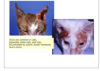
What is the morphologic diagnosis of these images?

Etiologic diagnosis?

Disease name?

A

Morphologic diagnosis: Acute severe diffuse necrotizing/ proliferative dermatitis

23
Q

What species can get chronic wasting disease? Is this a wild animal problem?

A

White tailed Deer, Mule Deer and Moose.

Can be found in captive and domestic species.

24
Q

What are the diffences between benign and malignant tumors in the following categories?

  • Differentiation
  • Growth rate
  • Local invasion
  • Metastisis
A

Differentiation

Benign: Well differentiated morphologic features and function. Structure similar to tissue of origin, little or no anaplasia.

Malignant: Poorly differentiated, morphologic features and function. Tissue of origin sometimes unclear. Variable degrees of anaplasia

Growth Rate

Benign: Slow, progressive, expansion, rare mitotic figures, notmal mitotic figures, little necrosis.

Malignant: Rapid growth, frequent mitotic figures, abnormal mitotic figures, necrosis if poor blood supply.

Local Invasion

Benign: No invasion, cohesisve and expansile growth. Capsule often present

Malignant: Local infiltrative growth, capsule often absent or incomplete

Metastisis

Benign: No metastisis

Malignant: Metastasis sometimes present.

25
Q

What is tumor heterogeneity?

A

• Generated during tumor growth
◦ By progressive accumulation of heritable changes in tumor cells
• Generation of
◦ subclones &
◦ successful subclones
• can give rise to cells that can become metastatic.

26
Q

Can tumors be emboli?

A

Yes, tumors can induce proliferation of fibroblastic tissue and can travel via lymphatics/ vascular tissue to distant areas of the body.

27
Q

What are teratomas? Is it a mixed tumor? Where can they be seen? Are they benign or malignant?

A

• Teratomas - usually in horses/ young animals
◦ Usually is incidental finding.
◦ You can find teeth, hair, cartilage ect in this mass within the testicle. They arise from totipotent germ cells.
◦ Occasionally you can find it in the ovaries of mares.

28
Q

What is the morphologic diagnosis of this histological slide? What is a likely etiologic diagnosis?

A

Acute severe heterophilic necrotizing multifocal hepatitis

Etiological diagnosis: Bacterial hepatitis

29
Q

What is likely the cause of this lesion?

A

Canine TVT

30
Q

The image on the left is an image of the lymph node of the cat that the subsection of mammary tissue is from? Wjat can be seen with the blue arrow?

A
  • Sample of regional lymph node was taken as well to look for metastasis.
  • Arrow points to areas of lymphatic metastisis.
  • 90% of tumors in cats are malignant
31
Q

What is a concern for lymphocystis that can change the prognosis?

A

The presence of secondary bacterial infections.

32
Q

What is involved in the staging of a tumor? Which is more helpful, staging or grading?

A

• Staging: It is based on the size of the primary tumor, its extend of
spread to regional lymph nodes, and the presence or absence of
hematogenous metastases.
• Staging from a clinical point of view has proved to be more useful than grading.

33
Q

What are the 8 changes in cell
physiology that together determine malignant phenotype?

A
  1. Self-sufficiency in growth signals (continue to grow)
  2. Insensitivity to growth-inhibitory signals (dont respond to inhibitory signals)
  3. Evasion of apoptosis (can avoid apoptosis)
  4. Defects in DNA repair
  5. Limitless replicative potential
  6. Sustained angiogenesis
  7. Ability to invade and metastasize
  8. Ability to escape from immunity and rejection
34
Q

What animals are at risk of transmissable veneral tumors? What kind of disease is this? What is its route of transmission.

A

Strays/ dogs allowed to roam, mostly in southern US/ Mexico. Not very common in north america.

Route of transmission: Sexual transmission

STD.

35
Q

What are characteristics of normal avian skin?

A
  • Normal avian skin (thin)
  • Keratinized
  • Basement membrane -> 3-4 layers of epidermal skin -> and stratum corneum
36
Q

What are the differential diagnosis’ to rule out with hypercalcemia?

A

hyperparathyroid, renal failure, hypoadrenocorticism, hypervitaminosis D

37
Q

What kind of virus is avian pox?

A

Genus Avipoxvirus of the Poxviridae
- DS DNA enveloped

38
Q

What occurs to the cells in lymphicystis? What kind of inflammation is present?

A

Non suppurative inflammation, lymphoplasmacytic

Eventual cell death, can infect adjacent cells.

Has macrophage/ lymphocyte inflammation.

39
Q

What are the main signs of lymphocystis?

A
  • Cytomegaly - cell enlargement
  • Karyomegaly -> nucleus enlargement
40
Q

What can occur in lymphocystis if the fish does not die from complications associated from the lesion?

A

It will either remain the same or eventually regress.

41
Q

What is the brown around this cell indicating? What kind of stain is this? What is it usually used in?

A

This is IHC. The brown is the target which shows your histology is positive. This is positive for psittacosis.

42
Q

How many chromosomes do the normal host cells have in dogs with TVT? What about in the neoplasia cell? What kind of transfer is occuring with this tumor? What is the likely cell type?

A
  • 59 chromosomes in neoplastic cell. vs 78 in normal dog cells
  • It is transfered via xenograft, and is proably histiocytic.
43
Q

What are signs of hypercalcemia?

A
  • Muscle weakness
  • Cardiac arrhythmia (rare)
  • anorexia
  • Vomiting
  • Renal Failure
  • Polyuria/ polydipsia
44
Q

What kind of bacteria is Chlamydophila (Chlamydia) psittaci? How does it survive?

A
  • Gram (-) bacterium
  • Obligate intracellular (depends on the host’s ATP)
    Needs hosts ATP to proliferate.
45
Q
A
46
Q

What is seen in this image? What is the cause?

A
  • Mechanical damage can cause hemorrhage/ ulceration. This is from avian pox (dry) (cutaneous form)
47
Q

Is there transcoelimic spread in this image?

A
  • Peritoneal implantation (transcoelomic spreading -> peritoneal carcinomatosis
  • Nodules attached to the mesentary.
48
Q

What is occuring in this image?

A

• Multifocal tumor - can be primary or metastatic ( that arrised from somewhere else and then eventually made it to the liver tissue). This is a pancreatic carcinoma that spread to liver tissue

49
Q

What are the characteristics of successful subclones?

A

· high proliferative rate,

· evade host immune response,

· can stimulate development of independent blood supply,

· are independent of exogenous growth factors,

· can spread to distant sites

50
Q

Why is the accumulation of prions so problematic?

A

No one knows exactly why it is such a big deal. It is alot of accumulated prion protein “ not goof to have alot of junk in your cytoplasm” and a loss of the needed proteins.

51
Q

What are the names of the malignant and benign tumors of epithelial origin?

  • Squamous epithelial cell
  • Adenexal cells
  • Melanocyte
  • Transitional epithelium
  • Uterine columnar epithelium
  • Lining of glands/ ducts
  • Hepatocyte
  • Renal tubular cell
  • sertoli cell
  • germ cell ( testicle)
  • stromal cell ( ovary)
  • Germ cell (ovary)
A
52
Q

What are epigenetic changes? What else are seen in tumor cells aside from epigenetic changes?

A

Epigenetic changes: Refers to heritable changes in gene expression in somatic cells resulting from something other than a change in the DNA sequence (most common ones are DNA methylation and histone modification)”.

• DNA mutations, epigenetic changes & chromosomal alterations are also observed in tumor cells.
Some people have genetic predisposition to cancer
Epigenetic changes- No DNA damage but, molecular changes can alter some functions of the tumor.

53
Q

What is diptheric membranes?

A

They are the term used to describe necrosis of the lumen of a tubular organ.

54
Q

What is occuring in this image? What is the way its appearing called? What could occur as a potential side effect or issue with this kind of neoplasm?

A
  • Benign tumor: lipoma, common in mesentary of most horses.
  • Usually pedunculated, doesnt produce any clinical problems, but dependent on location it can change if it results in clinical sings/ death.
  • If lipoma twists around the peduncle, it could cause ischemic damage or necrosis of the lipoma, and this could cause issues.
  • If the lipoma is large enough/ the peduncle has enough give it is possible that the peduncle may wrap around intestines and can cause volvulus.
55
Q

What is evident in this section of gastric tissue? What is circled by this red circle?

A
  • these areas of blue are mucous secreting neoplastic epithelial cells, characterized by bluish foamy cytoplasm, infiltrate the muscle layer of the stomach.
  • The morphology is similar to goblet cells. These are the cells that became neoplastic, which infiltrated the wall of the stomach.
  • This is mucinous gastric carcinoma.
56
Q

What are undifferentiated tumors?

A
  • undifferentiated tumors
  • Mixed tumors (multiple cell types derived from a single or multiple germ cell layer - pluripotential or totipotential.
57
Q

What is anaplasia usually characterized by?

A
  • pleomorphism- different shape
  • abnormal nuclear morphology
  • high mitotic rate
  • loss polarity (disorganization)
58
Q

Why is psittacosis significant? What Birds are known to be the highest infected?

A
  • Zoonotic disease
  • May be fatal in immunocompromised
  • Pigeons are highest reported, followed by raptors.
59
Q

What are the preneoplastic changes? Why is mitotic division very important to the neoplasticity of a cell? What happens if you take out the inciting agent that induce the preneoplastic change?

A

Preneoplastic changes:
Hyperplasia, metaplasia, dysplasia

Hypertrophy can be considered preneoplastic change.

• Mitotic division is very important to the neoplasticity of a cell. If the cell is rapidly dividing, their is more chance of DNA damage and if they are allowed to replicate, eventually they can aquire the ability to proliferate outside the growth capacity.

If you can take out inciting agent that is inducing this preneoplastic changes than things should be fine, but it is not always.

60
Q

What is the cause of the lesion seen in this image?

A

Lymphocystis

61
Q

What are the advantages of using immunohistochemistry in tumor diagnosis?

A

• 1. Categorization of undifferentiated malignant tumors
◦ Use of antibodies against specific intermediate (cytoskeletal) filaments: Cytokeratins, Vimentin, Desmin etc.
• 2. Categorization of leukemias/ lymphomas 2. Determination of site of origin of metastatic tumors
• 3. Determination of molecules that have prognostic or therapeutic significance:
◦ e.g.: determination of estrogen/ progesterone receptors in
breast cancer cells -> receptor positive breast cancers have a better prognosis/ susceptible to anti-estrogen therapy (e.g. Tamoxifen -> antagonist of the estrogen receptor in breast tissue)

62
Q

What is cancer cachexia? What percentage of people get it? Is it common in animals ? Why/ Why not?

A
  • 50 % of people that develop cancer get cancer cachexia -> loss of muscle and fat.
  • Not as common in animals, usually diagnosis-> death is short so its hard to see these chronic changes.
63
Q

What is seen in this histologic slide of avian skin?

A

Thickened skin with large cells. Cells are not empty, pink things inside cells are inclusion bodies. This is typical presentation of pox virus. Not another virus will typically give you this presentation.

64
Q

What is the cause of many of the familial cancer syndromes? What is the role of carcinogens? Are they common?

A

Many of the familial cancer syndromes are due to mutation
in recessive tumor suppressor genes

Carcinogens are cancer causing compounds

Chemical carcinogens are widespread in the environment.
E.g. The toxin of the bracken fern plant causes urinary
bladder cancer in cattle grazing pastures containing the
plant. • To b a c c o smoke contains potent carcinogens.

65
Q

What is the p-53 gene? What does it do?

A

• p53 gene- cancer cop
◦ Growth inhibiting tumor supressor gene.
• Considered guardian of the genome.

66
Q

What is occuring in this image? Is it common? Is it benign?

A
  • Common in dogs and cats: meningiomas, benign, well confined, proliferative lesion,
  • No such thing as benign tumor in brain, due to compression it will always be problematic, just depends on growth rate prognosis.
67
Q

What kind of electron microscopy must be used to see avian pox?

A

Transmission.

68
Q

What are the main mechanisms that regulate tissue growth? What cells are not very mitotic? Which are highly mitotic?

A
  1. Rate of cell proliferation (fraction of cells in the replicative pool !cells undergoing mitotic activity)
  2. Rate of programed cell death (apoptosis)
  • hemapoeitic tissues are highly mitotic, as well as gastric mucosa, so important to remember.
  • cardiac and nervous tissue dont replicate (heart and cns dont change after birth, you got what you got)
  • Mutation in important gene can lead to neoplasm.
69
Q

What are the effects tumors have on the host?

A
  • Focal & hormonal effect
  • Paraneoplastic syndromes
70
Q

What is the condition seen in this image?

A

Dry Avian Pox

71
Q

What is seen in this subsection of feline mammary tissue?

A
  • section of skin in mammary area. Purple/ blue areas of hypercellularity is seen. This indicates neoplasia, since they are not clearly demarcated/ boundries are blurred it is likely malignant.
  • Malignant tumor of epithelial origin -> carcinoma
72
Q

What is occuring in this image? What are the yellow arrows pointing at? The black arrows?

A

Pink areas pushing nucleus aside are inclusion bodies, this is typical apperence of avian pox.

Yellow arrows -> individual large cells

Black arrows -> Inclusion bodies -> millions of them.

73
Q

How does canine TVT evade immune destruction? What occurs with the tumor over time?

A
  • These cells make enough MHC-I to avoid being killed by natural killer cells, (self) as well as not enough to be picked up by cytotoxic T cells/ lymphocytes (non self) and make no MHC-II to avoid detection.

Eventually lymphocytes catch up and they produce IL-6 and INF-y and then they start to produce more MHC- I and can be recognized as non self.

Tumors can regress over time, but usually they keep growing and growing, causing severe lesions.

74
Q

What is the abnormal lone cell this image?

A

Mitotic figure of neoplastic cell. Has abnormal mitosis since it is neoplasia.

75
Q

What is notable about the round cells found with TVT?

A

Enlarged nucleus

76
Q

What is occuring in this histological image of a bird liver? What is the condition associated with it?

A

Psittacosis - Necrosis ( coagulative necrosis -> still has some cell shape/ sinus outlines)

77
Q

What are the macro environmental and microenvironmental causes of tumors?

A
    1. Macro-environmental (extrinsic) causes: e.g.: UV light, ionizing radiation, chemical carcinogens, oncoviruses. (Example: FELV cat with lymphoma-> lymphoma caused by FELV (oncovirus))
  • 2.Micro-environmental (intrinsic) causes: e.g.: heritable genetic changes, byproducts of normal metabolism including reactive oxygen species.
78
Q

What is the scientific name of Pisttacosis?

A

Chlamydophila (Chlamydia) psittaci

79
Q

What occurs to cells that have dna damage in the presence of P-53 and in the absence?

A

• Cell exposed to DNA damage -> activated p53 gene -> binds to DNA -> stops cell division until problem is correct/ DNA repaired so damaged cells do not proliferate.
• p21 and GADD45 -> mechanics of cell
◦ If repair fails then cell is normal, if not then apoptosis.

• BAX -> hitman (induces apoptosis)

Cell with loss or damage to P53 then the mutant cells will expand and continue mutating.

80
Q

What does the brain look like grossly in patients with CWD? What about Histologically?

A

Grossly brain looks normally so you must look histologically. You will have the spongiform areas of vacuolated neurons within the brain. You must do IHC to be sure targeting the prion protein.

81
Q

Will inflammation occur with any of the possible cell outcomes?

A

Inflammation may occur along with many cell outcomes.

82
Q

What are the major cells involved in immune survailence for cancer cells? What are the main defense mechanism? Is humoral response also effective?

A
  • CD8 + Cytotoxic T lymphocytes (CTLs) are the mayor immune defense mechanism against tumors.
  • Natural killer cells (specific type of lymphocytes) and macrophages also play a role. Interferon-gamma (IFN-γ), a cytokine produced by T-cells and NK cells, is a potent activator of macrophages.
  • Antibodies against tumor antigens are also part of the defense mechanisms of the host but there is little evidence that humoral immunity is effective against tumors.
83
Q

Does anaplasia represent reverse differentiation of mature normal cells?

A

The truth is that in most cancers anaplasia does not represent reverse differentiation of mature normal cells; anaplastic cells arise from less differentiated stem cell- like (totipotent cells) present in tissues.

84
Q

What forms of avian pox are there? What is most common?

A
  • Dry and wet form
  • Dry form typically affects area of skin that is less feathered or unfeathered.
  • Wet form affects the pharynx and esophagus. Causes diptheric membrane in trachea/ esophagus

Dry form is the most common

85
Q

What testing can determine if this is TVT vs another kind of cancer cell?

A
  • The dna of tumor will have different DNA than host cell. The tumor also has less chromosomes than the host cell
86
Q

TRUE or FALSE: You can look for TSE on a brian that has been sitting around

A

FALSE

You must have fresh brain tissue. Any decay/ autolysis will cause vacuoles so you will not be able to tell if it is a spongiform disease or if it is just a normal post mortem change.

87
Q

What are the 7 steps in microbial disease?

A
  1. ) Access body through a port of entry
  2. ) Contact target cell (tissue/organ)
  3. ) Colonize & amplify in target cell (tissue/organ)
  4. ) Spread locally, neighboring cells (tissues/organs)
  5. ) Spread systemically (blood, lymph, PNS)
  6. ) Colonize LN and/or distant tissues
  7. ) Injure or kill [distant] target cells (tissues/organs)
88
Q

What occurs if a meningioma is located on the ventral aspect of the brain?

A

can be difficult to treat, ( ventral aspect of brain), affect cranial nerves, ect.

89
Q

What percent of mammary tumors in cats are malignant? Is this the same for dogs?

A
  • 90% mammary tumors in cats are malignant : mammary carcinomas
  • Not the case in dogs
90
Q

What is the likely cause of the abnormalities in this image? If told this is a 3 year old moose what would be your concern? What about if it was a 1 year old cow? A mink?

A

TSE

  • Caused by prions

Moose: CVD (Chronic wasting disease)

Bovine: BSE ( Bovine Spongiform Encephalitis)

Mink: TME (Transmissible Mink Encephalitis) Mink Spongiform Encephalitis

91
Q

What is this stain and what is it highlighting? What in this stain is the bright pink areas?

A

This is a gram stain, and the bright pink ares are showing the gram negative bacteria in this sample of tissue.

92
Q

What is the morphologic diagnosis of this gross image? What condition has gross morphology like this?

A

Acute, severe, multifocal necrotizing hepatitis.

You can also say -> acute diffuse severe fibronous pericarditis.

Psittacosis

93
Q

How do neoplasias avoid apoptosis?

A

• Many neoplasms may present alterations in apoptotic pathways -> further genomic instability -> cells that may have suffered significant DNA damage are allowed to replicate (no cell -cycle arrest), eventually this may lead to the formation of increasingly aggressive subclones .

94
Q

What is an example of an oncovirus?

A

FELV in cats

95
Q

What animals are susceptible to TVT? What areas of the body can be infected?

A

Dogs, coyotes, foxes, and wolves

  • Sexual transmission
  • Urogenital / other mucosae (ocular, nasal, oral)
96
Q

What are the components of neoplasms? What effects the stroma and how?

A

Components of Neoplasms:
• Paranchyma
• Stroma ( connective tissue/ blood vessels that support the neoplasticism cells. Some tumors induce desmoplasia (development of collagen- rich fiberous connective tissue stroma))
- Some things tumor produces will effect the stroma and vice versa. Cytokines in stroma can modify biological effects/ response of tumor.
• Breast cancer can cause a lot of desmoplasia ( tumor may be very hard, a lot of fibrosis) malignant epithelial tumor indication sometimes

97
Q

What is seen in this histological slide? What does it look like? What can this cause ?

A
  • Vacuoles being produced within neurons ( vacuolation ) due to prions.
  • Looks like a sponge.
  • Makes the animal continously lose weight and become emaciated, if you do not euthanize the animal will die from this emaciation.
98
Q

What is being indicated by the yellow arrows? The blue arrow?

A

Yellow arrow: macrophages with unicellular debris.

Blue arrow: Dead heterophils

99
Q

What birds are affected by avian pox?

A

Any bird species can be affected by Avian pox.

> 200 bird species are susceptible

  • Some species specificity
  • Domestic fowl & canaries particularly susceptible
100
Q

What is transcoeleomic spreading? What is it called usually when this occurs?

A
  • Transcoelomic spreading- small scattered nodules
  • usually termed carcinomatosis.
101
Q

What is the pathogenesis of avian pox?

A
  • Virus enters target cell ( usually epithelium)
  • Replications in cytoplasmvia cell mechanism
  • Viral gene encodes epidermal growth factor causing HYPERPLASIA
  • Large Inclusion bodies formed (Bollinger bodies)
  • Virus release via Budding OR cell necrosis (most likely)
102
Q

What are the 4 classes of normal regulatory genes that are the main targets of genetic damage and play a significant role in carcinogenesis? What happens if you lose repair cells?

A
    1. growth-promoting proto-oncogenes
    1. growth-inhibiting tumor suppressor genes
    1. genes that regulate programmed cell death (apoptosis), and
    1. genes involved in DNA repair

If you lose repair cells, you can have abnormalities printed in the genomes.

103
Q

What is a neoplasia? Neoplasm? Oncology?

A
  • Neoplasia-> The process of tumor formation.
  • Neoplasm: “New growth” - “composed of cells originally derived from normal tissues that have undergone heritable genetic changes that allows them to become unresponsive to normal growth controls and expand beyond their normal anatomic boundary”
  • Oncology: Study of tumors or neoplasms.
104
Q

What is the mechanisms of injury for psittacosis?

A

Mechanisms of injury:

  • Cell death (necrosis)
  • Persistent or latent infection
105
Q

What is hyperplasia? What can occur in a mammary gland? What does insitu mean? What should you do if you have a neoplasm in situ?

A

Hyperplasia: excess of cell growth.
Normal mammary duct-> hormonal changes can cause intraductal hyperplasia-> which will with chronigincity cause intraductural hyperplasia with atypical-> later it can become intraductal carcinoma in situ -> invasive ductal cancer

In situ- means still confined to basement membrane and has not infiltrated adjacent tissues.
• If you do something right away, there is a high chance you can prevent worsening.

106
Q

IMPORTANT SLIDE TO GO OVER

A
107
Q

What is ocular squamous cell carcinoma in cows? What is the cause? who is more susceptible? Is it malignant? What happens if a cow sent to slaughter has a tumor? What is the way to get around thi?

A

• Ocular squamous cell carcinoma
• Normally cattle that have white faces -> more susceptible to UV light.
◦ Within conjunctiva
◦ Can metastisize.
◦ When sent to slaughter, the entire carcass is condemed.

  • “UV radiation causes dimerization and protein cross- links in DNA molecules. Also UV light induces formation of a carcinogen (cholesterol alpha oxide) from natural sterols in unpigmented skin.
  • Can also occur in horses.

If they preform enucleation and let it heal carcass will not be condemed at slaughterhouse.

108
Q

What is the main issue with meningiomas and the damage they cause?

A

you can see there is no infiltration but significant compression.
• Tumor causing compression of brain/ brain stem, act quickly

109
Q

What is the specialized stain for mucus?

A
  • Sometimes using specialized stain is important.
  • In this case alcian blue helps with cell differentiation.
110
Q

What is a common name for Bovine Spongiform Encephalitis?

A

Mad Cow disease

111
Q

What is metaplasia? What is the tissue usually changed to?

A
  • metaplasia -> reversible change in which one adult cell type is replaced by another of the same germ line. Usually specialized epithelium is replaced by less specialized epithelium.
  • Metaplasia is often but not always an adaptive response.
112
Q

What mechanisms can tumors use to evade the immune system?

A

Mechanisms by which tumors evade the immune system
• Failure to produce tumor antigen
• Mutation in MHC genes or genes needed for antigen processing
• Production of immunosuppressive proteins.

113
Q

What is the morphologic diagnosis for TSE? Etiological Diagnosis? Is there any inflammation?

A

Chronic moderate diffuse, neuronal vacuolation, spongiform, with degeneration, and necrosis encephalitis

No there is not any inflammation

Etiologic Diagnosis: Prion spongiform Encephalitis

Name of disease: Transmissible Encephalitis

114
Q

UNDERSTAND THIS SLIDE

A
115
Q

What is the name of the inclusion bodies of avian pox?

A

bollinger bodies

116
Q

What is the number one mechanism of cellular injury for avian pox?

A

Cell proliferation (REMEMBER HYPERPLASIA)

117
Q

What is the pathogenesis of psittacosis?

A

Elementary Bodies enter cells via endocytosis or phagocytosis

  • They Blocks fusion of endo/phagosome + lysosome (so it can replicate)
  • Elementary Bodies turn into Reticular Bodies ( in order to reproduce)
  • Reticular bodies replicate by binary fission
  • Reticular Bodies turn back into Elementary Bodies to leave the cell.
  • Elementary Bodies leave cell:
  • Via Lysis or exocytosis
118
Q

What is this black arrow indicating about the cells of this muscle tissue?

A
  • Pleomorphic- varying shapes
  • Black arrow shows rhabdomyosarcoma,
  • nuclei of some cells very large, variation of cytoplasm quantity, ect.
119
Q

What is the idea of how cells can become malignant?

A
  • One particular cell may undergo a bunch of alterations to allow it to become malignant
  • specific clones of cells.
  • Successful clones- high proliferation and decreased immune response.
  • When they are successful they can metastisize.
120
Q

What are the 3 types of prion TSE’s focused on? Which is zoonotic?

A

Transmissible Spongiform Encephalopathies:

  • Bovine Spongiform Encephalitis – zoonotic (vCJD)
  • Scrapie (sheep & goats)
  • Chronic Wasting Disease (deer & elk)

Scrapie and CWD not known to be zoonotic.

121
Q

What can be seen in this image?

A

The characteristic dumbell shape of pox virus on Electron microscopy

122
Q

What is a way that psittacosis can be aerosolized?

A
  • If someone is sweeping infected feces it can aerosolize infection and cause pneumonia.
123
Q

In this image, is there transcoelmic spread?

A

yes this is transcoelimic spread ( peritoneal implantation)

124
Q

what are the ways we can diagnose tumors in the lab?

A

• Histologic & Cytologic Examination
◦ Clinical data is quite valuable for diagnosis
◦ “Laboratory evaluation can be only as good as the specimen available for examination”

• Immunohistochemistry. The availability of monoclonal antibodies has greatly facilitated the identification of cell products or surface markers.

125
Q

What is directed cell dysfunction?

A
  • virus takesover cell and has it make proteins for its capsid.
126
Q

What occurs with a carcinoma in situ?

A
  • entire thickness of the epithelium is replaced by dysplastic cells.
  • This occurs a lot in cats.
  • Cats will develop dermal keratosis from sunbathing, chronic irritation of the skin with dysplasia. The layers of epidermis becomes a bunch of disorganized cell, and eventually can become squamous cell carcinoma.
  • White cats/ white areas more prone.
127
Q

What are the possible cell outcomes / mechanisms of injury?

A
  • Directed cellular disfunction
  • Malignant transformation
  • Structural injury
  • Cell death (necrosis)
  • Persistent / latent infection
  • Cellular proliferation
128
Q

Can they cure the individual cancer genes?

A

No there are too many.

129
Q

What is the most frequently observed paraneoplastic syndrome? What is the cause? What should you do if a patient comes in and has high calcium on their bloodwork?

A
  • Most frequently observed paraneoplastic syndrome – primarily in dogs is hypercalcemia.
  • Due to the production of calcemic humoral substances (parathyroid hormone-related protein, PTHRP) by neoplastic cells from extra- osseous neoplasms.
  • Hypercalcemia due to osteolysis by skeletal metastases is not a paraneoplastic syndrome

If a dog comes in and has high calcium, and then returns and repeat blood shows hypercalcemia, you have to think there is the potential of an occult tumor,

130
Q

What is gastric carcinoma? What can be seen in tumors of this kind? Where do these tumors arise from, who is it common in? What tissue is involved in this? Ect?

A
  • Large animals also get tumors.
  • Gastric carcinoma: arrises from esophageal region of gastric mucosa. It is not uncommon in horses.
  • This area is lined with stratified squamous epithelium. (Squamous cell carcinoma) Causes ulcerations, proliferatice lesions, ect)
  • Infiltrative to tissue so areas of this stomach would be thickened.
131
Q

What is a signs of nodular dermatofibrosis in german shepards? Will their be only one cutaneous lesion? What are they often associated with? What other benign tumors can be paraneoplastic syndrome in german shepards? What phenotype is nodular dermatofibrosis?

A
  • Affected dogs develop multiple benign cutaneous lesions almost always associated with underlying bilateral renal disease: polycystic kidneys, renal cystadenomas or cystadenocarcinomas (most common). Occasionally reported in other breeds.
  • Fibromas-> benign tumors, this can be a paraneoplastic syndrome in germanshepards, this can be associated with tumors in the kidneys.
  • Malignant tumor of glandular epithelium with fluid within the kidney (Renal Cystadenocarcinoma)
  • Autosomal Dominant
132
Q

What is seen in this image? What kind of elecronmicroscopy is used to obtain this view?

A

In the blue you can see the typical viral hexagonal appearence)

  • Tranmission EM
133
Q
A
134
Q

What is seen in this image? What are its characteristics? Is it benign or malignant?

A
  • proliferative lesion in mammary gland,
  • malignant, ulceration and necrosis, not typical for benign.
  • most benign tissues are mobile, malignant tumors are usually well attached
135
Q

What is a nephroblastoma? Is it a mixed tumor?

Where can they be seen? Are they benign or malignant?

A

Nephroblastoma - more common renal tumor in pigs.
Benign tumor
• usually found in younger animals, normally in the slaughter house this is found.
• Mixed tumor because it has an epithelial component that form tubules, which give the appearance of fronds and look like primitive glomeruli.
• You can see nephroblastomas also in children.
• Can also be seen in chickens

These tumors arrise from totipotent cells.
Can also be considered mixed types
• German Shepard may develop tumor in the thoracolumbar area in spinal cord and they are thinking it is a nephroblastoma.
• Present with neuro signs, ataxia, ect.

136
Q

What is the target cell for lymphocystis?

A

Fibroblasts

137
Q

What is the staging system for tumors? What does each part represent?

A

TNM System:
• T= Primary tumor, with increasing size: T1-> T4.
• T0= “in situ” lesion. (Malignant will begin this way)
• N: regional LN involvement.
• N0= No LN involvement.
• N1 to N3 would denote increase number and range of nodes. (increase number by number of nodes involved)
• M: Blood-borne metastases.
• M0= No blood-borne metastases,
• M1 or M2 indicates the presence of blood- borne metastases and some judgment as to their number.

138
Q

What is the routes of transmission for lymphocystis? Where are lesions usually located? What kind of fish species are affected? Is this disease malignant?

A

Route of transmission: ¡ Ingestion, direct contact, ‘inhalation’

Location: Skin & gills, but also internal organs

Affects Many teleost fish species
Has Low mortality (benign disease) unless affects the individual to a point they cant see or eat, swim, ect.

139
Q

What causes necrosis in malignant tumors? what is important about the borders of malignant tumors?

A

malignant tumors can outgrow their blood supply which causes ischemic damage, and necrosis. Borders arent well circumscribed and is infiltrating surrounding tissue, this is more indicative of malignancy.

140
Q

What can be seen in this image?

A

• Peritoneal carcinomatosis ( peritoneal spreading )

141
Q

What is indicated by these black arrows?

A

Black arrows are showing more normal mitotic figures,.

142
Q

What are the names of malignant and benign tumors of nervous tissue?

  • astrocyte
  • oligodendrocyte
  • microglial
  • schwann cell
  • Neural cells (PNS)
  • Neural Cells (CNS)
A
143
Q

What is lymphocystis? Who does it affect? What kind of virus is it?

A
  • Non cartilaginous fish
  • DNA virus
  • Iridoviridae
  • Non-enveloped
144
Q

What percentage of patients (human) with malignant disease have paraneoplastic syndromes?

A

n humans they occur in approximately 75% (10% if cachexia is
not included) of patients with malignant disease.

145
Q

What occurs to normal cells that cause benign or preneoplastic lesions? What will cause them to potentially become malignant?

A

• In the presence of a promoter, these initated cells expand to form a preneoplastic lesion or benign tumor. With further genetic and epigenetic alterations, a malignant tumor emerges from a subclone of cells within the benign precursor lesion.

  • Initiated cells have growth advantage against other cells if they are in the presence of a premotor.
  • Formation of benign tumor can become malignant tumor. If it becomes one, it will have the ability to infiltrate surrounding tissue.
146
Q

What is the mechanism of cell injury for TSE?

A
  • Progressive degeneration nervous system
  • Directed cellular disfunction
  • Structural injury
  • Persistent or latent infection
147
Q

What is metastisis? What does it indicate? What characteristic increases with malignancy?

A

• Metastases are tumor implants discontinuous with the primary tumor
• Metastasis is the hallmark of malignancy since benign neoplasms do not metastasize.
◦ The more aggressive, the more rapidly growing and the larger the primary neoplasm, the greater the likelihood that it will metastasize or that has already metastasized

148
Q

What is seen in this section of mammary tissue?

A
  • Abnormal: cells are anaplastic, they have degree pleomorphism, cell nuclei enlarged, ect.
  • 90% mammary tumors in cats are malignant : mammary carcinomas
  • Not the case in dogs
149
Q

What are examples of paraneoplastic syndromes?

A

• Examples of paraneoplastic syndromes in domestic
animals are:
• Cachexia
• Hypercalcemia of malignancy.
• Hypoglycemia
• Thrombotic disease
• Peripheral neuropathy
• Nodular dermatofibrosis (German Shepherd dogs)
• Hypertrophic osteoarthropathy (hypertrophic osteopathy)

150
Q

Is this tumor benign or malignant? What are some identifiers/ characteristics that let you know what it is?

A

• Proliferative lesion, no exudate, well demarcated, likely be benign ( solid tissue, likely tumor)

151
Q

What is occuring in this image? Why?

A
  • Umbilicated appearence of the lesion is often suggestive of carcinoma. Umbiliicated = like a naval.
  • Tumor is outgrowing its blood supply so there are these central areas of necrosis, which has a crater like or naval appearence.
  • There is some sarcomas that ocassionally also look like this.
152
Q

What is the main mechanisms of cell injury in lymphocystis?

A
  • Directed cellular disfunction
  • Cell proliferation
153
Q

What are the malignant and benign tumors of each of these Mesenchymal tissues?

  • Fat
  • Fibrous connective tissue
  • Cartilage
  • Bone
  • Blood vessel
  • Lymphatic Vessel
  • Synovium
  • Mesothelium
  • Meninges
  • Ovary
  • Lymphoid tissue
  • Bone marrow
  • Connective tissue
  • Smooth Muscle
  • Skeletal Muscle
A
154
Q

What is the biology of tumor growth, benign vs. malignant?

a) Differentiation/ anaplasia b) Rate of growth c) Local invasion ( expansile / infiltrative growth) d) Presence or absence of metastasis

A

A.) differentiation/ anaplasia: benign tumors are usually well differentiated , and you can tell cell of origin. Malignant normally are anaplastic and its usually undifferentiated)
B.) Rate of growth- Slow growth (usually benign)
• rapid growth ( usually rapid growth)
C.) local invasion (expansile/ infiltration growth)
• benign tumors grow by expansion ( producing compression of adjacent tissue) Malignant tumors are infiltrative growth. They grow into the tissue. Hard to get good margins some time)
D.) Presence or absence of metastasis
• 2 degree growth, benign never Mets, malignant Mets.

155
Q

What is the disease humans can get from BSE?

A

vCJD or

Variant Creutzfeldt-Jakob disease (vCJD) is a prion disease that was first described in 1996 in the United Kingdom. There is now strong scientific evidence that the agent responsible for the outbreak of prion disease in cows, bovine spongiform encephalopathy (BSE or ‘mad cow’ disease), is the same agent responsible for the outbreak of vCJD in humans.

156
Q

What are the pathways of malignant tumors?

A

• malignant epithelial tissues more often than not will spread via lymphatic route.
◦ I.e Breast cancer -> regional lymph nodes
◦ not always the case, sometimes bypasses regional lymph nodes and can spread somewhere else.
• Can also spread through veins ( since the walls are thinner), mesodermal tissues are the ones that typically spreads that way.
◦ metastisis may first occur in the liver, since itll go through the portal vein through the liver and liver is the first tissue it will encounter.
• Transcoelomic spread (Seeding of body cavities and surfaces): some neoplastic cells will exfoliate ( tumor clusters will exfoliate), and will go into the cavity, and in the cavity there is fluid, so they can survive and affect other organs. When you see little tumors in the peritoneum your likely seeing carcinomatosis. It occurs from a primary tumor but will have metastasis.
• Specific tumors tend to spread this way ( like pancreatic carcinoma)

157
Q

What pattern of growth can you see with malignant tumors?

A

Crablike

158
Q

What is the morphologic diagnosis for this histologic slide? Etiologic diagnosis?

A

Chronic Focal with karyomegaly, basophilic cytoplasmic inclusions and moderate lymphoplasmacytic dermatitis

Etiologic diagnosis:

Viral Dermatitis

159
Q

What are tumor antigens? What are the types? and What can they be used for?

A

• Tumor antigens:
– Tumor-specific antigens (presented by MHC molecules on surface, presented only on tumor cells)
– Tumor-associated antigens (presented on surface of tumor and nontumor cells)

• Tumor antigens can serve as the targets of effective immune
surveillance

• Tumor antigens can be used for diagnosis, monitoring or
immunotherapy
( you can design antibodies against these antigens)

160
Q

Is it a pathogen that causes TVT? What is being spread? What kind of cell is it?

A

Tumor cell causes the disease not a pathogens. It is not the dogs own cell that is becomeing neoplastic, they are being infected by a neoplastic cell ( imortal cell) that continues to replicate and create more of itself.

161
Q

Is this epidermal sample abnormal?

A

No it is normal bird skin

162
Q

What proinflamatory cells are involved in the pathogenesis of cancer cachexia?

A

Cancer cachexia: progressive loss of body fat and lean body mass, accompanied by profound weakness, anorexia and anemia” ->
TNF, IL-1, IL-6, IFN-gamma, prostaglandins and PIF (proteolysis inducing factor) are apparently involved in the pathogenesis of cancer cachexia.

163
Q

What is seen in this histological image?

A

This is showing alot of round cells ( round cell tumor, or TVT)

164
Q

What are the mechanisms of cellular injust in avian pox?

A

Mechanisms of cellular injury:

  • Cell death (necrosis)
  • Directed cellular disfunction
  • Persistent or latent infection
  • Cell proliferation
165
Q

TRUE or FALSE:The vacuole itself in TSE neural tissue contain prion protein.

A

FALSE

It does not contain the prion protein.

166
Q

With scrapie, what can be seen even before you begin to see signs of emaciation?

A

Before seeing emaciation you can see signs of paresthesia, due to abnormal sensation of skin without apparent casue.

167
Q

What are the tumor antigens recognized by CTL or Cytotoxic T lymphocytes?

A

Tumor antigens recognized by CTLs:
• Product of oncogene or mutated tumor suppressor gene.
• Mutated self protein
• Overexpressed or aberrantly expressed self protein
• Oncovirus ( i.e feline leukemia)

168
Q

What is another potential issue with cutaneous (dry) avian pox?

A

They are susceptible to secondary bacterial infection.

169
Q

What is occuring in this image? What condition is it? What is the red areas? What wis the yellow arrow pointing at?

A

This is a slide indicating CWD, the red areas are IHC stain targeting prion protien, showing the presence of prion proteins in red. The yellow arrow is showing the vacuole.

170
Q

What are animal models used in cancer research? Why are these animals used? What percentage of people will perish from some sort of cancer?

A

Animal Models
• Experimentally-induced (Nude Mice) – able to accept grafting: no rejection (no only allografts but also xenografts -> tissue from other species) (No T lymphocytes -> lack thymus)
• Naturally occurring
• some stats of 20-25% of individuals will perish from some sort of cancer.

171
Q

What can occur with chronic spirocercosis?

A
  • 4 year-old dog with a history of chronic regurgitation, vomiting and weight loss associated with esophageal spirocercosis. This caused hypertrophic osteopathy since their was a space occupying lesion found in the esophagus. It was an inflammatory lesion caused by parasite ( spirocera lupi) but then it underwent malignant changes and became a tumor. You can see this highlighted in the next slide.
  • Ended up having osteosarcoma from this.

SPACE OCCUPYING LESION IN THORAX? ESOPHAGUS -> CAUSED HYPERTROPHIC OSTEOPATHY AND THIS CAUSED TUMOR FORMATION.

172
Q

What can occur in cats with nonpigemented ears/ faces? What can be the chain of events that lead to the formation of that tumor? Can it metastisize?

A
  • Cats with nonpigmented ears/ faces can get squamous cell carcinoma -> locally invasive
  • Usually will develop chronic dermatis, which will cause aplasia, and eventual Squamous cell carcinoma in situ -> which can metastisize.
173
Q

What is the reason for 90% of deaths related to cancers?

A

Metastisis

174
Q

What is more hardy, an enveloped or non eneveloped virus?

A

Non enveloped virus

175
Q

What is indicated by the purple lines, and what is indicated by the blue lines in this histological slide?

A

Purple is vacuolated neurons

blue is normal neurons,

176
Q

What are the names of mixed tumors of the mammary gland, testicle, and ovary?

A
177
Q

What is feline hepaocellular carcinoma?

A
  • Cat, hepatocellular Carcinoma.The liver appears diffusely infiltrated by the tumor. Note peritoneal metastases.
  • Malignant tumor - growing by extension not expansion, it is causing alot of damage. There is also some metastisis, and since the liver is completely involved it is likely a primary liver tumor not a tumor that spread from other areas.
178
Q

What are some examples of tumors that include hypercalcemia? 2 most common?

A
  • Apocrine gland carcinomas of anal sacs or lymphosarcomas.
  • 2 of the most common that present with hypercalcemia.
179
Q

What is the purpose of elementary bodies? Reticular bodies?

A

Elementary bodies are more hardy, can be used to infect or leave cells. They cannot reproduce though, that is only done by the reticular bodies.

180
Q

What is seen in this image? Is it malignant or benign?

A

Pituitary tumor, but is benign, clearly demarcated ( round), but since location is in brain it will cause significant issues.

181
Q

How can you obtain prions? What is the pathogenesis of TSEs?

A
  • ingestion is method of transmission ( maybe inhalation/ mucosal contact)
  • M cells in intestines are where prion particle begins infection. It is picked up by dendritic cells/ macrophages. -> It replicates in lymphoid tissues -> spreads to other lymph nodes -> Systemic spread -> gets to brain ( mechanism unclear, thought is via leukocyte trafficing or could be retrograde) -> once in brain creates these CNS lesions.
182
Q

Are prions alive? Can they survive in the environment?

A

Prions are not alive, they can be in the environment since they aer not alive and just sit there.

183
Q

What are teleost fish?

A

Non cartilaginous fish

184
Q

What is one indicator of a squamous cell tumor?

A

Causes areas of necrosis and ulceration. Can also infiltrate the orbit and cause raised nictated membranes.

Locally invasive, eats away at tissue. :(