Microbiology Introduction Flashcards
Vertical transmission
from mother to child before, during or shortly after birth.
fomite
An object that transmits an infection
Horizontal transmission
From one person to another
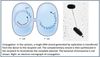
Zoonoses
infections acquired from vertebrates. They can occur by direct contact or in animal products
vector
an arthropod that transmits an infection
Adhesion to Host
Large parasites (e.g. worms) adhere by mechanical means, e.g. biting mouth parts. •
Microscopic organisms bind to host through receptor-ligand interactions. Ligands are normal host structures. Binding to a cell surface is the first step in entering host cell for intracellular pathogens
Intoxication
Due to ingestion of a toxin that bacteria have produced in food. None of the microbe need be ingested or invasive in order to be intoxicated.
Surface infection
Infection of the skin or mucosal surfaces
Invasive infection
Extend into normally sterile spaces in the body.
Routes of spread of pathogens in the body include along the axons of nerves, within the blood or lymphatics, and by direct extension within tissues.
antigenic variation
When a species of pathogen exists with different surface antigens (and expresses only some or one at a time) or when individuals within that species can change their surface antigens over time.
An effective immune response against one antigenic type does not prevent subsequent or prolonged infection with another antigenic type of that pathogen.
Opportunistic pathogens
Cause infections in hosts with compromised defenses. Most opportunistic pathogens are environmental organisms that have not evolved to evade the immune response.
virulence
The ability of a pathogen to cause damage
Ways by which a pathogen may damage the host
- Killing host cells by intracellular microbes. This usually happens after the microbe replicates and lyses the host cell.
- Secreted toxins
- Effector molecules injected into host cells
- The host immune system
- Transformation of host cells
- Mechanical impairment of host organs
- Malnutrition
The presence of such organisms at sites where they are part of the microbiota. . .
does NOT indicate an infection
Most bacteria can devide every ___.
Most bacteria can devide every 20-30 minutes.
At least, during the logarithmic growth phase
Role of peptidoglycan in bacterial cell walls
There is a high concentration of solutes within the bacterial cell, so water tends to move into the bacterial cell when they are in a hypotonic environment. This could lyse (pop) the bacteria. Bacteria must protect themselves from this osmotic stress.
Mycobacterium
Has a thick waxy layer surrounding the cell instead of a conventional layer of peptidoglycan.
This includes Mycobacterium tuberculosis, the cause of tuberculosis. Mycobacteria are the only “acid-fast” bacteria, meaning they retain stains even when decolorized with strong acids. This is due to the thick waxy coating. Another characteristic of mycobacteria is that they replicate more slowly than most other bacteria.
The Gram Stain
Outer Membrane of Gram-Negative Bacteria
Have second membrane surrounding the cell, in addition to the cytoplasmic membrane. The outer membrane encircles the peptidoglycan. This gives rise to a periplasmic space.
The outer membrane contains a unique molecule called lipopolysaccharide or LPS.
Usually contains porins to facilitate diffusion of imporant molecules. Loss of porin expression is an important mechanism of development of antibiotic resistance, as antibiotics utilize porins to enter the cell.
LPS
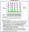
Porins
Trimeric proteins that form a pore in the outer membrane. The inside of the pore is lined with charged amino acids, which facilitates small, charged molecules moving across the outer membrane. Charged molecules ~700 Daltons, or about the size of a tripeptide, can cross this membrane via porins.
Most antibiotics act on Gram negative bacteria in the periplasmic space or in the cytosol. Loss of porin expression is an important mechanism of development of antibiotic resistance.
How hosts get around bacterial capsules
Antibodies!
But a few kinds of bacteria produce capsules that are identical to host molecules, and the body will not produce antibodies against such capsules. This may be considered another mechanism of immune evasion.
How capsules enable immune evasion
First, capsules are charged, making them difficult for phagocytes to engulf. Second, some capsules downregulate activation of complement, reducing opsonization of bacteria.
mechanisms of horizontal gene transfer in bacteria
conjugation, transformation and transduction.