Metabolic & Endocrine Disorders Flashcards

1
Q

Normal bone quality, decreased bone quantity =

A

Osteoporosis

2
Q

Females are affected by osteoporosis __:__ over males

A

4:1

(After 80, females = males)

3
Q

Secondary causes of ____ include metastasis, multiple meyloma, alcoholism, endocrine disorders, etc.

A

OSteoporosis

4
Q

Clinical presentation of ____ include:

  • Clinically silent
  • Increased kyphosis as disease progresses
  • Symptoms become apparent when there are fractures
A

Osteoporosis

5
Q

Radiographs are ____ (good/not good) are grading osteoporosis

A

Not good

6
Q

Radiographic findings of ____ include:

  • Osteopenia (on radiographs this is a descriptive term ie. less dense, DEXA scan it is diagnosis)
  • Loss of 2° stress trabeculae, accentuation of 1° trabeculae
  • Vertical striations in VBs (pseudohemangiomas)
  • Pencil thin cortices
  • Compression fractures (anterior wedge, fish vertebra)
A

Generalized Osteoporosis

7
Q

What are is the likely cause of these findings?

(Normal on left)

A

Generalized Osteoporosis - CT w/ Fish Verterbra

8
Q

What is the likely cause of these findings?

A

Generalized Osteoporosis - Accentuated Vertical Striations (pseudohamangiomas)

9
Q

What is the likely cause of these findings?

A

Generalized Osteoporosis - Accentuated Vertical Striations, anterior wedging, and gas bubble

10
Q

What is the likely cause of these findings?

A

Generalized Osteoporosis - Anterior compression fracture as a result of hyperkyphosis

11
Q

What is the likely cause of these findings?

A

Generalized Osteoporosis - Ward’s Triangle

12
Q

What is the likely cause of these findings?

A

Generalized Osteoporosis - Pencil thin cortices

13
Q

What is the likely cause of these findings?

A

Generalized Osteoporosis - Insufficiency Fracture

14
Q

____ is gold standard for osteoporosis diagnosis, ____ is most accurate but expensive

A

DEXA scan

Quantitative CT

15
Q

____ is the most common cause of regional osteoporosis

Other causes are ___ & ____

A

Disuse (common following immobilization)

Complex Regional Pain Syndrome (CRPS) & Transient osteoporosis of hip (TROH)

16
Q

Regional osteopenia can be seen within _____ in disuse regional osteoporosis, _____ following CRPS

A

7-10 days

3-4 months

17
Q

What is the most likely cause of these findings?

A

Regional Osteoporosis - Disuse

18
Q
  • Relatively normal bone quantity, poor bone quality
  • Results in soft bones

Imaging findings include:

  • Generalized osteopenia
  • Trabecular coarsening & indistinctness
  • Fracture deformites (fish vertebrae, Triradiate pelvis, kyphoscoliosis, etc.)
  • Pseudofractures (insufficiency fractures in unusual locations)
A

Osteomalacia

19
Q

What is the most likely cause of these findings?

A

Osteomalacia - but NOT associated specifically, can be seen in any condition with decreased bone density (quantity, quality or both)

20
Q

What is the most likely cause of these findings?

A

Osteomalacia - Pseudofractures

(Can also be seen in Padgets or Fibrous dysplasia)

21
Q
  • Vitamin D related
  • Primarily a disease of growth plates (seen w/ osteomalacia)
  • Clinically, may see bowing deformities, overgrowth of cartilage near joints, “rachitic rosary”
A

Rickets

22
Q

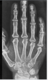
Image findings of ____ include:

  • “Paint brush” metaphysis
  • Splayed/cupping of metaphysis
  • Non-calcified zone of provisional calcification → widened growth plate
A

Rickets

23
Q

What is the most likely cause of these findings?

A

Rickets - Non-calcific zone of provisional calcification & cupped appearance

24
Q

What is the most likely cause of these findings?

A

Rickets - Paint brush metaphysis

25
What is the most likely cause of these findings?
Rickets - Rachitic Rosary
26
What is the most likely cause of these findings?
Rickets - Bowing Deformities
27
What is the most likely cause of these findings?
Scurvy
28
Dense zone of provisional calcification in scurvy =
White line of Frankel/scurvy
29
Well defined ring around epiphysis seen in scurvy =
Wimberger’s ring
30
- Arise from metaphyseal margins - Due to collapse of the scorbutic zone - Seen in Scurvy
Pelken’s spurs
31
Lucent region adjacent to the zone of provisional Ca++ seen in scurvy
Scorbutic/Trummerfeld zone of attrition
32
What is the most likely cause of these findings?
Scurvy
33
- Osseous changes seen following long term megadoses of Vitamin A - Vitamin A regulates osteoclastic and osteoblastic activity - Clinical findings include dermatitis, puritis, alopecia, and hepatosplenomegaly Imaging findings include: - Osteopenia - Solid periosteal reaction
Hypervitaminosis A
34
What is the most likely cause of these findings?
Hypervitaminosis A (but there are many other differentials)
35
- Soft tissue changes seen following long term mega doses of Vitamin D Imaging Findings include: - Periarticular calcification (referred to as tumoral calcinosis bc large mass of calcification) - Renal & vascular calcification
Hypervitaminosis D
36
What is the most likely cause of these findings?
Hypervitaminosis D - Tumoral Calcinosis
37
Clinically can see abdominal pain, encephalopathy & paralysis Imagining findings include: - Dense linear bands along the zone of provisional calcification - Wide sutures from brain swelling
Lead poisoning
38
What is the most likely cause of these findings?
Lead poisoning - sclerotic metaphyseal bands
39
What is the most likely cause of these findings?
Lead poisoning - multiple sclerotic metaphyseal bands
40
- Secondary to excessive growth hormone secretion - In **adults** results In predominate soft tissue & membranous bone overgowth - In **adolescence** results in increased length of bone (gigantism) Clinical findings include: - Broad, large hands/feet - Prominent forehead - Nerve compression syndromes - Arthritis
Acromegaly
41
What is the most likely cause of these findings?
Acromegaly - Enlarged Sella Turcica
42
What is the most likely cause of these findings?
Acromegaly - Enlarged frontal sinus
43
What is the most likely cause of these findings?
Acromegaly - Spade like distal tufts
44
What is the most likely cause of these findings?
Acromegaly - increased heel pad thickness
45
Imaging findings of ____ include: - Generalized osteopenia is main finding - May see increase incidence of fractures - Physical findings include enlarged neck, rapid HR, weightloss, proptosis
Hyperthyroidism
46
Clinical findings include: - Coarse dry skin & hair - Farigue, lethargy Imaging findings include: - Delayed skeletal maturation (hallmark) - Wormian bones - Fragmented epiphyses - Sail vertebra w/ gibbous deformity
Hypothyroidism
47
What is the most likely cause of these findings?
Hypothyroidism - Sail vertebra
48
What is the most likely cause of these findings?
Hypothyroidism - Delayed skeletal maturation
49
- Females affected 3:1 over males - Peak age 30-50 years - Increased osteoclastic activity w/ fibrous tissue replacement - Hypercalcemia and hypophosphatemia
Hyperparathyroidism
50
\_\_\_\_\_ hyperparathyroidism = due to overproduction of parathormone 2º to parathyroid adenoma
Primary
51
\_\_\_\_\_ hyperparathyroidism = due to overproduction of parathormone 2º to chronic renal disease (most common)
Secondary
52
\_\_\_\_ hyperparathyroidism = seen in dialysis patients, long term renal disease; parathyroid gland acts independent of calcium levels
Tertiary
53
Imagining findings of ____ include: - Osteopenia - Salt & pepper skull (granular appearance) - Rugger jersey spine - Resorption of lamina dura of mandible - Endplate irregularities - Widened SI joints & symphysis
Hyperparathyroidism
54
What is the most likely cause of these findings?
Hyperparathyroidism - Salt & Pepper skull
55
What is the most likely cause of these findings?
Hyperparathyroidism - loss of lamina dura
56
What is the most likely cause of these findings?
Hyperparathyroidism - Rugger Jersey Spine (Also small vessel calcification which = hyperparathyroidism or diabetes)
57
Which is rugger jersey vs. sandwich vertebra?
Left = sandwich, Right = Rugger jersey
58
What is the most likely cause of these findings?
Hyperparathyroidism - Subchondral bone resorption
59
Subperiosteal resoprtion on the radial aspect of 2nd & 3rd middle phalanges is diagnostic for \_\_\_\_
Hyperparathyroidism
60
Brown tumors (osteoclastoma) - Soap bubbly & expansile are associated with what condition?
Hyperparathyroidism
61
What is the most likely cause of these findings?
Hyperparathyroidism - Distal tuft resorption
62
What is the most likely cause of these findings?
Hyperparathyroidism - Subperiosteal resorption
63
What is the most likely cause of these findings?
Hyperparathyroidism
64
What is the most likely cause of these findings?
Hyperparathyroidism - Browns tumor
65
What is the most likely cause of these findings?
Hyperparathyroidsm - Small vessel calcification (differential = diabetes)
66
What is the most likely cause of these findings?
Hyperparathyroidism - Tumoral Calcinosis (w/ small vessel calcification)
67
What is the most likely cause of these findings?
Hyperparathyroidism - Chondrocalcinosis (depositional w/ secondary degenerative changes)
68
- Manifestations are due to decreased vascular integrity, particularly in lower extremitiy Imagining findings: - Cellulitis, osteomyelitis & septic arthritis - Neuropathic arthropathies - Osteopenia - Vascular calcification (Monkeberg's arteriosclerosis) - Vas deferens calcifcation
Diabetes
69
What is the most likely cause of these findings?
Diabetes - Neuropathic Arthropathy (Septic Arthritis is differential)
70
- Can take on the follow patterns: - Hypertrophic - Atrophic - Mixed hypertrophic & antrophic pattern
Neuropathic anthropathy
71
What is the most likely cause of these findings?
Diabetes - Neuropathic Arthropathy
72
What is the most likely cause of these findings?
Diabetes - Smal vessel calcification (differential = hyperparathyroidism)
73
What is the most likely cause of these findings?
Diabetes - cellulitis
74
What is the most likely cause of these findings?
Diabetes - Vas Deferens calcification
75
Identify the findings present and diagnosis or differential diagnosis or category as appropriate
76
Identify the findings present and diagnosis or differential diagnosis or category as appropriate
77
Identify the findings present and diagnosis or differential diagnosis or category as appropriate
78
Identify the findings present and diagnosis or differential diagnosis or category as appropriate
79
Identify the findings present and diagnosis or differential diagnosis or category as appropriate
80
Identify the findings present and diagnosis or differential diagnosis or category as appropriate
81
Identify the findings present and diagnosis or differential diagnosis or category as appropriate
82
Identify the findings present and diagnosis or differential diagnosis or category as appropriate
83
Identify the findings present and diagnosis or differential diagnosis or category as appropriate
84
Identify the findings present and diagnosis or differential diagnosis or category as appropriate
85
Identify the findings present and diagnosis or differential diagnosis or category as appropriate
86
Identify the findings present and diagnosis or differential diagnosis or category as appropriate
87
Identify the findings present and diagnosis or differential diagnosis or category as appropriate
88
Identify the findings present and diagnosis or differential diagnosis or category as appropriate
89
Identify the findings present and diagnosis or differential diagnosis or category as appropriate
90
Identify the findings present and diagnosis or differential diagnosis or category as appropriate
91
Identify the findings present and diagnosis or differential diagnosis or category as appropriate
92
Identify the findings present and diagnosis or differential diagnosis or category as appropriate
93
Identify the findings present and diagnosis or differential diagnosis or category as appropriate
94
Identify the findings present and diagnosis or differential diagnosis or category as appropriate