Hematologic Diseases Flashcards

1
Q

Clinical findings of ___ include:

  • Doesnt manifest until after 6 months of age
  • Weakness, pallor, jaundice
  • Abdominal crisis of bowel infarctions
  • Organomegaly
  • Hand-foot syndrome (children under 2 years)
A

Sickle Cell Anemia

2
Q

Bony changes in sickle cell anemia are primarily due to _____

A

ischemia/osteonecrosis

3
Q

Imaging findings of ____ include:

  • Fish vertebrae
  • “H” vertebrae (Reynolds)
  • Hair-on-end skull (uncommon)
  • Osteopenia/osteosclerosis
A

Sickle cell anemia

4
Q

What condition is likely the cause of these findings?

A

Sickle cell anemia - Hair-on-end

(differential = thalassemia should be first thought)

5
Q

What condition is likely the cause of these findings?

A

Sickle cell anemia - H vertebra

(differential = thalassemia should be 2nd thought)

6
Q

What condition is likely the cause of these findings?

A

Sickle cell anemia - H vertebra & fish shaped

7
Q

What condition is likely the cause of these findings?

A

Sickle cell anemia - osteosclerosis

(differential = osteopetrosis, padgetts, blastometaphysis)

8
Q

Erlenmeyer flask deformity is associated with osteopetrosis & _____

A

Sickle cell anemia

9
Q

What condition is likely the cause of these findings?

A

Sickle cell anemia - hand-foot syndrome

10
Q

What condition is likely the cause of these findings?

A

Sickle cell anemia - osteomyelitis

(aggressive lesion)

11
Q

What condition is likely the cause of these findings?

A

Sickle cell anemia - Erlenmeyer flask

12
Q

What condition is likely the cause of these findings?

A

Sickle cell anemia - multiple bone infarcts

13
Q

The following are soft tissue imaging findings associated with ____:

  • Gallstones (up to 65%)
  • Bowel infarction
  • Organomegal
  • Auto splenectomy (secondary to infarction and fibrosis)
  • Extramedullary hematopoiesis (uncommon)
A

Sickle cell anemia

14
Q

What condition is likely the cause of these findings?

A
15
Q

Clinical findings of ____ include:

  • Pallor, lethargy, retarded growth
  • Organomegaly
  • Rodent facies
  • Repeated transfusions → hemochromatosis → cardiac failure/death
A

B-Thhalessemia major

16
Q

What condition is likely the cause of these findings?

A

B-Thalassemia Major - hair-on-end (most commly occurs in this tradition)

17
Q

What condition is likely the cause of these findings?

A

B-Thalassemia - hair-on-end

CT

18
Q

What condition is likely the cause of these findings?

A

B-Thalassemia Major - Honeycomb trabecular pattern

19
Q

What condition is likely the cause of these findings?
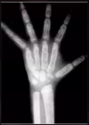
A

B-Thalassemia - Honeycomb trabecular pattern

20
Q

What condition is likely the cause of these findings?

A

B-Thalassemia Major - Extramedullary hematopoiesis

21
Q

What condition is likely the cause of these findings?

A

B-Thalassemia Extramedullary Hematopoiesis

22
Q

H-shaped vertebral bodies most common w/ ___ but can be seen with (just less common)

A

Sickle cell anemia

Thalassemia

23
Q

Most common childhood malignancy (peak age 2-5 years)

A

Acute childhood leukemia (acute lymphocytic)

24
Q

Imaging findings of ____ include:

  • Osteopenia
  • Lucent metaphyseal bands
  • Periostitis
  • Lytic bone lesions
  • Splenomegaly
  • Chloroma a soft tissue tumor of myeloid tissue (rare)
A

Acute childhood leukemia

25
Q

What condition is most likely the cause of these findings?

A

Acute childhood leukemia - lucent metaphyseal bands (other differentials such as rickets)

26
Q

What condition is most likely the cause of these findings?

A

Acute childhood leukemia - lytic bone lesions (differentials = Ewings, osteomyelitis, etc.)

27
Q

Deficiency of clotting factor VIII =

A

Hemophilia A (most common)

28
Q

Deficiency in clotting factor IX =

A

Hemophila B

29
Q

Clinical manifestations of ____ include:

  • Spontaneous bleeding or bleeding from minimal traum
  • Bleeding into joint (hemarthrosis) initiates and inflammatory response in the joint
  • Joint changes visualized on imaging are a result of this inflammatory process and that is why juvenile idiopathic arthritis is a differential diagnosis
A

Hemophilia

30
Q

Imaging findings of ___ include:

  • Overgrowth of epiphyses
  • Squared patella•Widened intercondylar notch
  • Tibiotalar slant
  • Osteopenia
  • Increased soft tissue density around joints (due to hemosiderin deposits into synovium)
  • Hemophilic pseudotumor (most common in femur and ilium)
A

Hemophilia

31
Q

What condition is most likely the cause of these findings?

A

Hemophilia - Widened intercondylar notch

32
Q

What condition is most likely the cause of these findings?

A

Hemophilia (differential = juvenile idiopathic arthritis)

33
Q

What condition is most likely the cause of these findings?

A

Hemophilia - tibiotalar slant (differential = juvenile idiopathic arthrophy)

34
Q

What condition is most likely the cause of these findings?

A

Hemophilia - pseudotumor (not specific)

35
Q
  • Overload of iron within tissues (liver most common)
  • Primary (Inherited) - autosomal recessive deficiency of hepatic xanthene oxidase
  • Secondary (Acquired)-secondary toalcoholicsiderosis, cirrhosis, repeated iron transfusions
A

Hemocromatosis

36
Q
  • Articular abnormalities occur in 20% to 50% of cases
  • Due to iron or calcium pyrophosphate crystals deposited into joints
A

Hemochromatosis

37
Q

The following triad can be seen in ____

  • Bronze skin
  • Diabetes
  • Cirrhosis
A

Hemochromatosis

38
Q

Hook-like or beak-like osteophytes at metacarpal phaleangeal joints indicate

A

Hemachromatosis

39
Q

What condition is most likely the cause of these findings?

A

Hemachromatosis - hook-like osteophytes

40
Q
  • AKA ischemic necrosis, avascular necrosis, bone infarction, bone infarct
  • Causes include spontaneous (most common), trauma, alcoholism, corticosteroids, hemoglobinopathies, systemic lupus erythematosus, Cushing’s, and radiation exposure
A

Osteonecrosis

41
Q

Term typically used for necrosis of bone in non-epiphyseal location, usually in medullary space of metaphysis

A

Bone infarction

42
Q

Term typically used for necrosis in epiphyseal location, usually in of subchondral bone adjacent to joint

A

Avascular necrosis

43
Q

What condition is most likely the cause of these findings?

A

Avascular necrosis of femoral head

44
Q

What condition is most likely the cause of these findings?

A

Avascular necrosis - Crescent sign

45
Q

What is this demonstrating the progression of?

A

Avascular necrosis

46
Q

___ is the best for diagnosis osteonecrosis early on as it is both sensitive & specific

A

MRI

47
Q

What condition is most likely the cause of these findings?

A

Avascular necrosis

48
Q
  • Appears as a region of serpentine calcification within medullary space
  • DDX: Enchondroma or Low grade chondrosarcoma
A

Bone infarct

49
Q

What condition is most likely the cause of thes findings?

A

Bone infarct - polyostotic

50
Q

What condition is most likely the cause of these findings?

A

Radiation necrosis

(irregular sclerosis)

51
Q
  • Affects the tibial tuberosity
  • Traction apophysitis from overuse
  • Clinical diagnosis
  • Radiographically, can see fragmentation of the tibial tuberosity apophysis, with overlying soft tissue inflammatio
  • Peak age = 11-15 years, males most common
A

Osgood-Schlatter

52
Q

What condition is most likely the cause of these findings?

A

Osgood-Schlatter (not necessarily an abnormality, need hx)

53
Q
  • Affects endplates
  • Most commonly in the thoracic spine
  • Hereditary factors play an important factor
  • Over stress of spine at young age, especially in athletes plays a role as well in some
  • Peak age = 13-16 years
A

Scheuermann disease

54
Q

Imaging of ____ includes:

  • Endplate irregularities
  • Anterior wedging of the vertebral bodies of 5 degrees of more involving three continues levels
  • Increased anterior to posterior width of the vertebral body
  • Increased kyphosis
A

Sceuermann disease

55
Q

What condition is most likely the cause of these findings?

A

Scheuermann Disease - Wedging of 3 Adjacent levels

56
Q

What condition is most likely the cause of these findings?

A

Scheuermann disease

57
Q

Identify the findings present and diagnosis or differential diagnosis or category as appropriate

A
58
Q

Identify the findings present and diagnosis or differential diagnosis or category as appropriate

A
59
Q

Identify the findings present and diagnosis or differential diagnosis or category as appropriate

A
60
Q

Identify the findings present and diagnosis or differential diagnosis or category as appropriate

A
61
Q

Identify the findings present and diagnosis or differential diagnosis or category as appropriate

A
62
Q

Identify the findings present and diagnosis or differential diagnosis or category as appropriate

A
63
Q

Identify the findings present and diagnosis or differential diagnosis or category as appropriate

A
64
Q

Identify the findings present and diagnosis or differential diagnosis or category as appropriate

A
65
Q

Identify the findings present and diagnosis or differential diagnosis or category as appropriate

A
66
Q

Identify the findings present and diagnosis or differential diagnosis or category as appropriate

A
67
Q

Identify the findings present and diagnosis or differential diagnosis or category as appropriate

A
68
Q

Identify the findings present and diagnosis or differential diagnosis or category as appropriate

A
69
Q

Identify the findings present and diagnosis or differential diagnosis or category as appropriate

A
70
Q

Identify the findings present and diagnosis or differential diagnosis or category as appropriate

A
71
Q

Identify the findings present and diagnosis or differential diagnosis or category as appropriate

A
72
Q

Identify the findings present and diagnosis or differential diagnosis or category as appropriate

A
73
Q

Identify the findings present and diagnosis or differential diagnosis or category as appropriate

A
74
Q

Identify the findings present and diagnosis or differential diagnosis or category as appropriate

A
75
Q

Identify the findings present and diagnosis or differential diagnosis or category as appropriate

A
76
Q

Identify the findings present and diagnosis or differential diagnosis or category as appropriate

A