Joint Disease Flashcards

1
Q

What are the 5 things that make up the checklist for evaluating joint disease?

A
  1. Joint Space
  2. Subchondral Bone
  3. Margins of joint
  4. Surrounding Soft Tissue
  5. Miscellaneous
2
Q

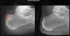
What is this showing?

A
3
Q

What is this showing & what condition it associated with?

A

Ankylosis of intercarpal joints

Uniform loss of radiocarpal joint

(Associated w/ inflammatory arthritis)

4
Q

What two conditions are demonstrated here?

A
5
Q

Deformities can be seen in what kind of joint disease?

A

Degenerative, Inflammatory or Deposition

6
Q

What is shown here?

A

Calcification of fibrocartilage (normally shouldnt be able to see meniscus)

7
Q

What is shown here?

A

Calcification of hyaline cartilage

8
Q

What is shown here?

A

Calcification of Intra-Articular Loose Bodies (not associated with any particular type of joint disease)

9
Q

Fibrocartilage or Hyaline Cartilage calcification are associated with what category of joint disease?

A

Deposition

10
Q

What is shown here?

A

Joint effusion (increased fluid) - Abnormal fat pads

Anterior sail sign, Posterior fat pad

11
Q

What is shown here?

A

Joint Effusion in knee

12
Q

What is shown here?

A

Joint Effusion ankle joint

13
Q

What is shown here?

A

Abnormal Air in the joint (disc)

Also osteophytes and thickened end plates

Associated with Degenerative joint disease

14
Q

What is shown here?

A

Normal gas in joint

15
Q

Increased density of subchondral bone =

A

Subchondral sclerosis; referred to as endplate sclerosis in the spine

16
Q

Decreased density of subchondral bone =

A

Periarticular osteopenia (aka juxta-articular osteopenia); referred to as endplate destruction in the spine
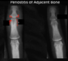
17
Q

What is shown here?

A

Subchondral sclerosis (non-uniform with non-uniform loss of joint space)

18
Q

Sclerotic changes (adding bone) is associated with what category of joint disease?

A

Degenerative

19
Q

Taking away bone (osteopenia) is associated with what category of bone disease?

A

Inflammatory

20
Q

What is shown here?

A

Endplate sclerosis

21
Q

What is shown here?

A

Endplate sclerosis

22
Q

What is shown here?

A

Periarticular osteopenia

23
Q

What is shown here?

A

Endplate destruction (periarticular osteopenia in the spine)

24
Q

What is shown here?

A

Subchondral cysts

25
Subchondral cysts are associated with category of joint disease?
Any of them (degenerative, inflammatory, deposition)
26
What is shown here?
Ill-defined Marginal Erosions (Also subchondral cysts and loss of joint space)
27
Ill-defined marginal erosions are associated with what category of joint disease?
Inflammatory
28
What is shown here?
Well-defined Marginal Erosions
29
What is shown here?
Romanus lesions (marginal erosions in the spine)
30
What is shown here?
Central joint erosions
31
What is shown here?
Osteophytes (traction)
32
What is shown here?
Osteophytes (claw)
33
Syndesmophytes are associated with what joint disease category?
Inflammatory
34
What is shown here?
Marginal Syndesmophytes
35
What is shown here?
Non-Marginal Syndesmophyte
36
What is shown here?
Non-Marginal Syndesmophyte
37
What is category of joint disease would the following condition fall under?
Degenerative
38
Increased soft tissue size/thickness is most often seen in what joint disease category?
Inflammatory (also can be deposition like gout if mass instead of swelling)
39
What is shown here and what joint disease category is it generally associated with?
Generalized increase in soft tissue size/thickness Inflammatory
40
What is shown here?
Calcification in soft tissue (supraspinatus tendon) (Deposition in this case)
41
What category of joint disease would this be associated with?
Inflammatory
42
This case appears to have a _____ distribution Associated with what type of joint disease category?
Unilateral Degenerative
43
This case appears to have ____ distribution Associated with what type of joint disease category?
Bilateral Asymmetric
44
This case appears to have ____ distribution Associated with what type of joint disease category?
Bilateral Symmetric Inflammatory
45
What is shown here?
Enthesophytes
46
What is shown here?
Enthesitis
47
What is shown here?
Non-articular bony erosions Likely depositional disease such as gout
48
What is shown here?
Periostitis of adjacent bone (Associated w/ inflammatory/arthritic changes)
49
Decreased or increased joint space (typically non-uniforrm) Osteophytes Subchondral sclerosis & cysts Enthesophytes Unilateral/asymmetric Joint effusion These are characteristics of what joint disease category?
Degenerative
50
Uniform loss of joint space is associated with what joint disease category?
Inflammatory
51
What joint disease category is this associated with?
Deposition
52
What conditions are associated with deposition category?
Gout CPDD (calcium pyrophasphate dehydrate deposition) HADD (hydroxyapatite deposition disease) Ochronosis