Mediastinum, Heart, Neck Imaging Flashcards
1
Q

A

2
Q
A
4 is the left anterior descending

3
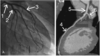
Q
Identify the structure marked “??” and identify the pathology.

A

4
Q
What is your diagnosis?

A
Calcific pericarditis
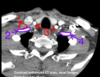
5
Q
What is your diagnosis?

A
Pericardial effusion

6
Q

A

7
Q
Identify the heart chambers and the pathology.

A
The thing at the yellow arrow is a thrombus.

8
Q

A
5 is a lymphoma in the cardiac wall

9
Q
Identify the heart chambers and the pathology (not marked).

A
Pathology is an atrial septal defect (yellow arrow)

10
Q

A
- SVC
- Aortic arch
- Pulmonary trunk
- Left ventricle
- IVC
- Right atrium
11
Q

A

12
Q
Identify the pathology (#1) and the structure labeled #2.

A
Thymoma, ascending aorta

13
Q
Identify the structures and the pathology labeled #3.

A
- SVC
- Aortic arch
- Lymphoma
- Trachea
- SVC
- Ascending aorta
- Main pulmonary artery
- Right pulmonary artery
- Descending aorta

14
Q
Identify the structures and the pathology.

A

15
Q
A
- Anterior scalene
- Right subclavian artery
- Common carotid arteries
- Left subclavian artery
- Internal jugular vein

16
Q

A

17
Q

A
- SVC
- Ascending aorta
- Left pulmonary artery
- Carina (bifurcation of the trachea)
- Esophagus
- Descending aorta
- Left pulmonary vein

18
Q
T4 level

A

19
Q
Identify the muscle group (#1) and the vessel (#2).

A
Scalenes and external jugular vein

20
Q

A

21
Q
What are those!?

A
Vertebral arteries
