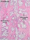
Male Reproductive System Flashcards
Tunica Vaginalis
2 layers:
outer parietal layer
inner viscera layer
Tunica albuginea
Directly on the testes
Dense CT
Form fibrous septa that divide testes into 250-300 pyramidal compartments called lobules

Lobule
1-4 seminiferous tubules (produce spermatozoa)
loose CT
nerves
blood & lymph
endocrine interstitial cells (Leydig cells)
Sperm Pathway
seminiferous tubules–> straight tubule–> rete testis–> 10-20 efferent ductules that connect to head of epididymis (head, body, tail)
Seminiferous tubules
250-1000 per testicle
200 million per day in adult male
Older in the center/lumen

Leydig cells
interstitial cells
secrete testicular androgens *testosterone

Spermatogenesis
Begins at puberty
Reduces ploidy & chrom number
Spermatocytogenesis- undiff spermatogonia beomes spermatogonia:
- Type A spermatogonia:
- Type A dark: A= type Ad or Type Ap
- Type A pale= type B spermatogonia
- Type B speratogonia=type B spermatogonia or primary spermatocytes
Meiotic divisions- allow primary spermatocytes to become spermatids
spermiogenesis- morphological diff of spermatids into mature sperm(atozoa)
Spermatogenesis continued

Spermiogensis
Transform round spermatids into elongated, free swimming spermatozoa capable of fertilization
4 phase: Golgi, Cap, Acrosome & maturation
Golgi Phase
first polarity
prominent golgi apparatus w/ proacrosomal granules to become acrosomal cap

Cap Phase
acrosome cap enlarges, then flattens & extends over the nucleus.
Acrosome is specialized lysosome, containing hydrolytic enz capable of dissociating the corona radiata & zona pellucida of oocyte
1 centriole acts to organize initiation of a flagellum

Acrosomal Phase
spermatids become oriented toward basement mem
nuclei become more elongated chromatin becomes more condensed
head cap begins to move toward tail
flagella continue to grow
Manchette= contains motor proteins!

Maturation Phase
Unneeded cytoplasm is shed as residual body
Mature sperm are released into lumen of seminiferous tubule

Spermiogenesis Histo
Myoid Cell= contracts to expel sperm into lumen
Spermatocytes largest more numerous cells in seminiferous tubule, have condensed chrom

Sertoli Cells
Columnar or pyramidal cells- base adheres to basal lamina
Apex extends into lumen of seminiferous tubule
Envelop spermatogenic cells
connected to ea other by numerous gap junctions
F as supporting (nurse) cells- ea contains 30-50 germ cells @ various stages of development
Function of Sertoli Cells
- support, protection, & nutrition of developing spermatogenic cells- because spermatocytes, spermatids & spermatozoa are isolated from blood supply by blood testis barrier, these cells depend on Sertoli cells to mediate nutrient & metabolite exchange
- exocrine- nutrients & androgen binding protein (ABP) which concentrates testosterone to a level required for spermiogenesis (promoted by FSH)
- Endocrine- secretion of inhibin & MIS (mullerian inhibiting substance)
- phagocytosis- residual bodies (cytoplasm) from spermiogenic cells is phagocytosed
Blood Testis Barrier
physical barrier between blood vessles & seminiferous tubule
formed b/t sertoli cells (tight junctions)
separates seminiferous tubule into a basal compartment & adluminal compartment
Prevents passage of cytotoxic agents into seminiferous tubules
*during spermatogenesis, cells after division squeeze through & transverse junctions to lie in adluminal compartment (above barrier)

Interstitial Tissue & Leydig Cells
Interstitial tissue- site of androgen production, CT containing mast cells, macrophages, n., lymphatics & blood vessels
Leydig cells- become apparent @ puberty, produce testosterone (stim by LH, important for male repro dev & spermatogensis)
Intratesticular Ducts
Straight tubules–> Rete testis–>Efferent ductules
carry spermatozoa & liquid from seminiferous tubules to duts of epididymis
Seminiferous tubules= arranged as loops, both ends join rete testis
Straigh tubules= char by gradual loss of spermatogenic cells, has initial seg of walls lined only by Sertoli cells, main seg is cuboidal epith supported by dense CT
Rete testis= interconnected network of channels lined by cuboidal epithelium
Efferent Ductules
15-20
lined by nonciliated cuboidal cells alternating w/ groups of taller ciliared cells
non ciliated= absorb most of fluid secreted by seminif tubules
ciliated= beat in direction of epididymus
Excretory Gential Ducts
Epididymis
Ductus (vas) deferens
Urethra
Accessory glands secrete into these
Essential for reproductive function
Ductus Epididymis
Single highly coiled tube
Forms head, body & tail
Sperm stored here & attain their final char including motility, mem R for zona pellucida proteins, maturation of acrosome & ability to fertilize
Ductus/Vas Deferens
long straight tube w/ thick muscular wall
Has narrow lumen & thick smooth m. (longitudinal outer & inner layers w/ middle circular layer)
mucosa folded longitudinally & is lined by pseudostratified columnar epithelium w/ sparse stereocilia
Pathway
Ductus deferens–> passes over urinary bladder–> then dilates to form ampulla (epith thick & more folded)–> at distal end of ampulla, seminal vesicles join the duct–> ductus deferens enter the prostate gland (as ejaculatory duct) & open into prostatic urethra