Lecture 4: Part 4 (the back) Flashcards
(36 cards)
Surface Landmarks of Back
- what is #13?
- how can it be identified?
- what vertebral level is this located at?
- site of what other structure?

Surface Landmarks of Back
-
Nuchal Groove
- divet you can palpate above C7
- where cervical vertebrae are, covered by nuchal ligament
- site of nuchal ligament

Surface Landmarks of Back
- what is structure #12?
- what vertebral structure is located here?

Surface Landmarks of Back
- Structure 12 - Vertebra Prominens: the spinous process of C7

Structure 8:

Erector Spinae

Structure 7:

Posterior Superior Iliac Spines (PSIS): dimples at bottom of back

Structure 5:

Site of Sacrum (below dimples of PSIS, above buttcrack)

structure 11:

medial borders of scapulae

Extrinsic Back Muscles:
- muscles you see first when you ______
- located ____, but ______
- act as _____ and control movment of ______ and ______
- in embryology, these muscles ______
- have unique _______
- -Superficial: (2)*
- -Intermediate: (3)*
- -Deep: (2)*
Extrinsic Back Muscles: “false back muscles”
- muscles you see when you first remove skin and subcutaneous tissue
- located in the back but do not act on the back (do not have a function or movement control of the vertebral column)
- act as accessory respiratory muscles and control movements of shoulder and arm
- in embryology, these muscles formed elsewhere and then rotated to the back
- have unique innervations
- -Superficial: Trapezius and Latissimus Dorsi*
- -Intermediate: Levator scapulae, Rhomboid minor, and Rhomboid major*
- -Deep: Serratus posterior superior and Serratus posterior inferior*

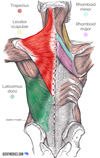
Extrinsic Back Muscles: Superficial Group
- 2 superficial back muscles (and what they look like)
Extrinsic Back Muscles:
- 2 superficial back muscles: Trapezius and Latissimus Dorsi
- the 2 large “sheet” muscles you encounter first in dissection

Trapezius:
- what type of back muscles? (extrinsic or intrinsic)
- subtype?
- goes from _____ down ______ until _____
- fiber direction: _______
- actions: (4)
- innervation:
Trapezius:
-
extrinsic back muscle
- superficial layer
- goes from external occipital protruberance down all spinous processes until T12
- fiber direction: fibers point down to the spine of the scapula
- actions: elevates, depresses, retracts and rotates the scapula
- innervation: spinal accessory nerve (CNX1)

Latissimus Dorsi
- What type of back muscle (extrinsic or intrinsic)?
- what subgroup?
- attaches to _____ and ______ (origin)
- fiber direction: points to ______
- inserts on _____
- has a sheet like tendon called _____ - allows latissimus dorsi to have ______
- action: (3)
- innervation:
Latissimus Dorsi
-
extrinsic back muscle
- superficial
- attaches to lumbar and some of lower thoracic spinous processes (origin)
- fiber direction: points to anterior surface of arm (near bicep tendon)
- attaches to lesser tuberosity of the humerus (insertion)
- has a sheet like tendon called an thoracolumbar fascia aponeurosis - this allows latissimus dorsi to have an expansive attachment
- action: extends, adducts, and medially rotates humerus
- innervation: thoracodorsal nerve (C6-C8)

Extrinsic Back Muscles: Intermediate Group
- 3 Intermediate extrinsic back muscles:
- all associated with _____
- all innervated by _____
Extrinsic Back Muscles: Intermediate Layer
- 3 Intermediate extrinsic back muscles: levator scapulae, rhomboid minor, rhomboid major
- all associated with the scapula
- all innervated by dorsal scapular nerve (C5)

Levator Scapulae:
- what type of back muscle (extrinsic or intrinsic) ?
- what subgroup ?
- Origin: ______
- Insertion (distal attachments): ______
- Action: _____ and ______
- Innervation: ______ and ______, and ______
Levator Scapulae:
-
Extrinsic back muscle
- intermediate group (the highest one)
- Origin: posterior tubercle of transverse process of cervical vertebrae 1 to 4.
- Insertion (distal attachments): Medial border of the scapula between spine and superior angle.
- Action: Elevates scapula and rotates its glenoid cavity inferiorly by rotating scapula
- Innervation: ventral primary rami of C3 and C4 and dorsal scapular nerve (C5)

Rhomboid Major:
- What type of back muscle (extrinsic or intrinsic)?
- what subgroup?
- Origin (Proximal Attachment): _____
- Insertion (Distal attachment): _____
- Action: 3 main actions:
- also assists serratus anterior with _____
- Innervation: _____
Rhomboid Major
-
Extrinsic back muscle
- intermediate layer (lowest one - “minor rides on top of the major”)
- Origin (Proximal Attachment): Spinous Processes T2-T5
- Insertion (Distal attachment): Medial border of scapula inferior to spine
- Action: Elevates and retracts scapula and rotate its glenoid cavity inferiorly; also assists serratus anterior to fix scapula to thoracic wall
- Innervation: Dorsal Scapular Nerve (C5)
Rhomboid Minor
- What type of back muscle (extrinsic or intrinsic)?
- what subgroup?
- Origin (Proximal Attachment): _____
- Insertion (Distal attachment): _____
- Action: 3 main actions:
- also assists serratus anterior with _____
- Innervation: _____
Rhomboid Minor:
-
Extrinsic back muscle
- intermediate group (rides on top of major)
- Origin (proximal attachments): Spinous process of C7-T1 vertebrae.
- Insertion (distal attachments): Medial border of scapula superior to spine (below levator scapulae)
- Action: Elevates and retracts scapula and rotate its glenoid cavity inferiorly; also assists serratus anterior to fix scapula to thoracic wall
- Innervation: Dorsal Scapular Nerve (C5)

Extrinsic back muscles: Deep layer
- 2 deep extrinsic back muscles: _______ and _______
- located underneath ______
- function:
- if robust, this means _____
Extrinsic back muscles: Deep layer
- 2 deep extrinsic back muscles: serratus posterior superior and serratus posterior inferior
- located underneath the rhomboids
- very thin muscles
- function: 2° respiratory muscles
- if robust, this means patient likely has a problem with inspiration (ie COPD, emphysema, ect.)

Serratus Posterior Superior:
- Extrinsic or instrinsic back muscle?
- what subgroup?
- Origin: _______
- insertion: ______
- innervation: _____
- Function: _______
Serratus Posterior Superior:
-
Extrinsic back muscle
- deep layer (underneath rhomboids)
- Origin: Nuchal ligament, spinous processes of vertebrae C7-T3
- insertion: Superior borders of ribs 2-5
- innervation: 2nd-5th Intercostal nerves
- Function: Elevates ribs (inspiration)

Serratus Posterior Inferior:
- Extrinsic or instrinsic back muscle?
- what subgroup?
- Origin: _______
- insertion: ______
- innervation: _____
- Function: _______
Serratus Posterior Inferior:
-
Extrinsic back muscle
- deep extrinsic back muscle (underneath latissimus dorsi)
- Origin: spinous processes of vertebrae T11-L2
- insertion: Inferior borders of ribs 9-12
- innervation: Anterior rami of spinal nerves T8-T12 (a.k.a. 8th-11th Intercostal nerves + 12th “subcostal” intercostal nerve)
- Function: Depresses ribs/ Draws ribs inferoposteriorly (expiration)

In a cross (axial) section:
- which extrinsic back muscles can we see and where are they located?
- where are the intrinsic back muscles located in relation to this?

In a cross (axial) section:
- can see the superficial extrinsic back muscles (trapezius and latissimus dorsi)
- located between the transverse process and the spinous process
- the intrinsic back muscles are located deep to the extrinsic back muscles
- located between the rib and spinous process in this cross-section

Intrinsic back muscles
- true or false back muscles? (and why)
- innervation?
- act to?
- layers? (and subtypes)
Intrinsic back muscles
-
true back muscles
- have a unifying innervation and act on the vertebral column (as well as where they attach - perhaps the head, perhaps the ribs, ect.)
-
innervated by dorsal segmental rami ONLY
- the dorsal segmental rami are going to leave the intervertebral foramen and push there way through all the intrinsic back muscles while dropping off motor innervation, and then keep going through the deep investing fascia and superficial fascia and finally up to the skin to supply a dermatome (because dorsal segmental rami are mixed system)
-
Action - move the vertebral column
- maintain posture and control movements of vertebral column and head
-
superficial layer
- splenius cervicis
- splenius capitus
-
intermediate layer = erector spinae muscles
- spinalis
- longissimus
- iliocostalis
-
deep layer = transversalis group
- multifidus

Intrinsic Back Muscles: Superficial Layer
- 2 muscles
- where they insert/attach
- innervation
- action
Intrinsic Back Muscles: Superficial Layer
-
Splenius capitus
- from spinous processes of C7-T4 to mastoid process
- moves neck
- when bilateral -> head and neck extension
- when unilateral ->ipsilateral head rotation
-
Splenius cervicis
- from spinous processes of T3-T6 to transverse processes of C1-C3
- moves neck
- when bilateral -> head and neck extension
- when unilateral ->ipsilateral head rotation
- Both innervated by dorsal segmental rami
*note that Splenius Capitis/Cervicis is different from semispinalis capitis/cervicis (semispinalis is deeper and note that capitis attached to cap/head while cervicis

Splenius Capitis
- true or false back muscle?
- what layer?
- origin:
- insertion:
- action:
- innervation:
Splenius Capitis
- true or false back muscle: intrinsic (true)
- superficial layer
- origin: spinous processes of C7-T4
- insertion: mastoid process (on occipital bone)
- action: moves neck
- when bilateral -> head and neck extension
- when unilateral -> ipsilateral head rotation
- innervation: dorsal segmental rami of middle cervical nerves
*note that Splenius Capitis/Cervicis is different from semispinalis capitis/cervicis (semispinalis is deeper and note that capitis attached to cap/head while cervicis attaches to the spine)

Splenius Cervicis
- true or false back muscle?
- what layer?
- origin:
- insertion:
- action:
- innervation:
Splenius Cervicis
- true or false back muscle? intrinsic (true)
- what layer? superficial
- origin: spinous processes of T3-T6
- insertion: transverse processes C1-C3
- action: moves neck
- when bilateral -> head and neck extension
- when unilateral -> ipsilateral head rotation
- innervation: dorsal segmental rami of lower cervical nerves
*note that Splenius Capitis/Cervicis is different from semispinalis capitis/cervicis (semispinalis is deeper and note that capitis attached to cap/head while cervicis attaches to the spine)

Spinalis
- true or false back muscle?
- what layer?
- origin:
- insertion:
- action:
- innervation:
Spinalis
- true or false back muscle? intrinsic (true)
- what layer? intermediate (erector spinae muscle group)
- origin: spinous processes of inferior vertebral level
- insertion: spinous processes of superior vertebral level
- action: (all erector Spinae muscles)
- when bilateral -> vertebral extension
- when unilateral -> ipsilateral lateral flexion
- innervation: dorsal segmental rami

Longissimus
- true or false back muscle?
- what layer?
- origin:
- insertion:
- action:
- innervation:
Longissimus
- true or false back muscle? intrinsic (true)
- what layer? intermediate (erector spinae muscle group; middle one)
- origin: transverse process below
- insertion: transverse process above
- action: (all erector Spinae muscles)
- when bilateral -> vertebral extension
- when unilateral -> ipsilateral lateral flexion
- innervation: dorsal segmental rami


