Histology Lab 1: The Cell (Lecture 3) Flashcards
(27 cards)
Which cell components are basophilic?
Nucleus (including chromatin and the nucleolus), ribosomes
Which cell components are acidophilic?
Cytoplasm (due to cytoplasmic proteins), and anywhere that proteins are concentrated (e.g., plasma membrane, secretory granules).
Question: Why is the nucleus basophilic?
It is largely composed of nucleic acids, which are attracted to basic dyes.
Question: In general, which macromolecule (i.e., protein, nucleic acid, lipid, carbohydrate) is colored purple by Nissl stain?
Nucleic acids
Question: What does this neuron’s nuclear morphology indicate about its protein synthesis?
This cell is actively synthesizing proteins. Euchromatin (as opposed to heterochromatin) is the form of chromatin necessary for transcription. The presence of a prominent nucleolus also indicates protein synthesis. The nucleolus is making rRNA, which is used to form ribosomes.
Question: Why are Nissl bodies basophilic?
Nissl bodies are basophilic because they are collections of rough endoplasmic reticulum. RER is studded with ribosomes, which are partly composed of rRNA. Basic dyes stain rRNA.
Question: Why is rough endoplasmic reticulum basophilic?
RER is studded with ribosomes, which are partly composed of rRNA. Basic dyes stain rRNA.
Question: Do all cells that secrete a relatively large amount of product always have a lot of secretory granules visible? Explain.
No. Some cells produce a lot of protein and secrete it immediately rather than storing it in secretory vesicles.
Question: Why does the location of glycogen appear to be white or poorly stained?
Glycogen is lost from the tissue during routine preparation of H&E-stained specimens. By the time the stains are applied, there is nothing left to stain.
Question: Which preparation technique could be used to better visualize glycogen and what color does it produce?
The periodic acid-Schiff (PAS) method since it stains carbohydrates. It produces a bright magenta color.
Question: Why does the location of the lipids appear to be white or poorly stained?
Lipids are lost from the tissue during routine preparation of H&E-stained specimens. By the time the stains are applied, there is nothing left to stain.
Question: How can you differentiate between lipid droplets and glycogen granules in H&E stained tissues?
Lipid droplets, even the smallest ones, have a relatively large round shape compared to glycogen. The white spaces that glycogen leaves behind are patchy and irregular.
Why do some adipocytes seem to lack a visible nucleus?
All of the adipocytes have a nucleus, but they may not all be in the plane of cut. Also, the nuclei are hard to see because they are squished over to once side of the cell by the lipid droplet.
Question: Adipocytes in real life are spherical, yet in the section some of them do not appear to be perfectly round. Why is this?
This is an example of artifact. Since lipid takes up most of the space in the cell, but it is removed during tissue preparation, there is little structure left and the cells can appear to be collapsed.
Question: Which special stain was used to color the myelin black?
Osmium tetroxide
Question: Where is the melanin in relation to the nucleus and why does it assume this cellular location?
It is next to the nucleus, specifically the side facing the surface of the skin where ultraviolet rays may enter the tissue. Melanin absorbs the UV rays to help protect the DNA against damage.

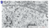
A: Nuclear membrane
B: Nucleolus
C: Euchromatin
D: Heterochromatin


A: Outer mitochondrial membrane
B: Inner mitochondrial membrane (forming a crista)
C: Matrix granule
D: Mitochondrial matrix


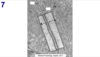
A: Lumen of rough endoplasmic reticulum
B: Ribosome


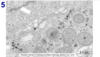
A: Lumen of smooth endoplasmic reticulum

(there is no A)

B: Golgi apparatus
C: Lysosome
D: Glycogen cluster/rosette
E: Mitochondrion

A: Line is on the nuclear envelope, outlining the nucleus
B: Mitochondrion

A: Plasma membrane. Yes, there is another plasma membrane next to it. There are two neighbor cells in this micrograph


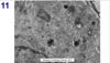
A: Mitochondrion
B: Golgi apparatus
C: Euchromatin
D: Heterochromatin
E: Rough endoplasmic reticulum