Lecture 2: Tools of Histology Flashcards
(34 cards)
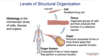
Histology:
Histology: microscopic study of cells, tissues, organs
Cell:
Cell: smallest Living Unit

Tissue:
Tissue: organized group of cells and their products that function in a collective manner

Organ:
Organ: structure composed of 2 or more tissue types that performs and specific function

Organ system:
Organ system: 2 or more organs that perform a common function
- Decimeter
- Centimeter
- Millimeter
- Micrometer (microns)
- Nanometer

Resolution (definition)
The smallest distance at which 2 points can be distinguished as seperate entities
- Smaller resolution = stronger
Resolution of:
- human eye =
- light microscope =
- transmission electron microscope =
- scanning electron microscope =
- Resolution of human eye: 100 um
- Resolution of light microscope: 0.2 um (200 nm)
- Resolution of transmission electron microscope: 3 nm
- Resolution of scanning electron microscope = 1 nm

Ultrastructure
Ultrastructure = cellular structures that can only been seen using an electron microscope
Light microscope:
- ____ travels through ____
- The ____ lens in the ____ (_x) and the ____ lenses (_x, _x, _x, _x) magnify the image
- Low = _x
- Medium = _x
- High = _x
- Oil immersion = _x
- Light travels through a thin section of tissue.
- The ocular lens in the eyepiece (10x) and the objective lenses (4x, 10x, 40x, 100x) magnify the image.
- Low = 4x
- Medium = 10x
- High = 40x
- Oil immersion = 100x

Virtual microscope (5 steps)
1) Slide collections
2) Slide Scanner
3) Servers
4) Virtual microscope software
5) Histology lab and mobile devices

Routine slide preparation for light microscopy (7)
1) Fixation
2) Dehydration
3) Clearing
4) Infiltratoin
5) Embedding
6) Sectioning
7) Mounting on slide, removal of paraffin,
hydration, staining.

Fixation
① Fixation = Preserve with formalin (so does not decompose)
- cross-linking of proteins and inactivation of enzymes (Formaldehyde polymerizes so we add something to it to to stop it - now called formalin)
- Macromolecules (like glycogen) get taken out of tissue

Dehydration with alcohol
② Dehydration = use of alcohol to remove all water
- To later embed in block of wax (so we can cut it into thin slices) we need to remove water with alcohol because water does not mix well with wax

Clearing
③ Clearing = use of organic solvent (e.g. xylol) to remove alcohol. The tissue is now saturated with organic solvent, which can dissolve paraffin used in the next step
- Called “clearing” because this step renders the tissue transparent
- Will also wash out lipids with clearing agent

Infiltration
④ Infiltration = melted paraffin penetrates the tissue

Embedding
⑤ Embedding = paraffin placed in a mold and hardens, Block is trimmed

Sectioning
⑥ Sectioning = tissue is sectioned into thin slices (5-10um)

Mounting on slide, removal of paraffin,
hydration, staining.
⑦ Tissue slice is mounted on a glass slide.
- Paraffin is removed and the tissue is rehydrated.
- Stain is applied to color the clear cellular components so that they may be seen.

Artefact
Artefact = structural abnormality not present in the living tissue as a result of the preparation process
- Something you see under microscopy that is not there in real life
- tear, fold, air bubble

Planes of cut (3)
- Cross section = the specimen was sectioned along its short axis (cut in top/bottom halves)
- Longitudinal section = the specimen was sectioned along its long axis (cut in left/right halves)
- Oblique section = the specimen was sectioned on an angle

4 chemical building blocks of tissues
1) nucleic acids
2) proteins
3) carbohydrates
4) lipids
* Tissue becomes colorless during “clearing”, so we need to color it. You can color (stain) any of the basic macromolecules*
Hematoxylin:
- behaves like
- has what charge
- stains what (generally)
- most commonly stains what (and why)
- causes what color change
Hematoxylin:
- behaves like a base
- has positive charge
- stains things that are basophilic
- stains nucleic acids (most common example)
- why: + charged in solution, attracted to - charge in cell (- phosphate in nucleic acids)
- So, anything that turns blue = contains nucleic acids
- stains nucleic acids (most common example)
(Base = Blue = Basophilic stains)

Eosin:
- acts like:
- what charge:
- what does it stain (generally):
- most commonly stains what:
- why?
- most commonly stains what:
- what color change:
Eosin:
- Acts like an acid
- has a negative charge
- Stains things that are acidophilic
- Stains proteins (most common example)
- why: - charge = acidic in solution, attracted to the + structures in cells like proteins (with ionized amino groups +)
- Stains proteins (most common example)
- Stains things pink


