Lecture 4: Histology of the Small and Large Intestine Flashcards
Where are Brunner’s Glands found?
Only in the duodenum (submucosa)
Where are Vili of the small intestine shortest and longest?
- Longest in Duodenum
- Shortest in Ileum
In regards to the Plicae Circulares, where are they most numerous and where are they absent?
Most numerous: Jejunum and Proximal Ileum
Absent: Proximal duodenum and distal ileum
What do golblet cells do, and where do they increase?
- Secrete mucus
- Increase in number more distally along the GI tract

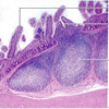
What is pictured here; what layer are they found in?

Peyer’s Patches; found in the Mucosa
What is encircled by the blue box; found most abundantely where?

Plicae Circulares; most abundant in jejunum and proximal ileum
Label A-C

A - Vili
B - Lamina Propria
C - muscularis mucosae
*At the bottom of the Vili lies the crypt with the bolded c.

Label the arrows from top to bottom

Top: Lumen of crypt
Stem Cell
Paneth Cell
Enterodendocrine cell

Label A-C

A) Circular layer
B) Myenteric Plexus
C) Longitudinal layer

What cell type are the arrows pointitng to?

Goblet Cells
What is the arrow labeled P; distinguishing feature?

Paneth Cells; Prominent Eosinophilic Apical granules

What are the role of Paneth cells?
Defensive funcion:
- Secrete lysozyme
- TNF-alpha
- Defensins
What is the structure denoted by E; distinguishing characteristic?

- Enteroendocrine (Neuroendocrine) Cells
- Cytoplasmic granules which are in a Subnuclear positon

What is the function of Enteroendocrine Cells?
Produce locally acting hormones that regulate GI motility and secretion:
- Gastrin
- CCK
- Secretin
What is the structure denoted by MF?

Stem Cell (Mitotic Factor)

What part of the small intestine is this and what are the distinguishing features?

- Duodenum
- Leaflike vili
- Brunner’s glands (submucosa)

What part of the small intestine is this and the distinguishing feature?

- Jejunum
- Finger-like vili

What part of the small intestine is this and what are the distinguishing features?

- Ileum
- Vili are short
- M cells
- Many Peyer’s patches in LP and submucosa

What are characterisitics of the large intestine mucosa?
- Simple columnar (microvili)
- NO vili
- Abundance of Enterocyttes and Goblet Cells
- Deep Crypts of Liebrkuhn
What are the arrows pointing to and where in GI tube are they found?

- Teniae Coli in Large Intestine (Muscuralis Externa)
Label these large intestine cell types from top to bottom.

- Enterocytes
- Goblet Cells
- Enteroendocrine Cells

What organ is this?

Veriform Appendix
What is this picture showing; how do you know?

Transition from the Retum –> Anal Canal
Rectum = Simple columnar w/ Goblet Cells
Anal Canal = Stratified Squamous
*Notice the tranisition at the pectinate line
Label all the arrows


What part of the GI tract is this, be specific?

Mucosa of the Large Intestine
What part of the GI tract is this and what is found here?

- Submucosa of the Large Intestine
- Location of Rectal Venous Plexus in the Distal Rectum
What is shown in this picture labeled A?

Taenia Coli

Microvili are only found where?
In the Small Intestine
What is the 1st, 2nd, 3rd, and 4th degree of folding within small intestine?
1st = Plicae Circularis
2nd = Villus
3rd = Crypts of Lieberkuhn
4th = Microvilli


