Histology Review Flashcards
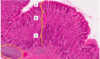
Identify this structure, what do they produce?

Mucous acini -> Mucous secretory product
What structure is the arrows pointing to and the yellow circles; what do each produce?

Arrows = Serous acini -> serous/watery secretory product
Yellow circle = Mucoserous acini -> Mixed secretory product

What organ is this?

Parotid gland

What organ is this?

Submandibular gland
*Both serous and mucus glands present

Acinus ↓
_________ (________epithelium) ↓
_________ (________epithelium) ↓
_________ (________epithelium) ↓
_________ (________epithelium) ↓
Main duct
Acinus ->
Intercalated duct (squamous-to-low cuboidal epithelium) ->
Striated duct (cuboidal-to-columnar epithelium) ->
Interlobular duct (pseudostratified columnar) ->
Lobar duct (stratified columnar) ->
Main duct
What structure is this; answer questions 1 and 2 as well.

- The esophagus
1) Outer layer of adventitia
2) Submucosa

- Identify this region
- What are the hallmarks

1) Pyloric stomach
2) Gastric pits and glands. The pits are wider and deeper as compared to other areas of the stomach

- What is this structure?
- What layer is it in?

- Myenteric plexus of Auerbach
- Muscularis Externa

Answer 1, 2, and 3?

- Muscularis externa
- Mucosa
- Gastroesophageal junctions

- Identify the organ (yellow arrows)
- Identify the structure (red arrow)

- Duodenum w/ Brunner’s glands
- Pyloric sphincter

- Identify the organ
- Identify the specific region of the organ shown

- Stomach
- Cardiac region indicated by tubular coiled glands

- Identify the structure in the red bracked
- Identify what the little red arrows are pointing to
- What lymphatic structure is found in the base of the structure indicated in questions 2

- Plicae circulares
- Vili
- Lacteals

Identify structure A and B, what type of epithelium in each?

A. Rectum proper w/ simple columnar epithelium
B. Anal canal w/ non-keratinized stratified squamous epithelium

- Identify the specific part of the GI tract
- What are the blue circles

- Duodenum
- Brunner’s Glands

- Identify the organ
- What specific region of the organ is shown

- Stomach
- Body/fundus

- Identify the organ
- How are the glands organized? What is the gland structure?

- Pancreas
- Branched tubuloacinar gland

- Identify the organ
- Identify the layer w/ red arrow, how does this layer change

- Large intestine
- Adventitia/Serosa -
- The ascending/descending colon and rectum = adventitia
- The transverse, sigmoid, and cecum = serosa

- Identify this organ, specific part
- How are the glands organized/gland structure

- Stomach (fundus/body region)
- Glands are simple tubular, branched glands
* Exception is the cardiac glands, which are simple tubular, coiled

- Identify the organ
- What are the red arrows pointing to

- Large Intestine
- Goblet cells

- Identify this organ
- What are the yellow circles showing

- Veriform appendix
- Lymphoid follicles/nodules

- Identify the organ
- What are the blue circles showing

- Ileum
- Peyer’s patches

- Identify regions A, B, and C + the type of cell in each region
- Identify the structure in the yellow circle

A) Pit = surface mucus cells
B) Neck = mucus neck cells and some parietal cells
C) Body = extensive parietal cells and stem cells
- Lower body region contains extensive chief cells and enteroendocrine cells
2. GALT
What is shown in the yellow circles?

Cardiac Glands!
Tubular w/ a coiled end

