Lecture 11 Flashcards

1
Q
A

Paramphistomosis

2
Q

Where do Paramphistomosis locate

A

Immature - duodenum/abomasum

Mature - rumen and reticulum

3
Q
  • Wha the intermediate host of Paramphistomosis
A

Snails from Planorbidae

4
Q

What is the distribution of Paramphistomosis

A

Depends on the presence of the planorbid snail

5
Q

How are animals infected with Paramphistomosis

A

Ingestions of grass with metacercaria

  • During rainy season
  • During dry ceason - snails take refuge in marshy areas
6
Q

Who is susceptible ot Paramphistomosis

A

Weaner cattle and lambs

7
Q

What is the pathogenesis of the immature stages of Paramphistomosis

A
  • Locate in the wall of the duodenum and the abomasum
8
Q

What do the immature stages feed on Paramphistomosis

A

Mainly tissue but also blood

9
Q

What damage do the immature stages of Paramphistomosis cause

A
  • Mechanic action/trauma by posterior sucker and movement
    • Erosion of the intestinal villi
  • Antigenic activity -> acute inflammatory reaction
10
Q

What do the immature stages of Paramphistomosis cause

A
  • Congestion and oedematous enteritis
  • Erosions and petechiae surrounded by necrotic zone
  • Atrophy of the villi
11
Q
A

Paramphistomosis

12
Q
A
13
Q

Where do the mature stages loacte of Paramphistomosis

A
  • In the rumen and reticulum
14
Q
A

Paramphistomosis

15
Q

What are the clinucal sings with Paramphistomosis

A
  • Heavy infection with immature parasites -> have clinical signs
  • Usually cilincal signs are seen on young animals
  • With immature parasites clinical signs can develop in 2-4 weeks
  • Anorexia, polydipsia, weight loss, fluid, foul smelling diarrhoea
  • Dehydration, weakness, anaemia, submundibular, oedema, death
  • Moderate infections will havr reduced weight gains or milk production or ill thrift
16
Q

How to diagnose Paramphistomosis

A
  • History
  • CS
  • Coproscopy: infection with immature parasites
    • Microscopic exam: NEGATIVE ( due to them being immature forms so they dont lay eggs)
    • Macroscopic exam: immature stages gan be fround in poo
    • Microscopic ecam: positive - sedimentation method
17
Q

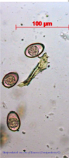
What do the eggs of Paramphistomosis look like

A

Ovoid with unequal poles, pale or geenish, smooth and thin shelled with an operculule at one end

18
Q
A

Paramphistomosis

19
Q

Treatment for Paramphistomosis

A
  1. Oxyclozanide
  2. Niclosamide
20
Q
A

Moniezia expansa

21
Q
A
22
Q

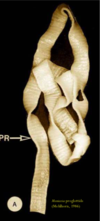
Explain the morphology of Moniezia spp

A
  • 4 suckers, no rostellum
  • 3-5m long and ip to 2cm wide
23
Q
A

Moniezia

24
Q
A

Moniezia

25
Q

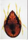
What is the lifecycle of Moniezia

A
  • Untermediate host: forage mites, commonly family Oribatidae
    • Defnitive host get infected by ingesting infected mites during grazing - PPT 4-6 weeks
26
Q
A
27
Q

Where is infection common with Moniezia

A

Young animals - the cyctocercoid larvae can survive as long as mites remain alive

28
Q

What are the CS in lambs with Moniezia

A

Lambs with heavy infection: diarrhoea, constipation, anaemia, wieight loss

29
Q

How to diagnose Moniezia

A
  • Finding proglottids/eggs in the faeces
30
Q

How to treat Moniezia

A
  • Praziquantal
  • Benzimidaziles
31
Q
A

Bovine Coccidiosis

32
Q

How many species of Eimeria

A

13

33
Q

How do you get infected with Eimeria

A
  • Ingestion of sporulates oocysts
34
Q

Explain the lifecycle of Eimeria

A
  • Infection by ingestion of sporulated oocysts
    • Asexual - immature schizont -> matture schizont with merozoites
    • Secual development
  • Sporogony/sporulation: in the environment needs moisture, proper remp and oxygen -> sporocysts and sporozites develop inside the oocysts -> oocysts become infective
35
Q

What are resistant with Eimeria

A

Oocysts

36
Q
A

Eimeria

37
Q

What does Eimeria usually affect

A

3 week to 6 month old

38
Q

What are the predisposing factors for Eimeria

A
  • Crowded animals
  • Poor nutrition and husbandry, stress, inter-current diseases - Clinical disease
39
Q

What is the source of infection for Eimeria

A
  • 2-3 monts infected calves: highesdt excretion rates
  • Adults with light infections migh be important source of infection with oocysts for young animals
40
Q

What is the pathogenesis of Eimeria

A
  • Species of the parasites
  • Infective dose
  • Stress, age
  • Pathogenesis assoicated woth gamonts and oocysts in the lower ilium, caecum and colon
41
Q

What is th epathology of Eimeria

A
  • Congested intestinal mucosa, catarrhal enteritis
  • Petechial hemorrhange -> diffuse hemorrhage
42
Q

What are the clinical signs with Eimeria

A
  • Diarrhoea, sometimes with blood, anaemia, weight loss, anorexia, dehydration, weakness
  • Tenesmus, rectal prolapse
  • Mild: yellow diarrhoea with blood
  • Nervous coccidiosis
43
Q

How to diagnose Eimeria

A
  • CS, history, age of animals
  • Detection of large number of oocysts in faeces
  • At early stages oocysts may not be present
44
Q

How to prevent Eimeria

A
  • Minimise the exposure of younf animals to sporulated oocysts: slatted floors, water tanks with overflow prevention control, elevates feed bunkers, dry bedding/environment
  • Avoid stress, overcrowding, maintain hygene
  • Penned animals should be of similar age
  • Administer a prophylactic drug
45
Q

How to treat Eimeria

A
  • Toltrazuril
  • Diclazuril
  • Lasalocid
46
Q

Where are clinical infections of Eimeria more common

A

Intensive husbandry -> husbandry deficiencies and stress -> infection with large numbers of oocysts -> clinical disease

47
Q
A

Eimeria - Unsporrulated oocysts are found in fresh faeces

48
Q

What are the CS signs of Eimeria in sheep

A
  • Profuse watery diarrhoea, sometimes containing blood or sheds of mucous membrane, tenesmus
49
Q
A
50
Q

Cryptosporidium spp location

A
  • Brush border of epithelial cells
  • Intracellular but extra-cytoplasmic
51
Q

Cryptosporidium parvum location

A
  • Ileum and proximal proximal portions of the large intestine
  • Parasites of neonatal ruminants
  • ZOONOTIC
52
Q

How do you get infected with Cryptosporidium parvum

A
  • Ingestion of oocysts
53
Q

What does Cryptosporidium parvum cause

A
  • Villous atrophy
  • Villous fusion
  • Crytp hyperplasia
  • Disruption of the intestinal microvilli
  • Infiltration of inflammatory cells
  • These changes result in malabsorption and secretory diarrhoea
54
Q

How to diganose Cryptosporidium parvum

A
  • CS
  • History
  • Detecton of oocysts in the faeces
  • Staining techniques
55
Q
A

Cryptosporidium spp

56
Q

How to treat Cryptosporidium spp

A
  • Halofuginone
57
Q
A