LEC 8: Connective Tissue - 08.21.2014 Flashcards

1
Q

From which primary germ layer does connective tissue originate

A

Mesoderm

2
Q

Connective tissue

A

connects, binds together, and supports other tissues and organs

3
Q

What are the two (2) components of connective tissue

A
  1. cells
  2. extracellular matrix
4
Q

What are the four (4) functions of connective tissue

A
  1. structural support
  2. medium of exchange
  3. defense and protection
  4. storage of fat
5
Q

What are the three (3) components of extracellular matrix

A
  1. ground substance
  2. fibers
  3. structural glycoproteins
6
Q

What are the two (2) components of ground substance

A
  1. glycoaminoglycans (GAGs)
    * old term: acid mucopolysaccharides
  2. Proteoglycans
7
Q

Proteoglycan

A

core protein to which molecules of glycosaminoglycans (GAGs) are covalently bound

8
Q

What is this structure and where is it found

A

Proteoglycan; found in ground substance of extracellular matrix

9
Q

Structural glycoprotein

A

globular protein molecules to which branched chains of monosaccharides are covalently attached

10
Q

Types of glycosaminoglycans (GAGs)

A
  1. Hyaluronic aicd
    * not sulfated or bound to a protein
  2. Chondroitin sulfate
  3. Dermatan sulfate
  4. Heparan sulfate
  5. Keratan sulfate
    * 2-5: highly negative charges on these molecules
11
Q

GAGs

A

GAGs are covalently bound to a core protein; together, the core protein plus the GAGs make up a proteoglycan

12
Q

Which GAG is **not **sulfated or bound to a protein

A

Hyaluronic acid

13
Q

What are the four (4) GAGs that have negative charges

A
  1. Chondroitin sulfate
  2. Dermatan sulfate
  3. Heparan sulfate
  4. Keratan sulfate
14
Q

What are the principal fiber types of connective tissues

A
  1. collagen fibers
  2. reticular fibers
  3. elastic fibers
15
Q

How many types of genetically distinct collagen are there

A

28

16
Q

What is the difference between Types I/II/III collagen and Type IV collage

A

Type I/II/III collagen form fibrils, while Type I does note

17
Q

Where is Type I collagen found

A
  • tendon
  • ligaments
  • bone
  • fibrous cartilage
  • dermis of skin
18
Q

Where is Type II collagen found

A
  • hyaline cartilage
  • elastic cartilage
19
Q

Where is Type III (reticular) collagen found

A
  • lymphoid organs
  • muscle cells
  • blood vessels
  • liver
  • endocrine glands
20
Q

Where is Type IV collagen found

A
  • basement membranes of epithelium, endothelium, muscle, and nerve axons
    • do not form fibrils
    • form mesh-like structure
21
Q

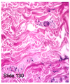
H&E stain of collagen fibers

A

Collagen fibers stained acidophilic (pink)

22
Q

Mallory Trichrome Stain of collagen fibers

A

Collagen fibers are stained blue (Type I and III collagens)

23
Q

Properties of collagen fibers (Type I/II/III)

A
  1. mechanical support
  2. confer great strength to the tissue
  3. resistance to stretching when pulled
24
Q

Fibroblasts have which two organelles in large quantities for synthesis of procollagen

A
  1. RER
  2. Golgi bodies
25
Procollagen
* Procollagen is produced and secreted into extracellular matrix * Procollagen then converted into tropocollagen * Tropocollagen is then polymerized into fibrils
26
Silver Stain of Type III collagen (reticular fibers)
* Type III collagen are stained black (agryrophilic) * Reticular fibers are thinner than Type I collagen * Reticular fibers are 6-12% hexose sugar residues and are PAS positive for this reason NB: Type I collagen is 1% hexose sugar residues
27
What composes basement membrane of an epithelial cell
1. type IV collagen 2. GAG (heparan sulfate) 3. laminin 4. entactin
28
Characteristics of Type IV collagen
* do not form fibrils, but a mesh-like structure * PAS positive
29
Two (2) components of elastic fiber
1. elastin (desmosine and isodesmosine -- amino acid derivatives) 2. microfibrils (fibrillin)
30
Describe this tissue
Elastic fibers (connective tissue spread preparation) * fibers are thin and branched, forming irregular networks
31
Properties of elastic fibers
* stretchable as rubber (amino acid derivatives desmosine and isodesmosine) * can stretch 150% of length without breaking and when tension released, snap back (eleastic recoil)
32
What three (3) structures are responsible for producing elastic fibers
1. fibroblasts (connective tissue) 2. smooth muscle cells (in blood vessel wall, especially of arteries) 3. chondroblasts and chondrocytes in elastic cartilage
33
Classification of Connective Tissues
A. Connective tissue proper 1. Loose connective tissue 2. Dense connective tissue * Dense irregularly arranged * Dense regular arranged B. Specialized connective tissue 1. Reticular tissue 2. Elastic tissue 3. Adipose tissue C. Embryonic connective tissue 1. Mesenchyme 2. Mucous
34
What are the two (2) types of connective tissue proper
1. Loose connective tissue 2. Dense connective tissue * Dense irregularly arranged * Dense regular arranged
35
What are the three (3) types of specialized connective tissue
1. reticular tissue 2. elastic tissue 3. adipose tissue
36
What are the two (2) types of embryonic connective tissue
1. mesenchyme 2. mucous
37
What type of tissue is this and what are its characteristics
Loose Connective Tissue (Connective tissue proper) * more cells than collagen fibers * most tissues and organs * lamina propria * largely Type III collagen
38
What type of tissue is this and what are its characteristics
Dense irregular connective tissue * more collagen fibers than cells * largely Type I and Type III collagens * some elastic fibers * found in dermis of skin
39
What tissue is this and what are its characteristics
Dense regular connective tissue * parallel bundles of collagen fibers separated by linear rows of fibroblasts * Type I collagen * Found in tendons, ligaments, aponeurosis NB: aponeurosis are layers of flat, broad tendons; primary function is to join muscles and the body parts the muscles act upon, whether it be bone or muscle
40
What type of tissue is this and what are its characteristics
Reticular tissue * composed of loose network of Type III collagen (remember, Type III collage = reticular fibers) * forms supportive stroma for tissues and organs
41
Where is reticular tissue most abundant
Reticular tissue is most abundant in lymphoid organs, like the lymph node or the spleen
42
What kind of tissue is this and what stain is used
Resorcin fuchsin stain, elastic arteries (elastic tissue)
43
What kind of tissue is this and what stain is used
Resorcin fuchsin stain, elastic cartilage (elastic tissue)
44
What kind of tissue is this
Adipose tissue (white/yellow); adipocytes (fat cells) contain one large single fat droplet
45
Characteristics of adipose tissue (fat)
* found in subcutaneous tissue, omentum (abdomen), mesenteries, breast, and bone marrow * thermal insulation -- poor heat conductor * storage of energy in form of triglycerides * release of free fatty acids to distant sites as energy source * visceral fats = obesity
46
What kind of tissue is this and where is it found
Mesenchyme * mesenchymal cells are stellate or spindle shape * delicate branching cytoplasmic processes surrounded by ground substance * unspecialized -- can differentiate into almost all cell types found in mature connective tissues
47
What kind of tissue is this and where is it found
Mucous (mucoid) connective tissue; Wharton's Jelly * mucuous connective tissue is a type of embryonic connective tissue * found in umbilical cord
48
stroma
the connective tissue within an organ
49
parenchyma
the cells supported by the stroma
50
What are the eight (8) major cell types in connective tissue (especially loose connective tissue)
1. fibroblasts 2. mast cells 3. macrophages 4. lymphocytes 5. plasma cells 6. neutrophils 7. eosinophils 8. adipocytes
51
What kind of tissue is this and what cells are found here
Connective tissue, with fibroblast cells * fibroblasts have spindle shape with elongated nucleus * have slightly basophilic cytoplasm (blue) * fibroblasts charged with continuous slow turnover of ECM (collagens, elastic fibers, ground substance)
52
What kind of tissue is this and what cells are noticeable
Connective tissue, mast cells * single nucleus * strongly acidic cytoplasm * finely granular
53
What are the abundant granules in the mast cell cytoplasm
1. heparin 2. histamine NB: Histamine released from mast cells trigger allergic reaction "hayfever" (rx: antihistamines)
54
monocytes
precursor of macrophages (hence, monocyte-macrophage system)
55
What are the six (6) cells that make up the monocyte-macrophage system
1. Macrophages 2. Microglia 3. Osteoclasts 4. Kupffer cells 5. Langerhans cells 6. Dendritic cells
56
Where are macrophages found
connective tissue, lungs, lymphoid organs, bone marrow
57
Where are microglia found
CNS (brain, spinal cord)
58
Where are osteoclasts found
bone
59
Where are Kupffer cells found
Liver
60
Where are Langerhans cells found
Skin
61
Where are dendritic cells found
Lymphoid nodes
62
What is this cell and what are its characteristics
Monocyte in blood circulation, precursor of macrophage * monocytes develop in the bone marrow and circulate in the blood * after leaving circulation into the connective tissue, monocyte differentiates into a macrophage
63
What are the characteristics of a macrophage
* lysosomes (primary, secondary, tertiary) * lysosomal enzymes like acid phosphatase * phagocytosis of cell debris, inert materials, bacteria, damaged ECM * secrete cytokines * antigen presenting cells * processing and presentation of antigents to lymphocytes (T cells); component of immune system
64
Describe this schema
Macrophage is an antigen presenting cell; processes antigen (Ag), presents to lymphocyte (T cell) and T cell activates
65
What kind of tissue is this and what cells are visible
Macrophages in connective tissue showing phagocytic particles in cytoplasm
66
What kind of tissue is this and what cells are visible
Lymphocytes in blood circulation * develop in the bone marrow * T lymphocytes * B lymphocytes * leave the circulation and enter the connective tissue
67
What kind of tissue is this and what cells are visible (X, Y); how are they related
Loose connective tissue * X = lymphocytes (B lymphocytes) that differentiate into plasma cells that make antibodies (live 1-2 yrs) * Y = Plasma cells make antibodies (live 2-3 wks)
68
What are antibodies and how are they made
(also called immunoglobulins) are glycoproteins * RER = synthesis of the glycoproteins * Golgi = glycosylation of the glycoproteins NB: antibodies do not aggregate into secretory granules
69
What kind of tissue is this and what cells are visible
Neutrophils in blood circulation * multi-lobed nucleus (3-5 lobes) * slightly pinkish cytoplasm containing barely visible granules * approx. 6 hours in blood circulation
70
What kind of tissue is this and what cells are visible
Neutrophils in loose connective tissue * acute infection * amoeboid movement * phagocytic cells * lysosomal enzymes * terminally differentiated cells (spend a few days in CT)
71
Example of dirty splinter
* in response to accute inflammation, increased number neutrophils leave blood circulation * migrate to site of bacterial infection * phagocytize bacteria, microorganisms, cell debris in poorly oxygenated environment (inflamed or necrotic tissue) * defunct neutrophils = pus cells
72
What kind of tissue is this and what cells are visible
Eosinophil in blood circulation * bilobed nucleus * strongly eosinophilic granules in cytoplasm * increased number in blood in parasitic infections and in allergy reaction
73
What kind of tissue is this and what cells are visible
Loose connective tissue, presence of eosinophils * bilobed nucleus * highly eosinophilic granules in cytoplasm * chronic inflammation * phagocytic Ag-Ab complex * Lysosomal enzymes * approx 2 weeks in tissue