Lab 10 and Chapter 14: Brain and Cranial Nerves Flashcards
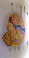
What do the various lobes of the brain do?
Frontal lobe: motor cortex (voluntary movement of skeletal muscle)
Occipital lobe: visual
Temporal: auditory and olfactory
Parietal: somatosensory (general senses-touch,pain, pressure, tempature)
Insula lobe: gustation
What is the white matter and the gray matter in the cerebral hemispheres?
White matter: tracts that connect the two hemispheres (corpus callosum)
- deep to cortical gray matter (opposite to spinal cord)
- Composed of tracts (bundles of axons) that connect one part of brain to another and to spinal cord
Gray matter: cell bodies (soma), dendrites, synapses, and unmyelinated fibers for decisions, memory, and sensation (and analysis of sensation)
- form cortex of cerebrum and cerebellum
- forms basal nuclei
- found in limbic system
What are the 6 main parts of the brain?
- Cerebrum
- conscious thinking
- Corpus Callosum
- white matter that connects the two cerebral hemispheres
- Cerebral cortex
- outer layer of the cerebrum
- Diencephalon
- Thalamus
- Hypothalamus
- Epithalamus (pineal gland)
- Optic chiasma
- Brain Stem
- midbrain
- pons
- medulla oblongata
- Cerebellum
- cerebral cortex (gray matter)
- arbor vitae (branched white matter)
What are the functions of the parts of the diencephalon?
- thalamus
- gateway to cerebral cortex
- fluid filled space between the two halves = 3rd ventricle
- hypothalamus
- connects to pituitary gland by infundibulum
- Autonomic control center: includes thirst, hunger, satiety centers and temperature regulator
- epithalamus /pineal gland
- secretes melatonin
- optic chiasma
- where cranial nerve II (optic nerve) crosses
What is the function of the medulla oblongata?
- includes the respiratory center (for breathing)
- and cardiovascular center (for HR and blood pressure)
Where is the olfactory nerve and what does it do?
Number I
Smell (sensory only)
Travels through ethmoid and cribriform plate
Where is the optic nerve and what does it do?
Number II
Vision
Sphenoid bone, optic canal
Where is the oculomotor nerve and what does it do?
NUmber III
Movement of eyeball
Superior orbital fissure (sphenoid bone)
Medial rectus, superior rectus, inferior rectus, inferior oblique muscles
What is the trochlear nerve and what does it do?
Number IV
Movement of eyeball
Sphenoid bone, superior orbital fissure
Superior oblique muscle
Where is the trigeminal nerve and what does it do?
Number V
Sensations of the face, chewing
Masseter, temporalis
Where is the abducens nerve and what does it do?
Number VI
Movement of eyeball (laterally)
Superior orbital fissure (sphenoid bone)
Lateral rectus
Where is the facial nerve and what does it do?
Number VII
Facial expression;taste
Frontalis, occipitalis, orbicularis oculi, obicularis oris, levator labii superioris, depressor labii inferioris, zygomaticus, risorius, mentalis, buccinator
Where is the vestibulocochlear nerve and what does it do?
Number VIII
equilirbrium; hearing
Where is the glossopharyngeal nerve and what does it do?
Number IX
Taste, movement of pharynx during swallowing and speech, secretion of saliva
Where is the vagus nerve and what does it do?
Number X
Taste, swallowing, coughing, parasympathetic stimulation of the heart and digestive tract
Where is the accessory (spinal) nerve and what does it do?
Number XI
Swallowing, movement of head and shoulders
Sternocleidomastoid; trapezius
Where is the hypoglossal nerve and what does it do?
Number XII
Movement of tongue during speech and swallowing
Genioglossus
What do the terms rostral and caudal mean?
Rostral: toward the forehead
Caudal: toward the spinal cord
What are the major components of the cerebrum?
- 83% of brain volume
- Longtudinal fissure: deep groove that separates cerebral hemispheres
- Gyri: thick folds
- Sulci: shallow grooves
- Corpus callosum: white matter that connects the hemispheres
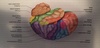
What are the major landmarks of the cerebellum?
- Also has gryi, sulci, and fissures
- 10% of brain volume
- over 50% of brain neurons
What are the components of the cranial dura mater?
- 2 layers
- outer periosteal
- inner meningeal
- layers separated by dural sinuses (collect blood circulating through brain)
- No epidural space in cranium
- Extensions of dura mater
- falx cerebri separates two cerebral hemispheres
- Tentorium cerebelli separates cerebrum from cerebellum
- Falx cerebelli separates right and left halves of cerebellum
WHat is meningitis?
- Inflammation of the meninges
- Serious disease of infancy and childhood (3 mo-2 years especially)
- Caused by bacterial or viral infection of CNS by way of nose and throat
What are the ventricles of the brain?
- Four internal chambers in the brain
- Two lateral ventricles: one in each cerebral hemisphere
- Interventricular foramen connects to 3rd ventricle
- Third ventricle is narrow medial space below corpus callosum
- Cerebral aqueduct runs through midbrain and connects third to 4th ventricle
- Fourth ventrilce is in chamber between pons and cerebellum
- connects to central canal that runs through spinal cord

What is choroid plexus? How do they relate to Ependyma?
- mass of blood capillaries on the floor of each ventricle
- Ependyma–neuroglia that line ventricles and cover chloroid plexus
- produces CSF
What is Cerebrospinal Fluid and how does it work?
- clear, colorless liquid that fills ventricles and canals of CNS
- Production begins with flitration of blood plasma through capillaries of brain
- CSF continually flows around the CNS
- 3rd and 4th ventricles add more CSF during cycle
- small amount fills central canal of spinal cord
- All CSF eventually escapes through three pores into subarachnoid space
- CSF is reabsorbed by arachnoid villi (granulations)
What are the functions of CSF?
- buoyancy
- protection
- prevents brain from striking the cranium
- shaken child syndrome and concussions do still occur
- chemical stability
- rinses metabolic waste, regulates chemical environment
- blood substitute
- rinses metabolic waste, regulates chemical environment
Why is blood important to the brain
- Brain is only 2% of adult body weight but receives 15% of all blood
- Neurons have high demand for ATP, and therefore oxygen and glucose, so blood supply is critical
- 10 second interruption of blood flow may cause loss of consciousness
- 4 minutes = irreversible brain damag
- 10 second interruption of blood flow may cause loss of consciousness
What is the brain barrier system and how does it work?
- System regulates what substances can get from bloodstream itno tissue fluid of the brain
- blood is crucial but can contain harmful agents
- Two points of entry must be guarded
- blood capillaries through brain tissue
- Consists of tight junctions between endothelial cells that form capillary walls
- capillaries of choroid plexus
- forms tight junctions between ependymal cells
- blood capillaries through brain tissue
- Anything leaving blood must pass through cells (not between) so harmful substances don’t get to brain tissue
What is permeable to the Brain Barrier system?
- Highly permeable to water, glucose, and lipid soluble substances
- coffee, alcohol, oxygen, carbon dioxide, nicotine, and anesthetics
Where is the brain barrier system vulnerable?
- trauma and inflammation an damage BBS and allow pathogens to enter brain tissue
- Circumventricular organs–in 3rd and 4th ventricles (no BBS)
- blood has direct access to brain
- enables brain to monitor and respond to fluctuations in blood glucose, pH, osmolarity, etc.
- CVOs afford a route for invasion by HIV
Describe the functions of the medulla oblongata.
- Begin at foramen magnum
- Slightly wider than spinal cord
- all ascending and descending fibers connecting to brain/spinal cord pass through here
- Contains pyramids (and their decussations): large motor tracts from cerebrum to spinal cord
- most neurons decuss here!!!
- Autonomic functions
- cardiac and vasomotor (blood pressure) center
- respiratory center
- reflex centers for swallowing, sneezing, couching, and vomiting
What are cerebellar peduncles?
Thick stalks on posterior pons that connect it (and the midbrain) to the cerebellum.
What is the function of the midbrain?
- regulates auditory and visual reflexes
- startle reflex
- Contains cerebral aqueduct
What is the reticular formation?
- Loose web of gray matter that runs vertically through all levels of the brain stem
- Has connections with many areas of cerebrum
- Functions:
- somatic motor control
- adjust muscle tension for tone, balance, posture
- central pattern generators for breathing and swallowing
- Cardiovsacular centers of medulla oblongata
- Pain modulation
- some pains signals ascend through here
- some descending analgesic pathways begin here
- sleep and consciousness
- alerts cerebral cortex to awaken from sleep; maintains alertness
- Habituation
- filters out reptitive and weak stimuli (99%)
- somatic motor control
What is the function of the cerebellum?
- highly important in motor coordination
- Aids in learning motor skills
- Maintains muscle tone and posture
- LEsions can cause ataxia
- clumsy, awkward gait
- WHat are the functions of the hypothalamus?
- controls and integrates activities of the Autonomic NS and the Endocrine System
- Controls the pituitary gland
- Produces hormones
- REgulates rage, aggression, pain, pleasure, and arousal
- Hunger, thirst, and satiety centers
- Thermoregulation
- Regulates daily patterns of sleep
What is the function of the pineal gland?
- secretes melatonin during darkness
- promotes sleepiness and sets biological clock
What is the function of the cerebrum?
- Seat of sensory perception, memory, thought, judgement, and voluntary motor actions
What is the limbic system and its components?
- Emotional or affective brain
- Cingulate gyrus
- aids in expressing emotions
- Hippocampus
- memory functions
- Amygdala
- emotion functions
- Compnents are connected through loop of fiber tracts
- somewhat circular pattersn of feedback
- Structures have centers for both gratification (pleasure/reward) and aversion (fear/sorrow)
- There is a limbic system in each cerebral hemisphere
What is the basal nuclei?
- Masses of cerebral gray matter buried deep in the white matter, lateral to the thalamus
- Control large automatic movements of skeletal muscles
- unconscious body movements and facial expressions)
- Uses dopamine
What are the integrative functions of the brain?
- Higher brain functions: sleep, memory, cognition, emotion, sensation, motor control, and language
- Involve interactions between cerebral cortex, basal nuclei, brainstem, and cerebellum
- Do not have easily defined anatomical boundaries
- Focus on the cerebrum, but involve action of multiple brain levels
What is an Electroencephalogram?
- monitors surface electrical activity of the brain waves
- used for studying normal brain functions such as sleep and consciousness
- diagnoses degenerative brain diseases, metabolic abnormalities, brain tumors, etc.
- lack of brain waves is common criteria of brain death
What are the 3 functional areas of the cerebral cortex?
- Motor areas: control voluntary movement
- Sensory areas: conscious awareness of sensation
- Association areas: integrate diverse information
- Conscious behavior involves the entire cortex working together
What is cognition?
- The range of mental processes by which we acquire and use knowledge
- sensory perception, thought, reasoning, judgement, memory, imagination, and intuition
- Accomplished by distributed association areas of the cerebral cortex
- about 75% of all brain tissue
What is emotion?
- Emotional feelings and memories are interactions between prefrontal cortex and diencephalon
- Prefrontal cortex: seat of judgement, intent, and control over expression of emotions
- fully matures at about age 25
- Feelings: (fear, etc.) arises from the hypothalamus and the amygdala
What makes up sensation in the brain?
- primary sensory cortex: sites where sensory input is first received and one becomes conscious of the stimulus
- Association areas: near primary sensory areas process and interpret that sensory information
- Multimodal association areas: receive input from multiple senses and integrate this into an overall perception of our surroundings
What are the special senses?
- senses that require their own special organ
- Vision
- primary cortex
- association area: makes cognitive sense of visual stimuli
- Hearing
- Primary auditory cortex
- association area: recognizes spoken words, familiar piece of music, or voice on the phonw
- Equilibrium
- signals for balance and sense of motion: to cerebellum
- Association cortex
- lateral sulcus near the lower end of the central sulcus
- consciousness of our body movements and orientation (balance) in space
What are the general senses?
- aka somesthetic, somatosensory, or somatic
- distibuted over entire body and employ simple receptors
- Touch, pressure, stretch, movement, heat, cold, and pain
- Primary somatosensory cortex: provides awareness of stimulus
- Somatosensory association area: makes cognitive sense of stimulus
- Brought in by cranial nerve (ascending tracts)> thalamus processes input from contralateral side > parietal lobe
What is motor control?
- Motor association area of frontal lobes
- where we plan our behavior (e.g. intention to contract a muscle)
- Where neurons compile a program for muscle contraction
- Program sent to the precentral gyrus (primary motor area)
- neurons send signals to the brainstem>descending tracts in spinal cord> skeletal muscles
- Basal nuclei and cerebellum also help
What are Wernicke’s and Broca’s areas?
- Wernicke’s area
- usually in left hemisphere
- permits recognition of spoken and written language
- when we intend to speak, formulates phrases and transmits plan of speech to Broca’s
- Broca’s area
- usually in left hemisphere (frontal area–goes to muscle movement)
- generates motor program for the muscles of the larynx, tongue, cheeks, and lips for speaking and hands when signing
- transmits program to primary motor cortex for movement
- Affective language area
- usually in right hemisphere
- lesions produce aprosody: flat, emotionless speech

What is cerebral lateralization in the human brain?
- Left hemisphere
- specialized for spoken and written language
- sequential and analytical reasoning
- math and science
- breaks info into fragments and analyzes it
- Right hemisphere
- seat of imagination and insight
- musical and artistic skill
- perception and patterns and spatial relationships
- comparison of sights, sounds, smells, and taste

What does the corpus callosum do in cerebral lateralization?
- allows two hemispheres to communicate and coordinate their functions
- transfers/integrates motor, sensory, and even cognitive information between the two hemispheres
- If damaged, info processed in one hemisphere cannot reach the other
- Symptoms: poor coordination, trouble with complex problem solving skills, seizures, developmental delays, trouble swallowing or feeding, social immaturity
What are the cranial nerve pathways?
- motor fibers of cranial nerves begin in nuclei of brainstem and lead to glands and muscles
- Sensory fibers begin in receptors located mainly in the head and neck and lead mainly to the brainstem
- most cranial nerves carry fibers between brainstem and ipsilateral receptors and effectors
- exceptions: optic nerve and trochlear nerve
What are the cranial nerve classifications?
- Sensory (I, II, and VIII)
- Motor (III, IV, VI, XI, and XII)
- Mixed (V, VII, IX, X)