L7 - Pelvis & Perineum Flashcards
The pelvic cavity is continuous with the abdominal cavity and divided into two regions
Name them and their contents:
False [greater] pelvis
- superior region related to upper parts of the pelvic bones
- generally considered part of the abdominal cavity
True [lesser] pelvis
- inferior parts of the pelvic bones, sacrum, and coccyx
- has an inlet and an outlet

What is the area inferior to the pelvic inlet?
A. True Pelvis
B. False Pelvis
A. True Pelvis

The Pelvis
- Pelvic inlet
- Pelvic walls (2 muscles)
- Pelvic floor (diaphragm)
- Pelvic Outlet (orfices coming out of pelvic diaphragm)
Contain Elements of the:
- urinary
- gastrointestinal
- reproductive systems
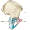
Name the Pelvic Bones
- Ilium
- Ischium
- Pubis
Converge into acetabulum (femur articulates there)
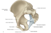
Name the purple starred structures

ASIS
Ischial Spine
Ischial Tuberosity

What is the sacral promontory?
In between the 5th lumbar and 1st sacral vertebrae
- More prominent in males

Where do the spinal nerves of the sacrum come out of?
Anterior sacral foramina
Posterior sacral foramina

What connects the sacrum to the L5?
zygapophysial joints
- connects adjacent vertebrae
- specifically between the L5 and the sacrum
Joints
- zygapophysial joints
- Sacro-iliac joints

Name the main ligaments of the pelvis
Main ligaments:
- Anterior Sacro Illiac ligament
- Lumbosacral ligament
- Iliolumbar ligament

What is the common site of fractures,
also known as
the weakest point in the pelvis
Superior/Inferior Pubic Ramus
Superior/Inferior Ischial Ramus
= Ishiopubic Ramus

Name the differences between women and men
pelvis size, shape, and projections
Female:
- Pelvic inlet is more circular
- Wider infrapubic angle (80-85 degrees)
- Less prominent Ischial Spine
- Less prominent Promontory
Male:
- Pelvic inlet is narrower and heart-shaped
- Narrower infrapubic angle (50-60 degrees)
- More prominent Ischial Spine
- Prominent Promontory (sacral promontory)

The pelvic inlet contains 5 structures, name them
PAAPP
- Promontory of sacrum
- Ala of sacrum
- Linea terminalis [Arcuate line]
- Pecten pubis/pectineal line
- Symphysis pubis

All of the following are a part of the linea terminalis EXCEPT:
A. Pubic Crest
B. Arcuate Line
C. Pectin Pubis
D. Ala of sacrum
D. Ala of sacrum
Linea terminalis = Arcuate line, Pecten pubis, Pubis crest
What are the contents of the Pelvic Wall?
- Sacrum (bone)
- Coccyx (bone)
- Piriformis (muscle)
- Obturator Internus (muscle)
- Sacrospinous Ligament & Sacrotuberous Ligament
- Pelvic bones inferior to linea terminalis

What are the ligaments that form the pelvic wall?
Sacrospinous Ligament:
- Sacrum to the ischial spine
Sacrotuberous Ligament:
- Sacrum to the ischial tuberosity
These ligaments stabilize the sacrum on the pelvic bones by resisting the upward tilting of the inferior aspect of the sacrum

What are the muscles that make up the pelvic wall?
Piriformis
- Piriformis goes through the GSF
Obturator internus
- Obturator Internus goes through the LSF
Both:
- External Rotators
- medial surface of greater trochanter of femur

How many Apertures are in the pelvic wall and what are they?
Three major apertures:
- the obturator canal
- Greater sciatic foramen
- Lesser sciatic foramen
Obturator canal –> obtrurator vessels
GSF –> Piriformis
LSF –> Obturator internus
What is the name of the following structure in green?
A. Promontory of Sacrum
B. Pubic Crest
C. Pectin Pubis
D. Arcuate Line
E. Margin of Ala

D. Arcuate Line

True or False
The superior gluteal nerve and vessels are inferior to the piriformis
False
The superior gluteal nerve and vessels are superior to the piriformis

Structures in relation to the Piriformis
Superior:
Superior Gluteal Nerve, Artery & Vein
Inferior:
Sciatic Nerve, Inferior Gluteal Nerve & Internal Pudendal Vessels
True or False
Obturator Vessels run through the obturator canal
True
Name the structure in green
A. Ischial Tuberosity
B. Ischial Spine
C. AIIS
D. PIIS

B. Ischial Spine

What is the Pelvic floor formed by?
- Pelvic diaphragm
- Perineal membrane
- Deep perineal pouch
The pelvic floor separates the pelvic cavity, above, from the perineum, below.

The pelvic diaphragm
Formed by funned shape muscles
Levator anni
- Contributes to the formation of the pelvic floor
which supports the pelvic viscera
- Maintains _90-degre_e angle between the rectum and anal canal
- Reinforces the external anal sphincter and, in women, functions as a vaginal sphincter
Coccygeus muscle
(not in levator anni)
- supports the pelvic viscera
- pulls coccyx forward after defecation
Name the levator anni muscles in the Pelvic Diaphragm
Levator Ani Muscles
- Puborectalis
- Pubococcygeus
- Iliococcygeus
Coccygeus Muscle

Where is the tendinous arch found?
Tendinous Arch:
Fascia of Obturator Internus and Levator Ani
(Specifically Iliococcygeus)

Which of the following muscles are within the pelvic wall?
A. Piriformis
B. Coccygeus
C. Obturator Internus
D. A and C
E. All of the above
D. A and C
Piriformis & Obturator Internus –> muscles within the pelvic wall
Coccygeus –> part of the pelvic diaphragm
Perineal membrane and Deep perineal pouch
Perineal membrane
- a thick fascial structure attached to the bony framework of the pubic arch
Deep perineal pouch
- contains a layer of skeletal muscle and various neurovascular elements

What does the perineal membrane and adjacent pubic arch
do for the roots of the external genitalia and the muscles associated with them?
The perineal membrane and adjacent pubic arch
provides attachment for the roots of the external genitalia and the muscles associated with them

True or False
The urethra penetrates vertically through a circular hiatus in the perineal membrane
True

skeletal muscle functions as a sphincter
Anteriorly
- muscle fibers surround the urethra and collectively form the
external urethral sphincter
(important in voluntary urinating)
1. Sphincter Urethrovaginalis
2. Compressor Urethrae
(found only in female)
= facilitate the closing of the urethra
deep transverse perineal muscle

The perineal body is on the side of the perineal membrane
The perineal body is on the posterior side of the perineal membrane
What are the muscles within the Deep Perineal Pouch?
Does the female and male have the same muscles within the deep perineal pouch?
Males:
Deep Transverse Perineal Muscle
External Urethral Sphincter
Females:
Deep Transverse Perineal Muscle
External Urethral Sphincter
Compressor Urethrae
Sphincter Urethrovaginalis
What structure is this?

Piriformis
- goes through the GSF
What structure is this?

Obturator Internus
- Goes through the LSF
What structure is this?

Coccygeus
What structure is this?

Levator Ani
What are the major arteries of the Pelvis: Internal Iliac
Anterior Trunk:
- up to 8 arteries
- differ in males and females
Supplies viscera: urinary bladder, internal and external genitalia, rectum, gluteal region
Posterior Trunk:
3 arteries
Supplies the parietal tissues: pelvic walls and gluteal region

True or False
Most of the pelvic organs are supplied by the anterior trunk, not the posterior trunk
True
What are the 3 arteries that come off of the Posterior Trunk?
P: Posterior Trunk
I: Iliolumbar Artery
L: Lateral Sacral Artery
S: Superior Gluteal Artery
Iliolumbar Artery
- One common trunk with two branches:
Iliac and Lumbar
Supplies:
Iliacus, Psoas major, quadratus lumborum muscles, and cauda equina
Lateral Sacral Artery
Supplies:
the piriformis muscle and vertebral canal
Superior Gluteal Artery
Exits pelvis through Greater Sciatic Foramen, superior to the Piriformis muscle
Supplies Gluteal muscles and Tensor Fascia Latae

What are the starred structures?

- Iliolumbar
- Lateral sacral
- Superior gluteal
Remember:
P: Posterior Trunk
I: Iliolumbar Artery
L: Lateral Sacral Artery
S: Superior Gluteal Artery
What are the 8 arteries that come off of the anterior trunk?

Anterior Trunk
- Pudendal Artery (Internal)
- Umbilical Artery –> Superior Vesical Artery (Lower Yield)
- Middle Rectal Artery
- Vesical Artery (Inferior)
- Obturator Artery
- Vaginal Artery (Females)
- Uterine Artery (Females)
- Gluteal Artery (Inferior)
Me: “A Person Used My Van On Vegas’s Unstable Grounds”

Anterior Trunk
Obturator artery
- Travels through the obturator canal, accompanied by the obturator nerve and vein. It supplies the muscles of the thigh’s adductor region.
Umbilical artery
- Gives rise to the superior vesical artery, which supplies the superior aspect of the urinary bladder.
Inferior vesical artery
- Supplies the lower aspect of the bladder. In males, it also supplies the prostate gland and seminal vesicles.
Vaginal artery (female)
- Descends to the vagina, supplying additional branches to the inferior bladder and rectum
Uterine artery (female)
- ascends along the lateral aspect of the uterus. At the fallopian tubes, it anastomoses with the ovarian artery.
- During its course, it crosses the ureters superiorly.
Middle rectal artery
- Travels medially to supply the distal part of the rectum. It also forms anastomoses with the superior rectal artery (derived from the inferior mesenteric) and the inferior rectal artery (derived from the internal pudendal)
Internal pudendal artery
- exit the pelvis via the greater sciatic foramen
- Accompanied by the pudendal nerve, it then enters the perineum via the lesser sciatic foramen
- It is the main artery responsible for the blood supply to the perineum.
Inferior gluteal artery
- The terminal branch of the anterior trunk.
- It leaves the pelvic cavity via the greater sciatic foramen, emerging inferiorly to the piriformis muscle in the gluteal region.
- It contributes to the blood supply of the gluteal muscles and hip joint.

Minor arteries of the pelvis do NOT come from the internal iliac
Gonadal Arteries
- The ovarian and Testicular.
- They arise from the abdominal aorta, distal to the origin of the renal arteries
Superior Rectal Artery
- terminal continuation of the inferior mesenteric artery.
Median Sacral Artery
- originates from the posterior aspect of abdominal aorta
All of these are branches off the posterior trunk of the internal iliac artery EXCEPT:
A. Iliolumbar Artery
B. Superior Gluteal Artery
C. Lateral Sacral Artery
D. Inferior Gluteal Artery
D. Inferior Gluteal Artery
Inferior Gluteal Artery comes off the anterior trunk
Somatic: Voluntary
Autonomic: Involuntary
Voluntary –> muscle
Involuntary –> heart rate

Sacral Plexus

These nerves provide somatic innervation to pelvic and lower limb structures
Pink: Ventral Division
This will become the tibial part of the sciatic nerve
Yellow: Dorsal Division
This will become the common fibular part of the sciatic nerve

What is the Pudendal Nerve of the Sacral Plexus formed by?
- Formed by anterior rami of S2-S4
- Innervates structures of the perineum
- provides sensory and motor function to the:
external genitalia
external urethral sphincter
external anal sphincters
S2,S3,S4 Keeps POOP Off the Floor

Pudendal Nerve
Dorsal Nerve of Penis:
Somatic ONLY
Deep Perineal Nerve:
Muscles
Superficial Perineal Nerve –>
Posterior Scrotal Nerve:
Mostly Sensory

Visceral plexuses
Paravertebral sympathetic chain
Via ventral rami
- lower limb and perineum
Via sacral splanchnic nerves
- Pelvic organs and erectile tissue
Sympathetic:
Sympathetic Trunk → Gray Rami Communicantes → Ventral Rami → Lower Limb and Perineum
Sympathetic Trunk → Sacral Splanchnics → Inferior Hypogastric Plexus → Pelvic Organs & Erectile Tissue
Superior Hypogastric Plexus → Inferior Hypogastric Plexus
Parasympathetic:
Ventral Rami → Pelvic Splanchnics → Inferior Hypogastric Plexus

Pelvic extensions of the prevertebral plexus
- carry sympathetic, parasympathetic, and visceral afferent fibers
Superior hypogastric plexus
Hypogastric nerves
pelvic splanchnic nerves carrying preganglionic parasympathetic fibers from S2 to S4
Inferior hypogastric plexus

- rectal plexus
- uterovaginal plexus
- prostatic plexus
- vesical plexus

Sympathetic fibers
- innervate blood vessels
- cause contraction of smooth muscle in the internal urethral sphincter in men and the internal anal sphincters in both men and women
- cause smooth muscle contraction associated with the reproductive tract and with the accessory glands of the reproductive system
- important in moving secretions from the epididymis and associated glands into the urethra to form semen during ejaculation.
Sympathetic = ejaculation
Parasympathetic fibers
- are generally vasodilatory,
- stimulate bladder contraction,
- stimulate erection
- modulate activity of the enteric nervous system of the colon distal to the left colic flexure (in addition to pelvic viscera, some of the fibers from the pelvic plexus course superiorly in the prevertebral plexus, or as separate nerves, and pass into the inferior mesenteric plexus of the abdomen).
Parasympathetic = erection
True or False
Parasympathetic = ejaculation
Sympathetic = erection
False
Sympathetic = ejaculation
Parasympathetic = erection
The inferior hypogastric plexus has sympathetic innervation coming from all EXCEPT:
A. Superior Hypogastric Plexus
B. Sacral Splanchnic Nerves
C. Pelvic Splanchnic Nerves
D. A and B
C. Pelvic Splanchnic Nerves
Pelvic Viscera
- Gastrointestinal
- Genitourinary
- Reproductive
Rectum
- most posterior element of the pelvic viscera
- anorectal junction is pulled forward (perineal flexure)
by the action of the puborectalis

Rectum
Rectum has three lateral curvatures:
- Upper and lower curvature to the right
- Middle curvature to the left
- Lower part of the rectum is expanded to form the rectal ampulla
Urinary system
The pelvic parts of the urinary system consist of the
- terminal parts of the ureters
- bladder
- Proximal part of the urethra

Urinary Bladder
Empty bladder is shaped like a three-sided pyramid
It has:
Apex
- directed toward the top of the pubic symphysis;
- Median umbilical ligament [urachus]
Apex of Bladder → Median Umbilical Ligament (Urachus)
Base
Trigone
- superior surface
- two inferolateral surfaces.
- Neck
Top corners of Trigone:
- Openings of the ureter
Bottom Corner:
- Internal Urethral Sphincter

True or False
Detrusor muscle contracts and pushes urine from the bladder into the urethra
True

Which of the following muscles are NOT a part of the Levator Ani Muscles?
A. Iliococcygeus
B. Pubococcygeus
C. Puborectalis
D. Coccygeus
D. Coccygeus
Neck of bladder
most “fixed” part.
It is anchored into position by a pair of tough fibromuscular bands
Pubovesical → female
Puboprostatic → male

Fascia of Denovilliers =
Rectoprostatic Fascia (Males)
Rectovaginal Fascia (Females)

Blood supply, lymph drainage, and nerve supply of bladder
Arteries
- Superior and inferior vesical arteries
Veins
- Vesical veins
Lymph drainage
- Internal and external iliac nodes
Superior Vesical Artery comes off
the Umbilical Artery
Sympathetic & Parasympathetic:
Inferior Hypogastric Nerves
Autonomic innervation of bladder
- The parasympathetic nerve (pelvic splanchnic nerve originating from S2–S4) stimulates to contract the musculature (detrusor) of the bladder wall, relaxes [inhibitory] to the internal urethral sphincter, and promotes emptying.
- The sympathetic nerve relaxes the detrusor of the bladder wall and constricts the internal urethral sphincter.
Visceral afferents follow the pelvic splanchnic into the spinal cord
Parasympathetic (Pelvic Splanchnic)
→ Contract Detrusor → Relax Internal Urethral Sphincter
Sympathetic
→ Relax Detrusor → Contract Internal Urethral Sphincter
Which curvature of the rectum is pointing to the LEFT?
A. Upper Curvature
B. Middle Curvature
C. Lower Curvature
D. A and C
B. Middle Curvature
Urethra in Males vs Females

Females:
- 4cm Long
- Anterior to Vaginal Orifice
- Surrounded by external urethral sphincter
Males:
- 20cm Long
Preprostatic:
- 1cm; Surrounded by Internal Urethral Sphincter

Male Urethra
Prostatic:
4cm; Urethral Crest turns into → Seminal Colliculus
Prostatic ducts secrete into → Prostatic Sinuses
Ejaculatory Ducts (2) secrete into → Seminal Colliculus
Prostatic Utricle located in Seminal Colliculus
Membranous:
Passes through deep perineal pouch
Surrounded by external urethral sphincter
Spongy:
Surrounded by corpus spongiosum
Forms bulb @ base; forms the navicular fossa at the glans penis
Which portion of the male urethra is surrounded by the internal urethral sphincter?
A. Preprostatic
B. Prostatic
C. Membranous
D. Spongy
A. Preprostatic
Membranous –> external urethtra


