Kolbe: Respiratory Pathophysiology 1 Flashcards
(17 cards)
1 kPa = 7.5 mmHg
What is the definition of Respiratory Failure
“hypoxic or Hypercapnic”
- When the lungs fail to adequately oxygenate the arterial blood and/or fail to prevent undue CO2 retention
In practical terms: 1kPa = 7.5mmHg
PaO2 < 8kPa (60 mmHg) (Hypoxic,Type I )
PaCo2 > 6.6 kPa (50 mmHg) (Hypercapnic, Type II )
What causes Hypoxaemia?
- Reduced PiO2
- Hypoventilation
- V/Q mismatch
- R-L shunt
- Diffusion

What determines your partial pressure of oxygen at each level?

- Partial Pressure of inspired oxygen?
- Dependent on altitude and fractional inspired O2
- Air = 0.21
- Athletes train in hypobaric chambers
- Supplemental O2 in clinical arena
- Dependent on altitude and fractional inspired O2
- What influences Palveolar O2 ? (If we disregard humidification of the air)
- Ventilation! Particularly alveolar ventilation
- Which can be determined by the partial pressure of pCO2 in the arterial blood
- Ventilation! Particularly alveolar ventilation
What is it that influences the gap between the pO2 alveolus and the pO2 arteriole blood ??

If we had a perfect lung these two values would have equilibration over time, and there would be no gap.
Therefore the gap gives us an indication as to how well (or not well) the lungs are working.
This is the A-a gradient that measures gas exchange
Geas exchange is measured as an A-a gradient and is influenced by?

- VQ mismatch
- R-L shunt
- Diffusion
if you have purely hypoventilation then you will have no Aa gradient as gas exchange is fine.
Hypercapnia is due to?
- (Alveolar) Hypoventilation
- PaCO2 is inversely proportional to alveolar ventilation, which is why alveolar hypoventilation is viewed as an increase in alveolar CO2
- Minute ventilation = alveolar ventilation + dead space ventilation (therefore you can have an increase in minute ventilation but a decrease in alveolar ventilation)
- (V/Q mismatch)

Causes of Hypoxaemia?
- Reduced Pi O2
- Hypoventilation
- V/Q mismatch
- Diffusion
- R-L shunt
What is the relationship of PaCo2 and alveolar ventilation
Hypoventilation = Raised PaCo2

PaCO2 is inversely proportional to alveolar ventilation, which is why alveolar hypoventilation is viewed as an increase in alveolar CO2
Case: A 17 year old NZ European woman is brought to the Emergency Department by ambulance.
She was previously fit and well.
She is unconscious and was found on the floor by a friend. You have no other history.
Her ABG shows PaCO2 = 7.5 kPa ( NR 4.5 - 6.0).
Why is she hypercapneic?
- Depressed Respiratory drive due to generalised cerebral depression as a result of narcotics/ drugs
Scenario: Mr Smith is now aged 75yrs. At the age of 20 yrs, he contracted poliomyelitis and spent many months in an “iron lung”. He subsequently developed marked kypho-scolosis.
He has put on 25kgs in the last 2 years.
He now presents with worsening shortness of breath, reduced exercise tolerance and morning headache. His ABG shows PaCO2 = 7.5 kPa ( NR 4.5 - 6.0).
- The poliomyelitis impacted the anterior horn cells that control the Respiratory muscles
- Level C3,4,5: the diaphragm
- Kyphoscoliosis: lateral distortion of the spine
- The Poliomyelitis caused decreased anterior horn cells impair NM transmission
- Twisted spine and obesity causes increased Respiratory load
- He has Fibrotic Lung disease from working in asbestos, where his interstitial tissue walls have thickened
- This fibrous tissue of additional collagen is not just laid down in the airways, but in the space between the alveolus and the capillary: ‘Alveolar-Capillary Block’
- Increased load due to decreased chest wall compliance, decreased drive (some from polio and may already have low drive and inc weight has exposed this)
What are the 3 components of things that impact alveolar ventilation?
How can these things cause hypercapnia?
- Respiratory drive: how much output from brainstem causing us to breathe, what are the adverse impactors on that
- Decreased Respiratory drive
- Neuromuscular transmission: need this to be uninterrupted, can be impacted at any point
- NM incompetence
- Muscle weakness/fatigue
- Load: in the form of resistive work and elastic work of breathing. These may be altered
- Abnormal load
In order to measure gas exchange, we measure the Alveoar-arterial Oxygen gradient, how is this done?

If perfect, PAO2 = PaO2

- PaO2 is measured
- PAO2 is estimated.
( at sea level, breathing air ∝ 20 kPa -PaCo2/0.8**
A-a Gradient = 20 - PaCO2/0.8 -PaO2)
(Normal A-a gradient = 1-2 kPa)
Pb = atmospheric pressure
**remember for every molecule of O2 there is not an equal number of CO2 molecules. Therefore PACO2 /0RQ is the Respiratory exchange ratio
What does Ficks Law tell us?

Fick’s Law: V gas = A/T . D . (P1 – P2) *pie sign* ( D = Sol/ MW )
That diffusion of a gas depends on the area, the thickness (alveolar-capillary block), the driving pressure and the solubility/molecular weight of the gas
Total difusion depends on a membrane component and a capillary component
Series Resistance: 1/ D Lco = 1/Dm + 1/ O . V capill

Fibrotic Lungs is a type of ___________ disease
Interstitial Lung Disease
- affects the loose CT of the lungs that surround alveoli, blood vessels
- in this case, thickening by collagen of the walls.
- Would potentially hear bilateral fine inspiratory crackles
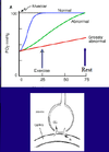
What does this graph show us and how does this explain Mr Smiths elevated symptoms during exercise?

- Top - - - - - line is the ‘perfect’ lung
- Mixed venous PO2 (churned up in the right heart) is our normal lung, which reaches equilibrium with the alveolar gas at a consistent rate over a fairly short amount of time (~0.75). it equilibrates with a large amount of time to spare
- With fibrotic lung disease like Mr Smith has, the time taken to reach equilibrium would have extended over time. Initially, he would have still reached equilibrium.
- As the disease progresses, he gets to a point where his PO2 is diminished/abnormal even at rest.
- When he exercises, or even walks across the room, and his O2 uptake increases (3-4x and more) and he need to increase his cardiac output. This increases the transit time of RBC across the capillary and his PO2 is extremely low.

What gas do we use to measure diffusion and why?
Carbon monoxide (CO)
Lung Function Labs use: DLCO
CO is
- Diffusion (and not perfusion) limited
- soluble
- avidly binds to Hb, so no back pressure (PCO in cap is zero)

If normal diffusion is dependent on (gas, diffusion distance/thickness, surface area, (Hb), capillary volume) then what can cause abnormal diffusion?
- Alveolar-Capillary Block
- Diffuse Lung disease (interstitial LD)
- Loss of Diffusing Surface
- emphysema (Elastase and other proteases destroy the fine tissue)
- Capillary Volume/ hemoglobin
- Pulmonary hypertension/ Pulm. vascular disease
- Anaemia


