Inflammation pt.2 (pictures) Flashcards
in what type of inflammation is this seen?
what happens in this type inflammtion?
is the fluid infected?

serous inflammation (type of acute inflammation)
exudation of cell poor fluid into spaces created by cell injury, or into body cavities
no, the fluid is not infected
what is this?

serous inflammation
Note: separation of epidermis from dermis to produce a blister cavity filled with serous (clear) fluid
what type of inflammation is seen here?
when is it seen?
what is seen ?

fibrinous inflammation (type of acute inflammation)
in severe injuries
increased vascular permeability and fibrinogen escapes while fibrin forms and deposits in extracellular space
what type of inflammation is seen here?

fibrinous inflammation (pink part is exudate)
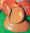
what type of inflammation is seen here?
what is this type of inflammation?
where do we see this type of acute inflammation?

Purulent (suppurative) inflammation in gallbladder
there is production of large amount of pus (neutrophils, necrotic cells and edema fluid)
examples: acute appendicitis, abscesses
what do we see in the picture?
what is the definition of what is in the picture?

ulcer
discontinuity of lining epithelium, tissue necrosis, and resultant inflammation
what is this picture?

Ulcer
Micro: note discontinuity in epithelial lining. Ulcer.

