Hemodynamic disorders pt. 2 (pictures) Flashcards
what is being shown?
Virchow triad: Endothelial Injury
- ulcerated plaques in atherosclerosis
- inflammatory vascular injury (vasculitis)
- heart chamber following MI
- exposes subendothelium
what is this?

Aneurysm: abnormal dilatation of blood vessel
what is this?
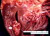
Arrow: adherent thrombi at the apex of (L) ventricle
what is this?

thrombus formed within heart or aorta, show lamination: ‘lines of Zahn’
what is this?

Pulmonary thromboembolism: note the presence of a large thrombus sitting astride the pulmonary bifurcation
what is this?

Chest X-ray in Pulmonary embolism: Hampton’s hump
what is this?

Chest X-ray in Pulmonary embolism: Hampton’s hump
what is this?

fat embolism
Note presence of fat microglobules within pulmonary vessel
what is this?
what stain is this positive for?

fat embolism
oil red o stain positive
what is this?

Pulmonary vessel filled with squames in amniotic fluid embolism
what is this?

Amniotic fluid embolism: Note: laminated squames within pulmonary vessels.
what is this?

Red Infarct in heart: This pattern of infarct is associated with reperfusion injury.
Remember: classically in MI, you find ‘pale’ infarct.
what is seen here?

Septic infarction: infective “vegetations” on heart valve
