Cell injury 3 (pictures) Flashcards
What stage of necrosis is this?

pyknosis
What stage of necrosis is this?

Karyorrhexis
What stage of necrosis is this?

karyolysis
what type of necrosis is this? (what is distinctive?)

coagulative necrosis
tombstone appearance
what type of necrosis is this?

coagulative
what type of necrosis is this?
Where?

coagualtive necrosis in heart (myocardial infaction)
what type of necrosis is this?
what is distinctive?
coagulative necrosis
infarction (localized coagulative necrosis)
what type of necrosis is this?

coagulative necrosis of the kidney
what type of necrosis is this?

coagulative necrosis of the myocardium (MI)
what type of necrosis is this?

liquefactive necrosis (lung showing abscess )
what type of necrosis is this?

Liquefactive necrosis: Cerebral abscess

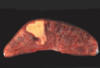
what type of necrosis is this?

Dry Gangrene: note “line of demarcation”
what type of necrosis is this?
Bowel ischemia: note darkened segments of bowel
(Wet gangrene)
what type of necrosis is this?

wet gangrene
what type of necrosis is this?

caseous necrosis
what type of necrosis is this?

caseous necrosis

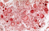
what type of necrosis is this?

fat necrosis
Red arrow: necrosis of fat cells. Black arrow: inflammatory cells.
what type of necrosis is this?

fat necrosis: pancreas

what type of necrosis is this?

fibrinoid necrosis
fibrinoid necrosis usually occurs on what body part? how?
blood vessels through antigen-antibody complexes that deposit
what type of necrosis is this?

fibrinoid necrosis
what type of necrosis is this?

Fibrinoid Necrosis: Autoimmune disorder: Rheumatoid Arthritis

where is this?
What is to be noticed here

Liver: apototic cells. Note, cell shrinkage, pyknotic nuclei. These cells may appear larger, due to retraction artifact (paleness around these cells). Retraction artifact happens while tissue processing.
what is being acummulated in this picture?
what stain can be used to visualize fat?
when do we have increased fatty tissue in liver?
tryglycerides in liver
oil red O
causes: Reye’s synd., acute fatty liver of pregnancy, Jamaican vomiting disease, drugs.