Histology-Female Repro Flashcards
Identify the structures indicated below

*

Identify the structures indicated below:

Note that the ovarian follicles are in the stroma of the cortex and surrounded by epithelial cells

What structures are indicated below?

Primordial follicles: note the squamous cells surrounding the ovum
What structures are indicated below?

Growing follicles: note the prominent nucleolus surrounded by the zona pellucida. Granulosa cells are cuboidal and become multi-layered in this phase.

What structures are indicated in the image below?

This is a vesicular ovarian follicle. Note the antrum full of hyaluronic acid.

What structures are indicated in the image below?

This is a mature/Graafian follicle

What structure is indicated below?

Atretic ovarian follicle: note that most follicles degenerate this way and it can happen anytime during development. Note pyknotic granulosa cell nuclei in the antrum and a hyaline (glassy) scar that replaces the atretic follicle.
What structures are indicated in the ovary below?

Note that granulosa cells and theca cells contribute to formation of the corpus luteum.

What would cells in this image look like on higher power?

Cells of the corpus luteum have the typical appearance of steroid secreting cells w/ mitochondria + tubular cristae, lipid droplets and abundant sER.

What are the layers of the uterine tube?
Note the many infoldings of the mucosa.

What is this?

Corpora hemorrhagica
What happens to the mucosa of the uterine tube as you approach the uterus?
They get smaller. Note the asterisk marks the lumen of the oviduct.

What cells are found in the uterine tube epithelium? How do these cells change during the cycle?
1) Ciliated cells (move oocyte down tube) and 2) Peg cells (secrete nutrients). The epithelium will be taller with more ciliary cells during the luteal phase.

What are the different layers of the uterus? What stage is this section from the uterus in?

Myometrium and endometrium (with stratum basalis and functionalis). Note large, straight endometrial glands and high amount of stroma indicating the uterus is in the proliferative stage. Note that during the secretory stage the endometrium has crinkled, dilated glands, edematous stroma and ragged shedding epithelium

What would this region of the cervix look like on higher power?

Zone of transition forms stratified squamous epithelium in vagina and simple cuboidal in cervix

What type of secretions come from these cervical clands in the periovulatory period?

Watery. Mucus post ovulatory b/c you don’t want sperm to enter if fertilization has already occurred.
What are the layers of the vagina?
Mucosa, lamina propria and muscularis

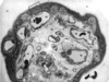
What would EMG of these structures look like?

These are all tertiary chorionic villi. You would have intervillous space (1), maternal blood (2), syncytiotrophoblasts (3), cytotrophoblasts (4) and fetal blood (5).
What is the difference between inactive and active breast tissue?
Inactive breast will have interlobular ducts with very few acini (top image). Active breast will have increased acini within each lobule leading to ducts (bottom image).

What type of gland is the ovary?
Endocrine & Exocrine
What about the follicular cells (granulosa cells) makes diffusion of nutrients to the ovum easy?
There are no tight junctions between them and they sit on the basement membrane, allowing nutrients to diffuse freely in and waste to diffuse freely out.

What structures are responsible for producing transudate in the vagina and erection during sexual intercourse?
CT indentations w/superficial blood vessels = transudate. Deeper vessels = erectile tissue.

What structures are indicated in the image below?

*

What structures are indicated in the image below?

*

