Histology - Cellular Structure & Biomechanics Block (I) Flashcards
What are the three basic visualization methods of histology?
Light microscopy,
fluorescent microscopy,
electron microscopy

What are three of the most common stains of light microscopy?
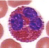
Hematoxylin and eosin (H&E);
periodic acid-Schiff (PAS);
Masson’s trichrome
What is the most common stain of light microscopy?
Hematoxylin and eosin (H&E)
What are the two principal colors of hematoxylin and eosin staining?
Blue and pink

What color is hematoxylin?
What color is eosin?
Blue-ish
pink-ish
In hematoxylin and eosin (H&E) staining, what are some structures that will be stained blue?
The nucleus;
keratohyalin granules and calcified material

In hematoxylin and eosin (H&E) staining, what are some structures that will be stained pink?
Cytoplasm, collagen, lewy bodies, and mallory bodies
The blue-pink differences in structures stained with H&E are largely due to what characteristic of the tissue being stained?
The relative pH
(acidic - nuclear components;
basic - proteins)
Hematoxylin and eosin staining is a common stain in what modality of histology?
Light microscopy
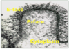
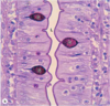
What is the periodic acid-Shiff (PAS) stain used for in light microscopy?
Staining specific cellular compartments, carbohydrates
(Note: the histology in the image is counterstained with hematoxylin)

What stain is often used as a counter-stain with PAS to illustrate the nuclei?
Hematoxylin
What are three substances often visualized with a periodic acid-Schiff (PAS) stain?
What color are they stained?
Mucins, glycogen, glycocalyx;
pink or magenta
(Note: the histology in the image is counterstained with hematoxylin)

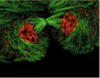
Which stain is especially good for connective tissue and collagen visualization?
Masson’s trichrome
What science is the 3D interpretation of 2D cross sections of materials or tissues?
Sterology

What is the most common histological stain?
What stain is useful for visualizing the nucleus in blue and surrounding cytoplasm/collagen in pink?
What stain is useful in visualizing cellular compartments and carbohydrates?
What stain is useful in visualizing muscle fibers as red, collagen as blue, cytoplasm as pink, and the nucleus as dark brown?
Hematoxylin & eosin (H&E);
hematoxylin & eosin (H&E);
periodic acid-Schiff (PAS);
Masson’s trichrome
For what structures is the hematoxylin & eosin (H&E) stain useful? What colors are these structures stained?
The nucleus (blue) surrounding cytoplasm/collagen (pink)
For what structures is the periodic acid-Schiff (PAS) stain useful? What colors are these structures stained?
Cellular compartments and carbohydrates (magenta)
(Note: Hematoxylin is often used as a counterstain when PAS is used)
For what structures is the Masson’s trichrome stain useful? What colors are these structures stained?
Muscle fibers (red),
collagen (blue),
cytoplasm (pink),
the nucleus (dark brown)

For what structures is the hematoxylin & eosin (H&E) stain useful? What colors are these structures stained?
For what structures is the periodic acid-Schiff (PAS) stain useful? What colors are these structures stained?
For what structures is the Masson’s trichrome stain useful? What colors are these structures stained?
The nucleus (blue) surrounding cytoplasm/collagen (pink)
Cellular compartments and carbohydrates (magenta)
Muscle fibers (red), collagen (blue), cytoplasm (pink), the nucleus (dark brown)
What histological stain is used in this image?

Hematoxylin & eosin (H&E)
What histological stain(s) is(are) used in this image?

Periodic acid-Schiff
(with hematoxylin counterstain)
What histological stain is used in these images?

Masson’s trichrome
What color are erythrocytes in H&E staining?
Intensely red
What study involves the interpretation of these 2D images to understand their 3D orientation and structure?

Sterology
What are the three main portions of the cell?
Cell membrane;
nucleus;
cytoplasm
What are the three main portions of the nucleus?
The nuclear envelope (and pores);
chromatin (eu- and hetero-);
the nucleolus
Are there lipids, proteins, and/or carbohydrates in the cell membrane?
All three
What are three common types of inclusions found in the cytoplasm?
Fat droplets;
glycogen granules;
lipofuscin granules
What is a common type of protein found in the cytosol?
Enzymes
(hundreds and hundreds of types)
What are the three main contributors to the cytoplasm?
Cytosol, organelles, and inclusions
What is the external surface of the cell membrane called?
What is the internal surface of the cell membrane called?
E-face
P-face
What two lipid types are found in high concentrations in lipid rafts?
Cholesterol;
glycosphingolipids
What is the function of lipid rafts in cell membranes?
To stabilize protein clusters and keep them in close proximity to one another
(this allows for efficient inter-protein interactions)
What structure stabilizes protein clusters within the cell membrane and allows for close, more efficient inter-protein interactions?
Lipid rafts
Where are glycolipids found in the cell membrane?
The E-face
What purpose does cholesterol serve in the lipid membrane?
Increased rigidity / stiffness
What are the three main colors of the Masson’s trichrome stain?
Red (keratin/muscle);
pink (cytoplasm);
blue or green (collagen)
On which surface of the cell membrane is spectrin found?
The P-face (protoplasmic)
On which surface of the cell membrane are the ECM proteins found?
The E-face (exoplasmic)
What term describes the bilipid cell membrane characteristic in which the associated lipids have hydrophobic and hydrophilic sections?
Amphipathic
What are the three main types of lipid found in the plasma membrane?
Phospholipids;
cholesterol;
glycolipids
How many main types of phospholipid are found in the lipid membrane?
5
Name the three phospholipid types found on the P-face of the plasma membrane.
Phosphatidylserine (PS)
phosphatidylethanolamine (PE)
phosphatidylinositol (PI)

Name the two phospholipid types found on the E-face of the plasma membrane.
Phosphatidylcholine (PC);
sphingomyelin (SPH)

What are the two types of cell membrane protein?
Integral and peripheral
Transmembrane and lipid-anchored proteins of the cell membrane are ________ proteins.
Integral
How do bilipid membranes appear on electron microscopy?
As trilaminar layers
(dark outer layers with a central light area)
The term glycocalyx encompasses what two types of structure?
Glycolipids and glycoproteins
What are some structures making up the dark layers of the plasma membrane as seen on electron microscopy?

Phospholipid heads;
integral proteins;
glycolipids
What are some structures making up the light layer of the plasma membrane as seen on electron microscopy?

Phospholipid tails;
integral proteins;
cholesterol
True/False. The E-face and P-face layers of the plasma membrane each have the same types of phospholipid in similar concentrations?
False
(the E-face has different phospholipids from the P-face)
What modality must be used to directly visualize the cell membrane?
Electron microscopy
Sphingomyelin is abundant on which surface of the cell membrane?
The E-face
Which enzyme breaks down sphingomyelin?
Where is this enzyme located?
Acid sphyngomyelinase;
the P-face
Acid sphingomyelinase breaks down sphingomyelin inside what structures?
Lysosomes
Sphingomyelin is found in high concentrations on which surface of the plasma membrane?
Acid sphingomyelinase is found in on which surface of the plasma membrane?
E-face;
P-face
Is pinocytosis clathrin-dependent or -independent?
Clathrin-independent
Is phagocytosis clathrin-dependent or -independent?
Clathrin-independet
Is receptor-mediated endocytosis clathrin-dependent or -independent?
Clathrin-dependent
Which of the following is(are) clathrin-dependent?
Which is(are) actin-dependent?
Pinocytosis
Phagocytosis
Receptor-mediated endocytosis
Receptor-mediated endocytosis;
Phagocytosis
What is unique about phagocytosis when compared to pinocytosis and receptor-mediated endocytosis?
It is actin-dependent
What is unique about receptor-mediated endocytosis when compared to pinocytosis and phagocytosis?
It is clathrin-dependent
Retrograde vesicular transport from the golgi apparatus to the ER is associated with what surface protein?
COP - I
(1)

Anterograde vesicular transport from the ER to the golgi apparatus is associated with what surface protein?
COP - II
(2)

What is V-snare?
What is T-snare?
A vesicle-specific membrane protein;
a target membrane -specific protein
What are some examples of non-membranous organelles?
Ribosomes;
proteasomes;
cytoskeleton
Ribosomes are present on which side of the RER cisternae (interior or exterior)?
The cytoplasmic (exterior) side
Cytochrome P450 is present in large concentrations in what organelle of hepatocytes?
The SER
Which organelle of myocytes is used for calcium sequestration?
The smooth endoplasmic reticulum (a.k.a. the sarcoplasmic reticulum)
What family of enzymes is involved in phase I detoxification in the hepatocyte SER?
What family of enzymes is involved in phase II detoxification in the hepatocyte SER?
Cytochrome P450;
UDP - glucuronosyltransferases

In what organ is the highest concentration of SER in the body?
The liver
Which side of the Golgi apparatus faces the RER and receives vesicles from it?
The cis face (convex)

Which side of the Golgi apparatus releases vesicles and faces the plasma membrane?
The trans side (concave)

In what direction do proteins enter the Golgi apparatus (from the RER) and leave it?
RER –> cis Golgi apparatus –> trans Golgi apparatus

What enzyme is added to synthesized proteins in the RER and cis Golgi apparatus to mark these proteins as future lysosomal enzymes?
Mannose-6-phosphate

What is the importance of mannose-6-phosphate?
Where is it added to select proteins?
It marks lysosomal proteins;
the RER and cis Golgi apparatus

What protein controls vesicle movement from the cis Golgi apparatus to the trans Golgi apparatus?
What protein controls vesicle movement from the trans Golgi apparatus to the cis Golgi apparatus?
COP-II (anterograde);
COP-I (retrograde)

What are four locations to which the Golgi apparatus sends synthesized products?
Lysosomes,
secretory granules,
plasma membrane,
back to the RER
Where do both sulfation of certain tyrosine residues and addition of sialic acid to sugars occur?
The trans Golgi apparatus

Besides COP-I and COP-II, what other protein is important in Golgi apparatus vesicle formation?
(Hint: this protein is often involved in vesicles heading to lysosomes or the cell membrane)
Clathrin
Which of the four locations to which the Golgi apparatus sends vesicles (the cell membrane, lysosomes, secretory granules, and the RER) receive clathrin-coated vesicles from the Golgi apparatus?
Lysosomes;
secretory granules
What factor decides the relative abundance of mitochondria in a cell?
The energy demands of the cell
What types of nucleic acid are present in the mitochondrial matrix?
DNA, tRNA, mRNA, rRNA
What percentage of the proteins of the mitochondria are coded for by mitochondrial DNA?
< 1%
The ‘elementary particles’ visualized on the matrix-side of the mitochondrial cristae refer to what structures?
ATP synthase
Where is mitochondrial DNA (and associated RNA and ribosomes) within the mitochondria?
The mitochondrial matrix
Why would mitochondria be found in the cell periphery near the inner leaflet of the plasma membrane?
The membrane active transport requires huge amounts of ATP
The enzymes of the electron transport chain are found in what structure?
The inner mitochondrial membrane
What is the order of endosome formation after pinocytosis or receptor-mediated endocytosis?
Early endosome –> late endosome –> lysosome

(early becomes late, then fuses with a lysosome)
At what stage do vesicles of pinocytosis and receptor-mediated endocytosis enter the following pathway:
Early endosome –> late endosome –> lysosome
At what stage do vesicles of phagocytosis enter the above pathway?
At what stage do vesicles of autophagy enter the above pathway?
Early endosome;
late endosome;
lysosome

What is a primary lysosome?
What is a secondary lysosome?
A lysosome that has nothing to digest;
a lysosome that has fused with with a late endosome or phagosome or cytoplasmic organelle
What are residual lysosomal bodies?
Of what are they typically full?
The non-digestible remains of whatever was initially in the lysosome to be digested;
lipofuscin
What term refers to the lysosomal digestion of materials brought into the cell via endocytosis?
What term refers to the lysosomal digestion of old cellular organelles?
Heterophagy;
autophagy
What membrane-bound organelles are the site of intracellular digestion?
What membrane-bound organelles break down fatty acids and contains both myeloperoxidase and catalase?
What non-membrane-bound organelles destroy ubiquinated proteins?
Lysosomes;
peroxisomes;
proteasomes
Via what two methods are intracellular proteins destroyed?
Lysosomes (autophagy);
proteasomes (following ubiquination)
What two substances are often found in intracellular inclusions as a form of energy storage?
Glycogen (clumped granules);
lipids (spherical, single monolayer droplets)
What is the most common pigment of the body?
What pigment is common in old cells?
What pigment is related to the phagocytosis of RBCs and hemoglobin?
Melanin;
lipofuscin;
hemosiderin
Melanin, lipofuscin, and hemosiderin all appear somewhat similarly in histology.
They be differentiated according to what?
The type of tissue in which they are found
What is the term for the images produced by microscopy?
Micrographs
What type of electron microscope created this image?

(Note: the electrons did not go through the structure)
Scanning electron microscope
What type of electron microscope created this image?

(Note: the electrons did go through the structure)
Transmission electron microscope
What cellular structure(s) is(are) responsible for cell polarity, intracellular vesicular movement, and organelle placement/anchoring?
The cytoskeleton
What cellular structure (broadly speaking) is responsible for cellular shape, cell motility, and cellular adhesion?
The cytoskeleton
How big are each type of cytoskeleton (on average, in nm)?
Microfilaments - 7 nm
Intermediate filaments - 8-12 nm
Microtubules - 25 nm
What are some functions of microfilaments?
Cytoplasmic transport, cellular motility, cell shape, microvili support
(microfilaments are basically just actin)
From where do microtubules radiate throughout the cell?
The centrioles and axonemes
Describe where microfilaments are found in highest concentrations in the cell.
Cell wall, microvili

Describe where intermediate filaments are found in highest concentrations in the cell.
Throughout the cytoplasm

What type of cytoskeleton maintains nuclear shape?
Intermediate filaments
Describe where microtubules are found in highest concentrations in the cell.
Radiating throughout the cytoplasm from centrioles and axonemes

Which type of cytoskeleton is the most stable?
Intermediate filaments
Describe the general cytoskeleton.

Which type of cytoskeleton is responsible for chromosomal movement during mitosis?
Which type of cytoskeleton is responsible for cellular cleavage during mitosis?
Microtubules;
microfilaments
Describe some microfilament organizational patterns that have different roles in the cell.

Is actin a globular or filamentous protein?
Both;
it is filamentous when polymerized, but the polymer is made of small, globular monomers
What does it mean that actin is polar?
It has a plus end and a minus end
What are a few examples of the many types of intermediate filaments?
Myosin, spectrin, vimentin, lamin
What cytoskeletal component gives microvili projections their form?
What cytoskeletal component makes up the terminal web that anchor the microvili?
Microfilaments (and some myosin connecting them);
intermediate filaments

What is another name for the intermediate filament anchoring the base of microvili in place?
The terminal web

What intracellular intermediate cytoskeletal protein is especially relevant to the P-face of erythrocytes?
Spectrin
How does intracellular spectrin maintain cell membrane integrity?
It forms tetramers and anchors actin
In erthrocytes, what protein anchors the spectrin-actin complex to the cellular membrane?
Ankyrin
What protein carries vesicles down towards the plus end of microtubules?
What protein carries vesicles down towards the minus end of microtubules?
Kinesin;
dynein
The plus end of a microtubules is closer to the centriole or cell periphery?
The minus end of a microtubules is closer to the centriole or cell periphery?
Periphery;
centriole
Which globular tubulin monomer is on the plus end of a microtubule filament?
Which globular tubulin monomer is on the minus end of a microtubule filament?

What intermediate protein is especially present in lymphocytes?
What intermediate protein is especially present in all types of muscle?
What intermediate protein is especially present in astrocytes?
Vimentin;
desmin;
GFAP
What intermediate protein is especially important to connective tissue?
What intermediate protein is especially important to epithelial cells?
What intermediate protein is especially important to neurons?
Vimentin;
cytokeratin;
neurofilament
What intermediate protein is especially important to nuclear membranes?
What intermediate protein is especially important to some Schwann cells?
What intermediate protein is especially important to erythrocyte cell membranes?
Lamins A, B, and C;
GFAP;
spectrin
What cytoskeletal structure provides the basis of the mitotic spindle?
What cytoskeletal structure provides the basis of the cellular cilia?
What cytoskeletal structure provides the basis of the cellular flagella?
Microtubules;
microtubules;
microtubules
What two structures make up a single centrosome?
The centrioles
(at right-angles to one another)

Where is the centrosome typically located?
What are some other locations?
Near the nucleus

What is a nucleosome?
A single histone unit and its associated double-stranded DNA
The nucleolus is largest and most well-developed in what type of cells?
Highly transcriptionally active cells
Which of the following cell structures would stain with a basic dye after treatment with RNAse?
Chromatin
Nucleolus
SER
RER
Golgi
Chromatin
(the basic dye is binding to the acidic structure)
Is hematoxylin basic or acidic?
Basic
(it binds acidic structures)
What gives the nucleolus its deep blue color when stained with hematoxylin?
High concentration of ribosomes (RNA)
Which of the following would stain blue when stained with hematoxylin and eosin?
Chromatin
Nucleolus
RER
All three
All three
Which of the following would stain the deepest blue when stained with hematoxylin and eosin after treating the cell with RNAse?
Chromatin
Nucleolus
RER
All three
Chromatin
(the ribosomes and other RNA types of the RER and nucleolus have been degraded)
Which protein is responsible for cilia and flagellar movement?
Axonemal dynein
Addition of what chemical structure to dynein causes cilia or flagellar movement?
Phosphate
(from ATP)
Does axonemal dynein interact with the outer, inner or both microtubules found in cilia or flagella?
Outer
What three subunit proteins make up the ankyrin protein?
Band-3, ankyrin, band-4
Describe the axonemal arrangement of cilia and flagella.
Axoneme: 9 microtubule pairs surrounding two central microtubule filaments
(with inner and outer dynein arms)

Alpha-tubulin binds to what structure in the centriole?
Gamma-tubulin
As what are the individual tubulin filaments in a microtubule referred?
Protofilaments
One microtubule is made up of how many tubulin protofilaments?
One mature ciliar/flagellar axoneme is made up of how many microtubules?
13;
20 (9 pairs m. surrounding 2 central m.)
A centrosome is made of two perpendicular centrioles. How many microtubules make up one centriole?
9 triplets (27 total m.)
How many tubulin protofilaments would be found in one centrosome?
702
(13 protofilaments / microtubule x 3 microtubules / triplet x 9 triplets / centriole x 2 centrioles / centrosome)
What happens to the centriole during the beginning of cell division?
It replicates
This is an electron micrograph of the cross-section of what structure?

The axoneme of a flagellum or cilium

Which microtubule of any axonemal pair has the complete 13 protofilaments?
Which one is not complete and shares some of the complete one’s protofilaments?
A;
B

Which microtubule of an axonemal pair has attached inner and outer dynein?
Which microtubule of an axonemal pair has attached radial spokes?
A;
A

What is the name of the specialized centriole at the base of a ciliar or flagellar axoneme?
The basal body
The basal body of a flagellum or cilium is made of how many microtubules?
A centriole is made of how many microtubules?
9 triplets;
9 triplets
How will a transcriptionally active cell’s chromatin appear?
Large areas of euchromatin
Chromatin is made of what structure(s)?
DNA and proteins
What is the site of ribosomal synthesis?
What is the site of ribosomal assembly?
The nucleolus;
the nucleolus
The outer membrane of the nuclear envelope is continuous with what structure?
The RER
How thick are euchromatin strands?
How thick are heterochromatin strands?
11 nm;
30 nm

What is the name of the DNA between nucleosomes?
Linker DNA

Chromatids are connected at what point?
The centromere

A cell with a dark, splotchy nucleus is likely transcriptionally _______.
A cell with a light, clear nucleus is likely transcriptionally _______.
Inactive (+heterochromatin);
active (+euchromatin)
What is indicated by G in this electron micrograph of a nucleous?
What happens here?

Granular material;
initial ribosomal assembly
What is indicated by F in this electron micrograph of a nucleous?
What happens here? What is contained here?

Fibrillar material;
sites of active transcription; rRNA
What is indicated by FC in this electron micrograph of a nucleous?
What is contained here?

Fibrillar centers;
the DNA sequences for the rRNA genes, transcription factors
Where is DNA synthesized?
Where is RNA synthesized?
Where is protein synthesized?
The nucleus
The nucleus
The cytosol
What lies between the inner and outer nuclear membranes?
This space is continuous with what?
The perinuclear cisternal space;
the RER cisternal space

The square lattice / meshwork that supports the inner nuclear membrane is known as what?
(of what is it composed?)
The nuclear lamina
(made of lamin A and lamin B - types of intermediate filament)

Active transport in and out of the nucleus occurs at what sites?
What makes up these sites?
Nuclear pores;
nucleoporins protein complexes

What targets proteins for transport into the nucleus?
What targets proteins for transport out of the nucleus?
Nuclear localization signal;
nuclear export signal
What phase of the cell cycle is portrayed in this image?

Interphase
(G1 - S [or G0] - G2)
What phase of the cell cycle is portrayed in this image?

Prophase
(M)
What phase of the cell cycle is portrayed in this image?

Metaphase
(M)
What phase of the cell cycle is portrayed in this image?

Anaphase
(M)
What phase of the cell cycle is portrayed in this image?

Telophase
(M)
How many centrioles are present during interphase?
How many centrioles are present during mitosis?
2 (1 centrosome);
4 (2 centrosomes)
Phosphatidylcholine is found in which leaflet of the plasma membrane?
The E-face

Sphingomyelin is found in which leaflet of the plasma membrane?
The E-face

Phosphatidylethanolamine is found in which leaflet of the plasma membrane?
The P-face

Phosphatidylserine is found in which leaflet of the plasma membrane?
The P-face

Phosphatidylinisitol is found in which leaflet of the plasma membrane?
The P-face

This slide of the pancreas has been stained with:

Hematoxylin
This slide of the pancreas has been stained with:

Eosin
This slide of the pancreas has been stained with:

Hematoxylin and eosin
This slide of the liver has been stained with:

Periodic acid-Schiff
(and hematoxylin counterstain)
What two types of structure are present in all types of tissue?
Cells and ECM
What are the four principal tissue types?
Muscle;
Epithelium;
Nervous;
Connective
(MENC)
The four principal tissue types are very different from one another in regards to their ratio of cells to ____.
ECM
What are some general functions of epithelial tissue?
Protection, absorption, secretion, sensory reception
An epithelial cell membrane (and to some degree, portions of the cell) can be divided into what two main domains?
Apical and basolateral

The apical side of an epithelial cell typically contacts what?
The lateral sides of an epithelial cell typically contacts what?
The basal side of an epithelial cell typically contacts what?
The external environment or a bodily cavity / lumen;
other cells;
the basement membrane

Are epithelial cells polar?
How do they interact with surrounding cells?
Are they tightly attached to a basement membrane?
Yes;
tight attachments;
yes
What is the longer, branching type of microvillous cellular projection found in the vas deferens?
Stereocilia (stereovili)
What is unique about the epithelium of the PCT of the kidney?
Microvili
(a brush border)
What protein connects the microtubule pairs to one another in the axoneme?
Nexin
From the apex towards the base, what junctions unite adjacent epithelial cells?
Zona occludens (tight junctions)
Zona adherens
Macula adherens (desmosome)
What junction connects epithelial cells to the basement membrane?
Hemidesmosomes
The zona occludens (tight junction) is anchored by what major transmembrane protein(s)?
The zona adherens is anchored by what major transmembrane protein(s)?
The macula adherens (desmosome) is anchored by what major transmembrane protein(s)?
Occludins, claudins;
E-cadherins;
cadherin family proteins (e.g. desmoglein, desmocollin)
Hemidesmosomes are anchored by what major transmembrane protein(s)?
Gap junctions are anchored by what major transmembrane protein(s)?
Integrins;
connexins
What cytoskeletal structures are associated with epithelial gap junctions?
None
What stain can illustrate the basement membrane?
Periodic acid-Schiff stain
(the basement membrane is made of glycoproteins)
What are the two layers of the basement membrane?
A basal lamina;
a reticular lamina
What specialized apical structure can be found on the epithelium of the small intestine?
What specialized apical structure can be found on the epithelium of the respiratory tract?
What specialized apical structure can be found on the epithelium of the kidney PCTs?
What specialized apical structure can be found on the epithelium of the vas deferens?
What specialized apical structure can be found on the epithelium of the oviducts?
Microvili (small intestine)
Cilia (respiratory tract)
Microvili (kidney PCTs)
Stereocilia/stereovili (ductus deferens)
Cilia (oviducts)
The zona occludens (tight junction) connects to what type of cytoskeletal component?
The zona adherens connects to what type of cytoskeletal component?
The macula adherens (desmosome) connects to what type of cytoskeletal component?
Actin
Actin
Intermediate filaments (e.g. keratin)
Hemidesmosomes connect to what type of cytoskeletal component?
Gap junctions connect to what type of cytoskeletal component?
Intermediate filaments
None
The zona occludens (tight junction) connects to what type of cytoskeletal component?
They are largely made up of what transmembrane protein(s)?
Actin;
occludins, claudins
The zona adherens connects to what type of cytoskeletal component?
They are largely made up of what transmembrane protein(s)?
Actin
E-cadherins
The macula adherens (desmosome) connects to what type of cytoskeletal component?
They are largely made up of what transmembrane protein(s)?
Intermediate filaments (e.g. keratin)
Cadherins family proteins (e.g. desmogleins, desmocollin)
Hemidesmosomes connect to what type of cytoskeletal component?
They are largely made up of what transmembrane protein(s)?
Intermediate filaments
Integrins
Gap junctions connect to what type of cytoskeletal component?
They are largely made up of what transmembrane protein(s)?
None
Connexins
What cell junction type creates a sealed barrier between epithelial cells?
Zona occludens (tight junction)
What cell junction type connects the microfilaments of adjacent epithelial cells?
What cell junction type connects the intermediate filaments of adjacent epithelial cells?
Zona adherens
Macula adherens (desmosome)
What cell junction type connects the cytoplasm of adjacent epithelial cells?
What cell junction type anchors epithelial cells to the basement membrane?
Gap junctions
Hemidesmosomes
By what three categories are epithelial cells classified?
Layer(s) (simple, stratified, pseudostratified, transitional)
Shape (squamous, cuboidal, columnar)
Keratinization (keratinized or non-keratinized)
What are the two main types of gland?
Exocrine (duct-ed)
Endocrine (duct-less)

Describe basic gland development (exocrine and endocrine).

What is the name of the epithelial lining of blood vessel lumens?
What is the name of the epithelial lining of lymph vessel lumens?
What is the name of the interior epithelial lining of body walls and cavities?
Endothelium
Endothelium
Mesothelium
What is the name of the epithelial lining of the respiratory tract?
What is the name of the epithelial lining of nasal cavity odor receptors?
What is the name of the transitional epithelial lining of the bladder?
Respiratory epithelium
Olfactory epithelium
Urothelium
Exocrine glands typically secrete their products in what two fluid types?
Serous
Mucous
How does an apocrine gland release products?
How does a merocrine gland release products?
How does a holocrine gland release products?
Apical portion of cell becomes the secretion;
secretory exocytotic vesicles;
the entire cell dies and becomes the secretion
What does it mean for an exocrine gland to be unicellular?
What does it mean for an exocrine gland to be multicellular?
Only one cell type is involved (e.g. goblet cells);
multiple cell types are involved (e.g. stomach mucosa)
What type of epithelial tissue lines the inside of the heart?
What type of epithelial tissue lines the outside of the heart?
Endothelium;
mesothelium
(both simple squamous endothelium)
How do endothelial and mesothelial cells appear on microscopy?
What type of epithelial cells are these (layers and shape)?
Thin, flat, single layer with prominent nuclei and little cytoplasm;
both simple squamous epithelium
Cells of the kidney tubules are primarily what type of epithelium?
(Layers and shape)
Simple cuboidal epithelium
Can microvili be seen on light microscopy?
No
What type of epithelium makes up the mucosal border of the jejunum?
(Layers and shape)
Simple columnar epithelium
What type of epithelium lines the tracheal lumen?
(Layers and shape)
Do each of these cells originate on the basement membrane?
Do all reach the lumen?
Pseudostratified columnar epithelium;
yes,
no
Are cilia visible on light microscopy?
Yes
What type of epithelium lines the epididymis?
(Layers and shape)
Psuedostratified columnar epithelium
What feature is found on the surface of the pseudostratified columnar cells of the epididymis?
Stereovilli
What type of epithelium lines the bladder?
Transitional epithelium
What shape are the epithelial cells of the bladder when the bladder is contracted?
What shape are the epithelial cells of the bladder when the bladder is distended?
Done-shaped;
flat
What type of epithelium lines the esophageal lumen?
Stratified squamous epithelium (nonkeratinized)
What type of epithelium makes up the epidermis?
Stratified squamous epithelium (keratinized)
What type of epithelium lines the duct of a sweat gland?
Stratified cuboidal epithelium
What type of epithelium lines the duct of a submandibular gland?
Stratified columnar epithelium
Besides occludins and claudins, what other notable two proteins contribute to the zona occludins?
The ZO1 and ZO3 proteins
Connective tissue, like all other tissues, is made of what two substances in particular?
Cells and ECM
The ECM of connective tissue is made of what two components in particular?
Fibers and ground substance
Are the cells or general tissues of connective tissue polarized?
No
How does the intercellular connectivity of epithelial cells relate to the intercellular connectivity of connective tissue cells?
Epithelial cells are highly interconnected;
connective tissue cells are typically independent of one another
The ratio of ECM to cells is very ____ in connective tissue.
The ratio of ECM to cells is ____ in epithelial tissue.
High;
low
The ECM of connective tissue is made of protein fibers and ground substance.
What are three major types of protein fiber found here?
Reticular, elastic, collagen
(REC)
The ECM of connective tissue is made of protein fibers and ground substance.
What are three major types of ground substance found here?
Proteoglycans, glycosaminoglycans, glycoproteins
(PG-GAG-GP)
The ECM of connective tissue is made of protein fibers and ground substance.
What are three major types of protein fibers found here?
What are three major types of ground substance found here?
Protein fibers (REC) -
Reticular, elastic, collagen
Ground substance (PG, GAG, GP) -
proteoglycans, glycosaminoglycans, glycoproteins
What connective tissue cell is primarily involved in allergic and inflammatory reactions?
Mast cells
(differentitated basophils)

The cells of connective tissues can generally be placed into what three categories based on activity?
Resident (permanent), immigrant, and wandering (transitory) cells
What are the resident, non-migratory cells of connective tissue (2)?
What are the immigrant phagocytes of connective tissue (3)?
What are the wandering phagocytes of connective tissue (3)?
Fibroblasts, adipocytes;
mast cells, macrophages, plasma cells;
leukocytes
Basophils enter connective tissue and differentiate into:
Mast cells

Mast cell granules primarily contain what three substances?

Histamine, heparin, eosinophil chemotactic factors
What cell balances the effects of mast cells?
How?
Eosinophils;
arylsulfatases, histaminase, heparinase

What type of cell is shown in this electron micrograph?

Mast cell
What type of cell is apparent in this light micrograph?

Eosinophil
(characterized by many eosinophilic granules)
What is the broad category of ‘wandering,’ migratory connective tissue cell?
Leukocytes (e.g. plasma cells)
(note: but not macrophages or mast cells)
What is the main connective tissue cell responsible for synthesizing collagen / elastic fibers and secreting ground substance?
Fibroblasts
The nucleus of what cell could be described as an eccentric clock-face nucleus?
A plasma cell

What type of cell is apparent in this light micrograph?

A plasma cell
What type of cell is apparent in this light micrograph?

A lymphocyte
Macrophages in the skin are known as:
Macrophages in the liver are known as:
Macrophages in the CNS are known as:
Macrophages in the bone are known as:
Histiocytes;
Kupffer cells;
microglia;
osteoclasts
Mast cells are derived from:
Basophils
What cell is degranulated by IgE cross-linking and is responsible for immediate allergic reactions?
This cell is derived from what other cell?
What cell is an ‘anti-allergy’ cell that cancels out the effects?
Mast cell;
basophil;
eosinophil
Describe the nucleus of an eosinophil.
Describe the nucleus of a neutrophil.
Describe the nucleus of a plasma cell.
Bilobed;
segmented, PMN;
clockface
What shape is a fibroblast?
Spindle-shaped
What is the most abundant protein type in the body?
Collagen
What is the largest diameter collagen fiber?
What is the smallest diameter collagen fiber?
Type I;
Type VII
(decrease in diameter as they increase in type number)
What is the main type of collagen banded fiber found in bones, tendons, ligaments, dentin, and skin?
What is its main property?
Type I;
tensile strength
What is the main type of collagen fiber found in hyaline and elastic cartilage?
Type II
What is the first type of collagen synthesized in wound healing?
It is synthesized as a portion of what type of fiber?
What stain is used for its visualization?
Type III;
reticular fibers;
silver stain
What type of collagen fiber attaches as single molecules to help make up the basement membrane?
To what basement membrane protein do they attach?
In which layer of the basement membrane?
Type IV;
laminin;
basal lamina
What type of collagen is known as an ‘anchoring’ collagen?
What does it anchor together?
Type VII;
the basal lamina and reticular lamina
What type of collagen is most abundant? Where is it found?
What types of cartilage contain large amounts of type II collagen?
What is another name for type III collagen?
Where is type IV collagen most abundant?
What is type VII collagen’s main role?
Type I, bone/scar tissue/tendons/dentin/dermis/organ capsules/ligaments/etc.;
hyaline/elastic cartilages;
reticular (net- or mesh-like) collagen;
the basal lamina (in the basement membrane);
as an anchoring structure
In wound healing, type ___ collagen is initially laid down and later replaced by type ___ collagen.
III; I
Procollagen is synthesized where?
What else happens to this strand of procollagen in this location?
The RER;
hydroxylation of proline and lysine,
glycosylation, disulfide bond formation

What type of collagen is most abundant?
Where is it found?
Type I;
bone/tendons/scar tissue/dentin/dermis/organ capsules/ligaments/etc.
What type of collagen is exocytosed from the cell?
How does it become the next type of collagen?
Procollagen;
cleavage of non-helical ends (it is now tropocollagen)

What extracellular enzyme cleaves the procollagen ends to form tropocollagen?
What enzyme then cross-links lysine residues to form collagen fibrils?
Procollagen peptidase;
lysyl oxidase

What two types of cartilage contain large amounts of type II collagen?
Hyaline and elastic cartilages
What is another name for type III collagen?
Reticular (net- or mesh-like) collagen
What dye can stain elastic fibers black or dark blue?

Orcein
Where is type IV collagen most abundant?
In what form?
Bound to what?
The basal lamina (of the basement membrane);
individual molecules;
laminin
What is type VII collagen’s main role?
As an anchoring structure
(between the basal lamina and reticular lamina)
Silver stain is useful in visualing what type of collagen?
Type III
Most connective tissue cells come from what two origin sources?
Mesenchymal cells;
hematopoeitic stem cells
What three proteins are synthesized and released into the extracellular space to form elastic fibers?
What protein is released with them to serve as a connecting bridge between the fibers?
Tropoelastin, fibrillins (1 and 2), and fibulin (1);
desmosine

Type IV collagen is found in which layer of the basement membrane?
The basal lamina
(as opposed to the reticular lamina)
What two types of collagen are found in the basement membrane?
Which one is in the basal lamina attached to laminin?
Which one anchors the basal lamina to the reticular lamina?
IV, VII;
IV;
VII
Describe the general make-up of a proteoglycan aggregate.
Liken it to a pine/Christmas tree if that helps.
A central hyaluronan protein (like the trunk of a Christmas tree);
core proteins extending out from the central protein (the branches of the tree);
glycosaminoglycans extending off the core proteins (e.g. keratan sulfate, condroitin sulfate) (the pine needles of the tree)

What part of a proteoglycan aggregate is responsible for the viscosity of this substance?
The core protein proteoglycans

(the Christmas tree branches)
What parts of a proteoglycan aggregate are made up of glycosaminoglycans?
The axial protein (often hyaluronic acid);
the GAG chains (e.g. keratan sulfate, chondroitin sulfate)

What is a proteoglycan?
The core protein with attached GAG chains

What type of fibroblast specifically makes large amounts of reticular fibers?
What type of fibroblast specifically makes bone stroma?
What type of fibroblast specifically makes dentin?
What type of fibroblast specifically makes cartilage?
A reticular cell;
an osteoblast;
an odontoblast;
a chondroblast
Smooth muscle cells can synthesize what types of fibers?
Collagen I and III;
elastin
Kupffer cells, histiocytes, microglia, and osteoclasts all derive from what common cell?
Reticular cells, osteoblasts, chondroblasts, and odontoblasts all derive from what common cell?
Macrophage;
fibroblast
What type of immature collagen do fibroblasts secrete out of the cell?
Procollagen
Procollagen is hydroxylated and glycosylated and then sent where?
The Golgi apparatus for packaging and exocytosis
What happens to turn collagen fibrils into collagen fibers?
Cross-linking by FACIT fibers and proteoglycans
What types of cells secrete elastic fibers?
Fibroblasts, chondroblasts, smooth muscle cells
Which collagen types form long fibers?
I, II, III
Which collagen type(s) forms a network?
IV
Which collagen type(s) forms anchoring fibrils?
VII
What gives collagen its impressive tensile strength?
Its triple helix structure
What are two major types of mast cell?
Which of the two is T cell -dependent?
Connective tissue mast cells,
mucosa mast cells (found largely in the lamina propria of the intestines and lungs);
mucosa mast cells
What type of leukocyte is found in large numbers in the lamina propria of the intestines and lungs and is T cell -dependent?
Mucosa mast cells
In discussing proteoglycan aggregates, where do adhesive glycoproteins come into play?
As small, globular proteins (with short carbohydrate chains) that are attached to the proteoglycan aggregate
(e.g. laminin, fibronectin)
What is another term for the proteoglycan aggregates made up of proteoglycans, glycosaminoglycans, and glycoproteins?
Ground substance
What type of dense connective tissue is found in ligaments and tendons?
What type of dense connective tissue is found in the dermis?
Dense regular connective tissue;
dense irregular connective tissue
Which is more prevalent in loose connective tissue, fibers or ground substance & cells?
Ground substance & cells
What category of connective tissue is bone marrow?
What category of connective tissue is the lamina propria of the GI tract?
Reticular
loose (areolar)
What are a few types of specialized connective tissue in the body?
Adipose, cartilage, bone, reticular (bone marrow), blood
True/False.
The loose connective tissue of the body is not very vascular.
False.
An osteoclast is basically a differentiated/specialized ____________.
An osteoblast is basically a differentiated/specialized ____________.
Macrophage;
fibroblast